The pharynx and parapharyngeal spaces Viktria Vereczki M


 M. salpingopharyngeus – cartilage")


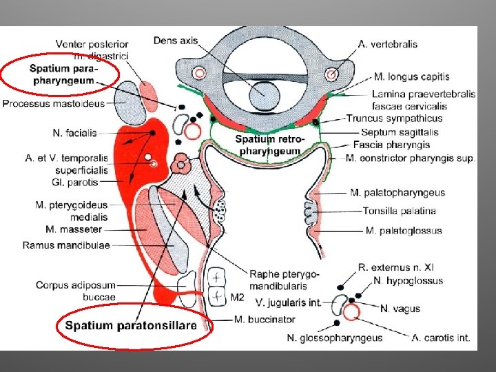
 Epipharynx Spatium parapharyngeum A. carotis interna. Tonsillectomia!!")


- Slides: 35
The pharynx and parapharyngeal spaces Viktória Vereczki M. D. , Ph. D.
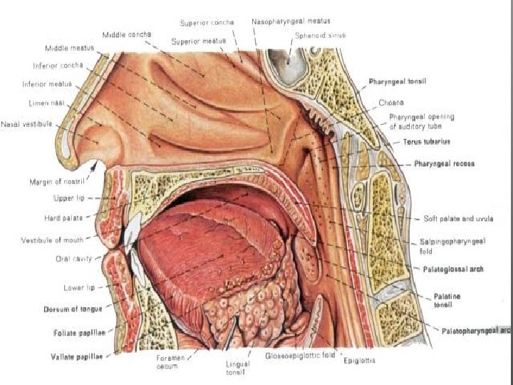
Pharynx choana Faucial isthmus C 6 Laryngeal inlet esophagus
Waldeyer’s lymphatic ring:
Blood supply of palatine tonsill:
Muscles of pharynx Raphe pharyngis Musculi constrictores pharyngis n. IX n. X Musculi levatores pharyngis –
Musculi constrictores pharyngis 1.
Musculi constrictores pharyngis 2. m. constrictor pharyngis sup. p. pterygopharyngea p. buccopharyngea p. mylopharyngea p. glossopharyngea
Musculi constrictores pharyngis 3. m. styloglossus m. stylopharyngeus m. constrictor pharyngis medius: p. chondropharyngea p. ceratopharyngea m. constrictor pharyngis inferior m. constrictor pharyngis inf.
Musculi levatores pharyngis M. stylopharyngeus – proc. styloideus (os temporale) M. salpingopharyngeus – cartilage of auditory tube, proc. pterygoideus lamina medialisa M. palatopharyngeus – velum palatinum
Arteries - a. vertebralis -ACI – -ACE: Au – a. auricularis post Oc – a. occipitalis Sty. M – a. stylomastoidea Te – a. temporalis superfic. Ma – a. maxillaris Fa – a. facialis a. Pha – a. pharyngea asc. Ly – a. lingualis Thy – a. thyroid sup.
Blood and nerve supply of the pharynx aa. pharyngea asc. Rami pharyngeales: aa. thyroidea sup. aa. thyroidea inf. vv. pharyngeales Plexus pharyngeus: Nervus glossopharyngeus (N. IX) Nervus vagus (N. X)
The topography of the pharynx
Pharyngeal muscles, posterior view
Connetive tissue spacesnaround the pharynx
Cervical organs in front of the pharynx
Cervical lymph nodes Superficial cervical lymph nodes along external jugular vein. Deep cervical lymph nodes near carotid sheet. Cervical lymph vessels reach the nodes along the blood vessels. Lymph vessels form the jugular trunk. Importance of retropharyngeal space: Between pretracheal and prevertebral layers of cervical fascia, communicates with mediastinum. Inflammation can spread progressively to cause mediastinitis.
1. Fa sci a c erv ica lis lam ina su pe rfic ial is 2. Fascia cervicalis lamina visceralis 3. F. cerv. lamina prevertebralis
SPATIUM PARAPHARYNGEUM (side view) Epipharynx Spatium parapharyngeum A. carotis interna. Tonsillectomia!!
Sp. retropharyngeale • behind pharynx and oesophagus • In front of fascia alaris (danger space) • From base of skull untill T 1 -T 2
Sagital plane
Danger space • Anterior border: fascia alaris Posterior border fascia prevertebralis • Between the base of rthe skull and the diaphragma
Sp. prevertebrale • • art. vertebralis, plexus brachialis, n. phrenicus Scalenus and prevertebral muscles, vertebrae
Pharnx Esophagus Danger space Mediastinum posterius
CT, MRI http: //rad. usuhs. mil/medpix_home. html#top
Cross section of the final exam: C 1 level A. carotis interna. Tonsillectomia!!
Cross section of the final exam: C 3 level
Cross section of the final exam: C 4 level
Cross section of the final exam: C 5 level
Cross section of the final exam: C 5 -6 level
Cross section of the final exam: C 6 -7 level
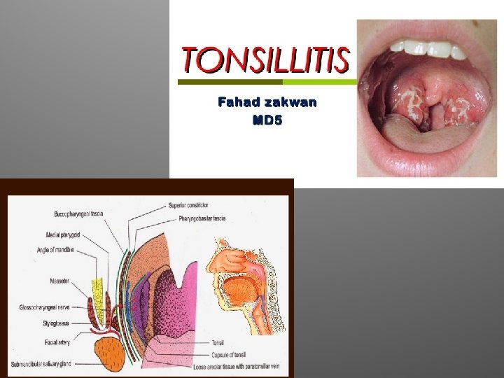
Pharyngitis <>
Radiographic picture of the esophagus