
The Peritoneum Omentum Mesentery and Retroperitoneal Space M



- Slides: 69
The Peritoneum , Omentum, Mesentery , and Retroperitoneal Space M. A. Kubtan 2
Learning Objectives To recognise and understand Ø The clinical features of localised and generalised peritonitis Ø The common causes and complications of peritonitis Ø The principles of surgical management in patients with peritonitis Ø The clinical presentations and treatment of abdominal /pelvic abscesses Ø The clinical presentations of tuberculous peritonitis Ø The causes and pathophysiology of ascites Ø The spectrum of mesenteric and retroperitoneal conditions M. A. Kubtan 3
Peritoneum Development The peritoneum develops ultimately from the mesoderm of the trilaminar embryo. As the mesoderm differentiates, one region known as the lateral plate mesoderm splits to form two layers separated by an intraembryonic coelom. These two layers develop later into the visceral and parietal layers found in all serous cavities, including the peritoneum. As an embryo develops, the various abdominal organs grow into the abdominal cavity from structures in the abdominal wall. In this process they become enveloped in a layer of peritoneum. The growing organs "take their blood vessels with them" from the abdominal wall, and these blood vessels become covered by peritoneum, forming a mesentery. M. A. Kubtan 4
Peritoneum In higher vertebrates, the peritoneum is the serous membrane that forms the lining of the abdominal cavity - it covers most of the intra-abdominal organs. (The corresponding serous membranes in the pleural and pericardial cavities of the thorax are called the pleura and the pericardium respectively. ) The peritoneum both supports the abdominal organs and serves as a conduit for their blood and lymph vessels and nerves. M. A. Kubtan 5
The peritoneum consists of two layers and a potential space between them: 1. The outer layer, called the parietal peritoneum, is attached to the abdominal wall. 2. The inner layer, the visceral peritoneum, is wrapped around the internal organs that are located inside the abdominal cavity. 3. The potential space between these two layers is the peritoneal cavity; it is filled with a small amount (about 50 ml) of slippery serous fluid that allows the two layers to slide freely over each other. M. A. Kubtan 6
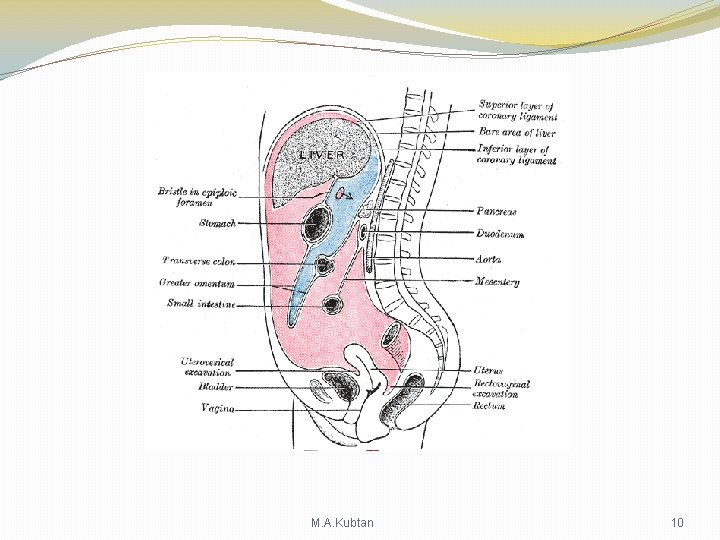
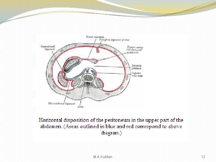
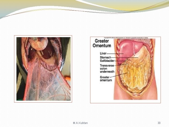
Peritoneum Structures There are two main regions of the peritoneum, connected by the epiploic foramen: • the greater sac (or general cavity of the abdomen), represented in red in the diagrams below. • the lesser sac (or omental bursa), represented in blue. The lesser sac is divided into two "omenta": • The lesser omentum (or gastro hepatic) is attached to the lesser curvature of the stomach and the liver. • The greater omentum (or gastro colic) hangs from the greater curve of the stomach and loops down in front of the intestines before curving back upwards to attach to the transverse colon. In effect it is draped in front of the intestines like an apron and may serve as an insulating or protective layer. M. A. Kubtan 7
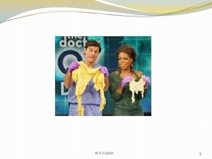
Omentum Definition: The omentum is a large fatty structure which literally hangs off the transevers colon and drapes over the intestines inside the abdomen. but it does reach every organ in the abdomen, draping over and attaching itself to areas of inflammation. It is called the Abdominal Scavenger so, as part of its function, it may act as a bandage in case of bad infection or intestinal rupture (such as appendicitis), limiting spread of infection. M. A. Kubtan 8
M. A. Kubtan 9
M. A. Kubtan 10
The mesentery is the part of the peritoneum through which most abdominal organs are attached to the abdominal wall and supplied with blood and lymph vessels and nerves. M. A. Kubtan 11
M. A. Kubtan 12
Functions of the peritoneum Ø Pain perception (parietal peritoneum) Ø Visceral lubrication Ø Fluid and particulate absorption Ø Inflammatory and immune responses Ø Fibrinolytic activity M. A. Kubtan 13
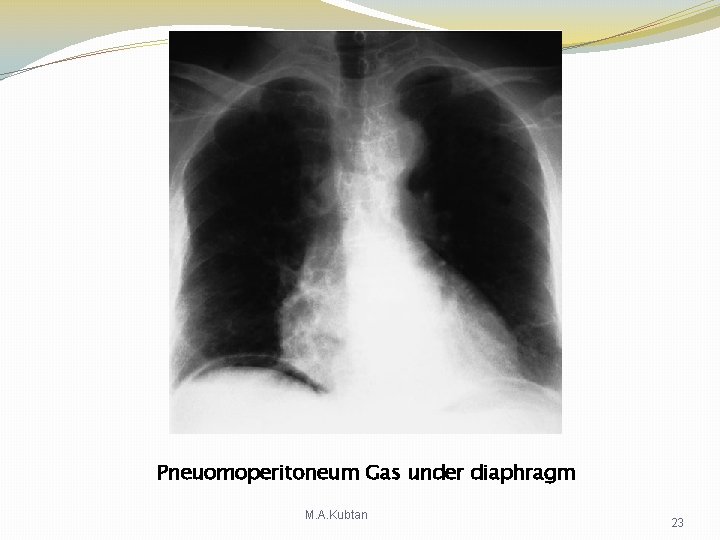
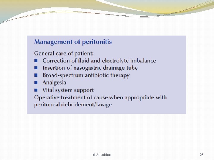
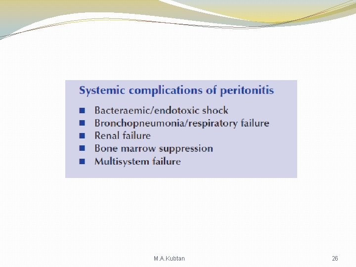
Disease states • Pneumoperitoneum is the presence of gas within the peritoneal cavity, as may occur when a perforation forms in the stomach or intestines, and heralds a perilous situation. • Peritonitis refers to inflammation of the peritoneal lining or cavity, as may occur with either a perforation or by spread of infection through the wall of one of the abdominal organs. This too is a serious condition, and often requires emergency surgery. • Ascites is an accumulation of excess fluid within the peritoneal cavity. M. A. Kubtan 14
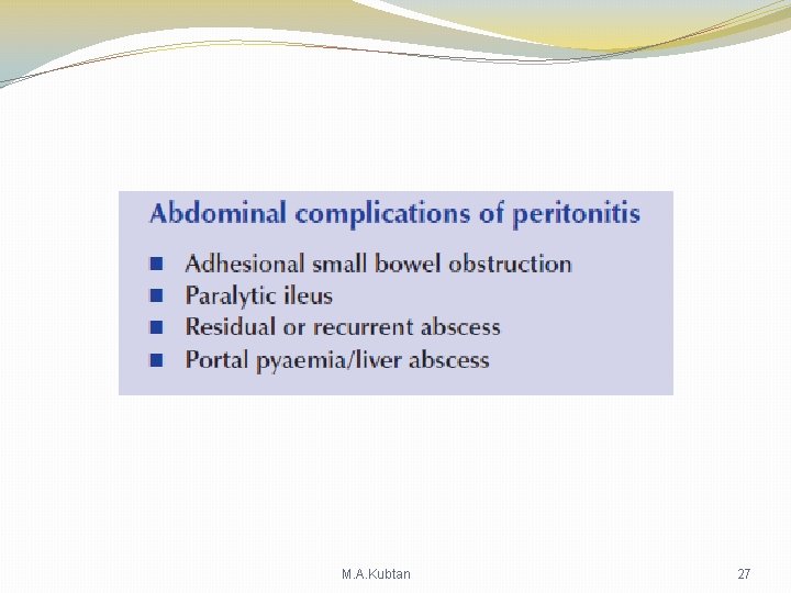
Causes of a peritoneal inflammatory exudate ØBacterial infection, e. g. appendicitis, tuberculosis Ø Chemical injury, e. g. bile peritonitis Ø Ischaemic injury, e. g. strangulated bowel, vascular occlusion Ø Direct trauma, e. g. operation Ø Allergic reaction, e. g. starch peritonitis M. A. Kubtan 15
Bacteria in peritonitis Gastrointestinal source � Escherichia coli � Streptococci (aerobic and anaerobic) �Bacteroides � Clostridium � Klebsiella pneumoniae � Staphylococcus Other sources � Chlamydia � Gonococcus � b-Haemolytic streptococci � Pneumococcus � Mycobacterium tuberculosis M. A. Kubtan 16
Paths to peritoneal infection Ø Gastrointestinal perforation, e. g. perforated ulcer, diverticular perforation Ø Exogenous contamination, e. g. drains, open surgery, trauma Ø Transmural bacterial translocation (no perforation), e. g. inflammatory bowel disease, appendicitis, ischaemic bowel Ø Female genital tract infection, e. g. pelvic inflammatory disease Ø Haematogenous spread (rare), e. g. septicaemia M. A. Kubtan 17
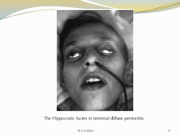
Clinical Features in Peritonitis Ø Abdominal pain , worse on movement Ø Guarding / rigidity of abdominal wall Ø Pain / tenderness on rectal / vaginal examination ( in case of pelvic peritonitis Ø Pyrexia ( may be absent ) Ø Raised pulse rate Ø Absent or reduced bowel sounds Ø Septic shock : systemic inflammatory response syndrome (SIRS) in later stages M. A. Kubtan 18
Acute Peritonitis Bacteriology Ø Bacteria within the lumen of the gastrointestinal tract is normally low until the distal small bowel is reached. Ø High concentration are found in the colon. Ø Intestinal obstruction , achlorohydria , diverticula of small intestine , may increase proxymal colonisation. Ø The biliary and pancreatic tracts are normally free from bacteria , it may be infected in disease ( GS). Ø Peritoneal infection usually caused by two or more bacterial strains. Ø Gram negative bacteria produce endotoxins( Lipopolysaccarides) in their cell walls that have multiple toxic effect , releasing tumour necrosis factor (TNF ) from host leucocytes. Ø Absorption of endotoxin may produce endotoxic shock with hypotension and impaired tissue perfusion. Ø Clostridia Welchii produce harmful exotoxins. Ø Bactroides Gramnegative none-sporing organisms. M. A. Kubtan 19
Non-Gastrointestinal causes of Peritonitis Ø Pelvic infection via fallopian tubes is responsiple for high proportion of non-gastrointestinal infections. Ø Immunodeficient ( HIV , Immunosuppressive treatment may present with opportunistic peritoneal infection. M. A. Kubtan 20
M. A. Kubtan 21
M. A. Kubtan 22
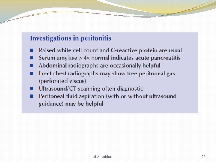
Pneuomoperitoneum Gas under diaphragm M. A. Kubtan 23
M. A. Kubtan 24
M. A. Kubtan 25
M. A. Kubtan 26
M. A. Kubtan 27
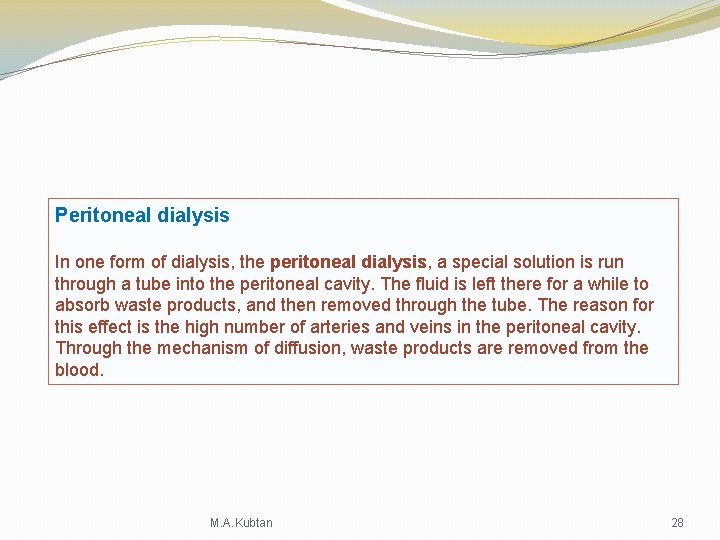
Peritoneal dialysis In one form of dialysis, the peritoneal dialysis, a special solution is run through a tube into the peritoneal cavity. The fluid is left there for a while to absorb waste products, and then removed through the tube. The reason for this effect is the high number of arteries and veins in the peritoneal cavity. Through the mechanism of diffusion, waste products are removed from the blood. M. A. Kubtan 28
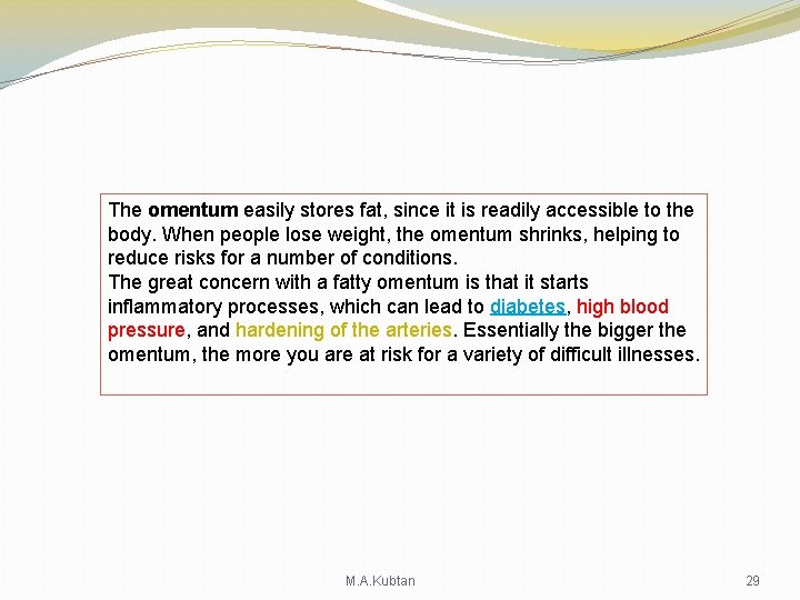
The omentum easily stores fat, since it is readily accessible to the body. When people lose weight, the omentum shrinks, helping to reduce risks for a number of conditions. The great concern with a fatty omentum is that it starts inflammatory processes, which can lead to diabetes, high blood pressure, and hardening of the arteries. Essentially the bigger the omentum, the more you are at risk for a variety of difficult illnesses. M. A. Kubtan 29
M. A. Kubtan 30
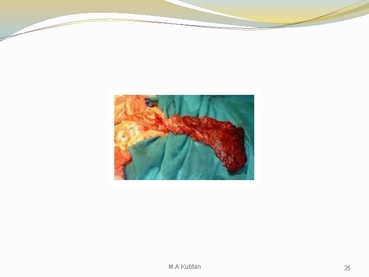
Torsion of The Omentum Definition : Torsion of the Greater Omentum is defined as a twist of the organ in it, s longitudinal axix around pedicle. It may be primary or secondary M. A. Kubtan 31
Aetiology The Primary torsion Ø Idiopathic torsion redundant , mobile segment of omentum rotates around a proximal fixed point in the absence of any associated intra abdominal pathology. It is relatively rare condition ( 200 cases reported in the literature ). Ø Factors predispose to torsion : Anatomical abnormalities of the omentum such as accessory omentum , bifid omentum , narrowed omental pedicle , anatomical arrangement of omental blood vessels ( Veins are larger , longer , and more tortuos than arteries ). Ø Incidence of torsion : on the right side of omentum is higher related to the greater size and mobility of the right side of the omentum. Ø Factors precipitating torsion : blunt trauma to the abdomen , coughing , straining , heavy exertion , sudden change in body position , hyperperistalsis in bowels M. A. Kubtan 32
Aetiology Secondary torsion More common than primary type. Associated with pre-existing abdominal pathology. Omentum usually twisted between two fixed points. Distal edge attached directly or by adhesions to intra abdominal lesion ( Cysts , Tumours , Foci of intra abdominal inflammation , Post surgical wound or scarring , Internal hernia , External hernia. ) M. A. Kubtan 33
Pathology Ø The omentum twists a number of times around a pivotal point , usually clockwise direction. Ø The venous return is compromised and distal omentum becomes congested and oedematous. Ø Haemorrhagic extravasation stimulate aseptic peritonitis with accumulation of serosanguinous fluid in the peritoneal cavity. Ø As the torsion proceeds , arterial occlusion leads to acute haemorrhagic infarction and eventual necrosis of the omental segment. Ø If the infarcted segment is not excised it becomes atrophic and fibrotic , on rare occasions it may even auto – amputated. M. A. Kubtan 34
M. A. Kubtan 35
Clinical Features 1. Occurs in the fourth or fifth decades of life. 2. Men are more affected twice as often as women. 3. The majority of patients are overweight. 4. The signs and symptoms reflected the underlying pathology. 5. The affected segment is usually on the right. M. A. Kubtan 36
Clinical Symptoms 1. 2. 3. Sudden onset of pain. Rebound tenderness. Guarding right sided often mistaken for acute appendicitis , or acute cholecystitis or twisted ovarian cyst. The clinical symptoms are not usually sufficient to allow an accurate pre operative diagnosis. The clinical finding warrant laparotomy even in the absence of definitive diagnosis. The finding of serosanguinous fluid in association with normal appendix , gallbladder , pelvic organs , and bowel should alert the surgeon to the possiblity of omental torsion. M. A. Kubtan 37
Treatment Ø This condition only becomes apparent once vascular thrombosis of omental vessels‘ has occurred and is irreversible even if the omentum is derotated. Ø Treatment consists resection of the affected portion of omentum. Ø Any associated disease with torsion should be dealt with. Ø Post operative recovery is usually rapid and morbidity is minimal. M. A. Kubtan 38
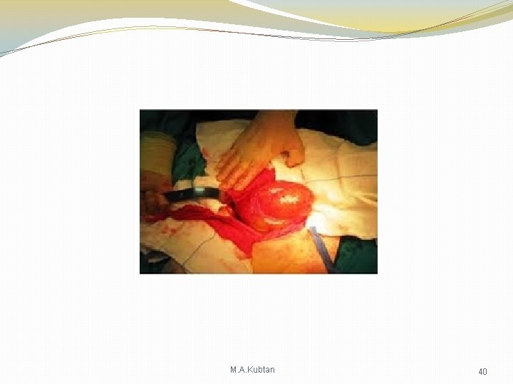
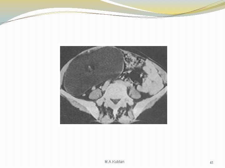
Cysts of The Omentum Pathology of Omental cysts Most cysts are of lymphatic or mesothelial origin , all are rare. Cystic Lymphangioma : In childhood , usually caused by development abnormalities of lymphoid tissue , such as obstruction of lymphatic channels or by growth of congenitally misplaced lymphatic tissue. They are variously called chylous cysts , cystic hygromas , cystic lymphangiomas , and are benign. They vary greatly in size many centimeters , can be unilocular or multilocular. Histologically the cysts contain foamy Macrophages , giving the fluid a milky appearance , and each cyst has endothelial lining. Cystic mesothelioma : Occur almost in adult life , usually in women under the age of 50 years , benign , recurrence after excision is expected. Dermoid Cysts and Pseudocyst. M. A. Kubtan 39
M. A. Kubtan 40
M. A. Kubtan 41
Clinical features and Treatment Cysts may be small and asymptomatic and may be discovered incidentally during surgery or CT scan or Ecko of the abdomen. Large cysts may present with diffuse abdominal distension , or smooth , mobile , palpable mass in lower midline. They are non-tender unless complicated by torsion. Clinical examination , plain abdomen xray , abdominal ecko , CT scan and multislices CT can be helpful in making the diagnosis. Differential diagnosis include mesenteric , peritoneal , retroperitoneal cysts and abdominal tumours. Treatment consist of surgical excision. M. A. Kubtan 42
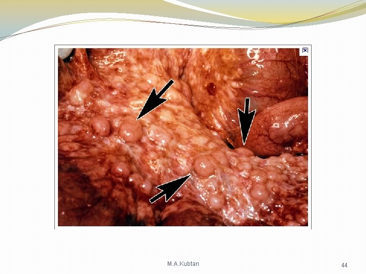
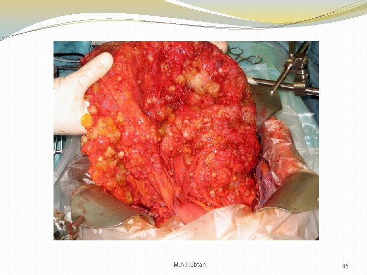
Solid tumours of the Omentum Pathology : Secondary tumour , Primary tumour Clinical features and treatment M. A. Kubtan 43
M. A. Kubtan 44
M. A. Kubtan 45
M. A. Kubtan 46
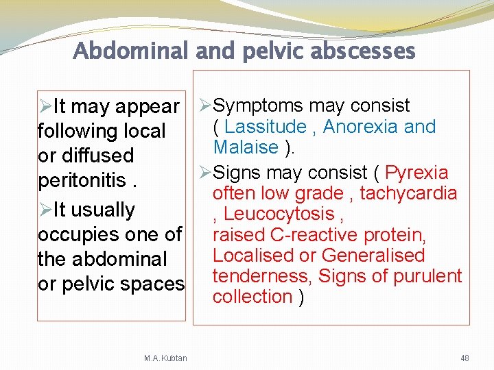
Abdominal and pelvic abscesses ØIt may appear ØSymptoms may consist ( Lassitude , Anorexia and following local Malaise ). or diffused ØSigns may consist ( Pyrexia peritonitis. often low grade , tachycardia ØIt usually , Leucocytosis , occupies one of raised C-reactive protein, Localised or Generalised the abdominal or pelvic spaces tenderness, Signs of purulent collection ) M. A. Kubtan 48
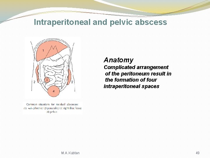
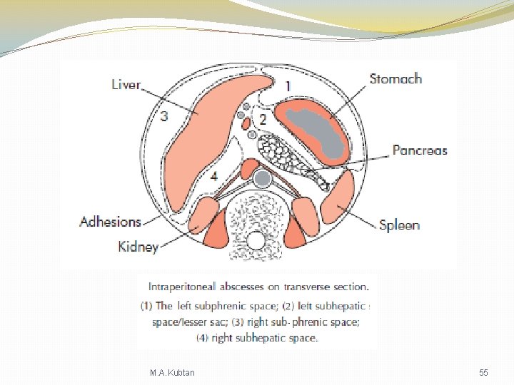
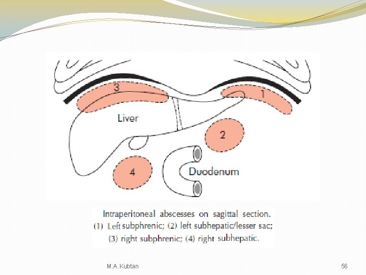
Intraperitoneal and pelvic abscess Anatomy Complicated arrangement of the peritoneum result in the formation of four intraperitoneal spaces M. A. Kubtan 49
Pelvic Abscess The pelvis is the commonest site of intraperitoneal abscess because : Ø Appendix is often in pelvic position. Ø Fallopian tubes are frequent sites of infection. Ø Because of dependency pelvic abscess can occur as a sequel to any case of diffuse peritonitis. Ø Common after anastomotic leakage following colorectal surgery. M. A. Kubtan 50
Related Symptoms & Signs Symptoms : Signs : Ø Diarrhoea. Ø Rectal examination reveals a bulging of the Ø Passage of mucus in the anterior rectal wall stool M. A. Kubtan 51
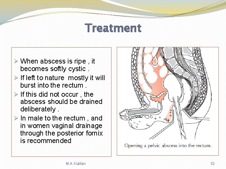
Treatment Ø When abscess is ripe , it becomes softly cystic. Ø If left to nature mostly it will burst into the rectum. Ø If this did not occur , the abscess should be drained deliberately. Ø In male to the rectum , and in women vaginal drainage through the posterior fornix is recommended M. A. Kubtan 52
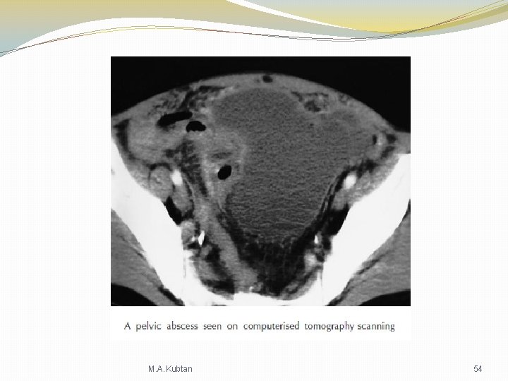
The diagnosis of pelvic abscess can be confirmed by Ultrasound or CT scanning and pus may be aspirated by needle through the rectum or vagina under ultrasound or CT guidance M. A. Kubtan 53
M. A. Kubtan 54
M. A. Kubtan 55
M. A. Kubtan 56
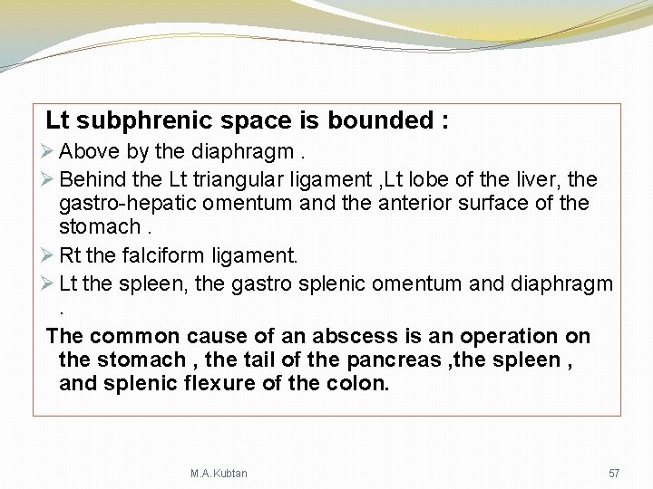
Lt subphrenic space is bounded : Ø Above by the diaphragm. Ø Behind the Lt triangular ligament , Lt lobe of the liver, the gastro-hepatic omentum and the anterior surface of the stomach. Ø Rt the falciform ligament. Ø Lt the spleen, the gastro splenic omentum and diaphragm . The common cause of an abscess is an operation on the stomach , the tail of the pancreas , the spleen , and splenic flexure of the colon. M. A. Kubtan 57
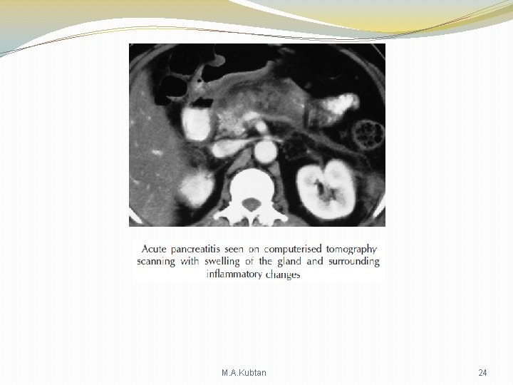
Lt subhepatic space/lesser sac : The commonest cause of infection is complicated acute pancreatitis , rarely perforated gastric ulcer. M. A. Kubtan 58
Rt subphrenic space : Lies between the Rt lobe of the liver and the diaphragm , it is limited posteriorly by the anterior layer of the coronary and the Rt triangular ligaments , to the Lt the falciform ligament. Common causes of abscess : perforated gall bladder, perforated duodenal ulcer , duodenal cap blow-out following gastrectomy , and appendisctis. M. A. Kubtan 59
Rt subhepatic space : Lies transversly beneath the Rt lobe of the liver ( Rutherford Morison, s pouch ). Ø Bounded above by the Rt lobe of the liver Ø It is bounded on the right by Rt lobe of the liver and the diaphragm. Ø To the left is the foramen of Winslow. Ø Below lies the duodenum , the transvers colon , and hepatic flexure. Ø In front are the liver and GB. Ø Behind are the upper part of Rt kidney and the diaphragm. The commonest cause of abscess : appendicitis , cholecystitis , perforated duodenal ulcer , upper abdominal surgery. M. A. Kubtan 60
Clinical features Symptoms and signs of subphrenic infection are frequently non-specific. It is well to remember the aphorism ( pus some where , pus nowhere else , pus under the diaphragm. M. A. Kubtan 61
Symptoms Common history Ø Infective focus in the abdominal cavity has been dealt with. Ø Patient improved temporarily. Ø After few days , weeks , the condition of the patient steadily , often rapidly deteriorates. Ø Sweating , wasting , anorexia , are present. Ø Some times epigastric fullness , and pain. Ø Shoulder pain on the affected side. Ø Persistent hiccoughs may be present. M. A. Kubtan 62
Signs Ø Swinging pyrexia is usually present . Ø Abdominal examination may reveal tenderness , rigidity , some times palpable swelling. Ø Some times liver is displaced downwards or fixed due to adhesions. Ø Chest examination > collapse of lung , or basal effusion , even empyema of the lung. M. A. Kubtan 63
Investigations Ø Blood tests >> Leucocytosis and raised C-reactive protein Ø A plain radiograph may demonstrates the presence of gas or pleural effusion. Ø On screening the diaphragm is seen to be elevated ( Tented diaphragm ) , and its movements impairede. Ø Ultrasound or CT scanning is the investigation of choice and permits early detection of subphrenic collection. Ø Radiolabelled white cell scanning may prove helpful. M. A. Kubtan 64
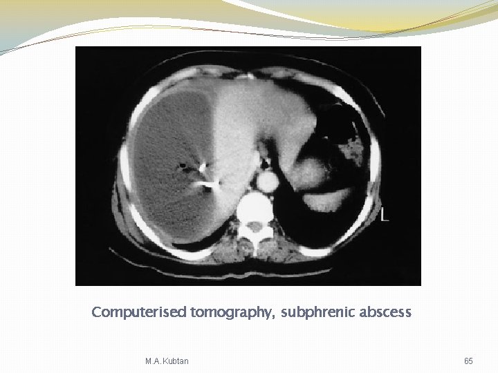
Computerised tomography, subphrenic abscess M. A. Kubtan 65
Differential diagnosis ØPyelonephritis. ØAmoebic abscess. ØPulmonary collapse. ØPleural empyema. M. A. Kubtan 66
Treatment Ø The aim is to evacuate the abscess cavity and drain it. Ø If Skilled help is available , it is possible to insert a percutaneous drainage tube under ultrasound or CT control. making sure that you would not spread the infection. Ø Operative intervention. M. A. Kubtan 67
The Retroperitoneal Space Ø Retroperitoneal hematoma , Ø Retroperitoneal abscess. Ø Retroperitoneal cyst ( unilocular or multi locular , congenital from wolffiian duct remenant) . Ø Idiopathic retroperitoneal fibrosis. M. A. Kubtan 68
Malignant retroperitoneal tumour Ø Lymphoma. Ø Sarcoma. Ø Carcinoid tumours. Ø Secondary deposits ( stomach , colon , breast , and prostate , kidneys and ureters , nervous system ). M. A. Kubtan 69