The Peripheral Nervous System Prepared by I Gede

The Peripheral Nervous System Prepared by I Gede Purnawinadi, S. Kep. , M. Kes.
 • Brain and spinal")
Organization of the Nervous System • Central nervous system (CNS) • Brain and spinal cord • Integration and command center • Peripheral nervous system (PNS) • Paired spinal and cranial nerves • Carries messages to and from the spinal cord and brain

• The PNS includes all neural structures outside the brain and spinal cord, that is, the sensory receptors, peripheral nerves and their associated ganglia, and efferent motor endings. • Sensory receptors are specialized to respond to changes in their environment; such environmental changes are called stimuli. • Sensation (awareness of the stimulus) and perception (interpretation of the meaning of the stimulus) occur in the brain.

Classification by Stimulus Type 1. Mechanoreceptors generate nerve impulses when they, or adjacent tissues, are deformed by a mechanical force such as touch, pressure (including blood pressure), vibration, and stretch. 2. Thermoreceptors are sensitive to temperature changes. 3. Photoreceptors, such as those of the retina of the eye, respond to light energy. 4. Chemoreceptors respond to chemicals in solution (molecules smelled or tasted, or changes in blood or interstitial fluid chemistry). 5. Nociceptors (no″se-sep′torz; noci = harm) respond to potentially damaging stimuli that result in pain. For example, searing heat, extreme cold, excessive pressure, and inflammatory chemicals are all interpreted as painful. These signals stimulate subtypes of thermoreceptors, mechanoreceptors, and chemoreceptors.

Sensory Receptor Locations �Exteroceptors �Stimuli outside the body �Skin and special sense organs �Interoceptors �Stimuli within the body �Chemical messengers, tissue stretch, and temperature �Proprioceptors �Internal stimuli �Monitor position and stretch of joints, tendons, and muscles
 division – transmits impulses from receptors")
Peripheral Nervous System: Afferent Division • Afferent (sensory) division – transmits impulses from receptors to the CNS.
 division – transmits impulses from the")
Peripheral Nervous System: Efferent Division • Motor (efferent) division – transmits impulses from the CNS to effector organs. Two subdivisions: • Somatic nervous system – provides conscious control of skeletal muscles • Autonomic nervous system – regulates smooth muscle, cardiac muscle, and glands

Nerves and Associated Ganglia • Ganglia are collections of neuron cell bodies associated with nerves in the PNS. • Ganglia associated with afferent nerve fibers contain cell bodies of sensory neurons. • Ganglia associated with efferent nerve fibers mostly contain cell bodies of autonomic motor neurons. glands CNS ganglion smooth muscle preganglionic neuron postganglionic neuron cardiac muscle

Cranial Nerves
 of smell, which")
Cranial Nerves I. Olfactory. These are the tiny sensory nerves (filaments) of smell, which run from the nasal mucosa to synapse with the olfactory bulbs. II. Optic. Because this sensory nerve of vision develops as an outgrowth of the brain, it is really a brain tract. III. Oculomotor. This nerve has a name meaning “eye mover” because it supplies four of the six extrinsic muscles that move the eyeball in the orbit. IV. Trochlear. This nerve’s name means “pulley” and it innervates an extrinsic eye muscle that loops through a pulley-shaped ligament in the orbit. V. Trigeminal. Three (tri) branches spring from this, the largest of the cranial nerves. It supplies sensory fibers to the face and motor fibers to the chewing muscles. VI. Abducens. This nerve controls the extrinsic eye muscle that abducts the eyeball (turns it laterally). VII. Facial. A large nerve that innervates muscles of facial expression (among other things). VIII. Vestibulocochlear. This sensory nerve for hearing and balance was formerly called the auditory nerve. IX. Glossopharyngeal. Its name, meaning “tongue and pharynx, ” reveals the structures it helps to innervate. X. Vagus. This nerve’s name means “wanderer” or “vagabond, ” and it is the only cranial nerve to extend beyond the head and neck to the thorax and abdomen. XI. Accessory. Considered an accessory part of the vagus nerve, this nerve was formerly called the spinal accessory nerve. XII. Hypoglossal. This nerve runs inferior to the tongue and innervates some tongue-moving muscles, as reflected by its name meaning “under the tongue. ”
 II OPTIC Vision (sensory) III OCULOMOTOR")
Numbers Cranial Nerve Function I OLFACTORY Smell (sensory) II OPTIC Vision (sensory) III OCULOMOTOR Eye movement (motor) (medial, inferior, superior rectus muscle & inferior oblique muscle) IV TROCHLEAR Eye movements (motor) (superior oblique muscle) V TRIGEMINAL Temperature, pain, crude touch of face (sensory) & mastication (motor) VI ABDUCENS Eye movement (motor) (lateral rectus muscle) VII FACIAL Taste (2/3 of anterior tongue) (sensory) Facial expressions (motor) VIII VESTIBULOCOCHLEAR Hearing & Equilibrium (sensory) IX GLOSSOPHRAYNGEAL Taste (1/3 of posterior tongue) (sensory) Pharynx (swallowing & gag reflex) (motor) X VAGUS Senses blood pressure (sensory) Stimulate heart rate and digestive organs (motor) XI ACCESSORY Head and neck movement (motor) e. g. trapezius, levator scapula XII HYPOGLOSSAL Tongue movement (motor)

Testing Cranial Nerves for Disorders • Olfactory • Facial • Optic • Vestibulocochlear • Oculomotor • Glossopharyngeal • Smell substances • Anosima • Eye chart • Anopsias • Follow object; pupil reflex • Strabismus, double vision, ptosis • Trochlear • See oculomotor • Make various faces; tasting substances • Bell’s palsy, loss of taste, can’t close eye • Tuning fork; distance of sound • Deafness, vertigo, tinnitus • Swallowing & gag reflex; say ‘ah’ • Vagus • See glossopharyngeal • Horseness, swallowing problems, death • Trigeminal • (Spinal) accessory • Abducens • Hypoglossal • Close/move jaws; touch face with objects • See oculomotor • Move head/shoulders against resistance • Stick out, retract, & move tongue to sides

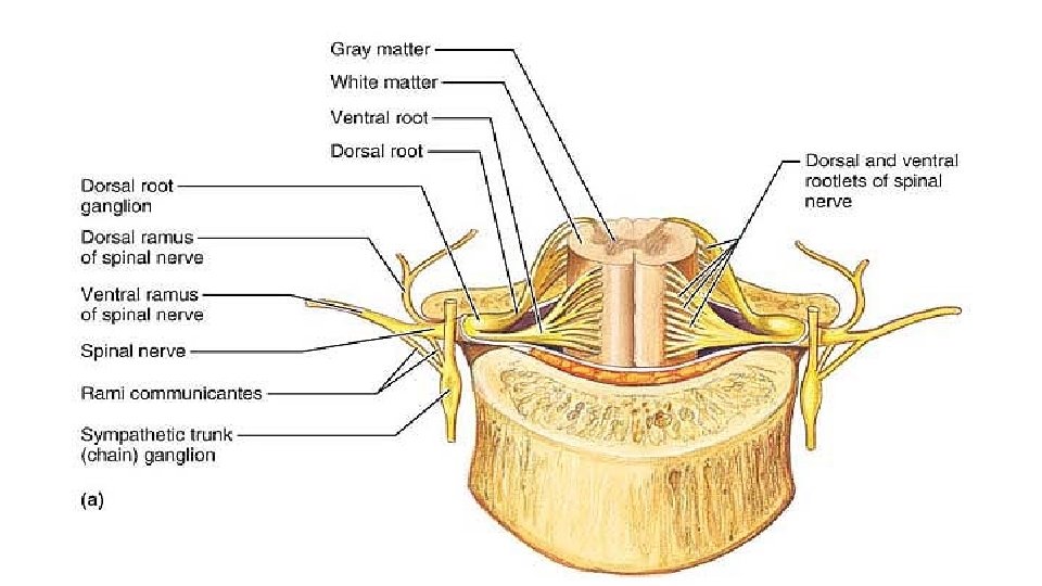
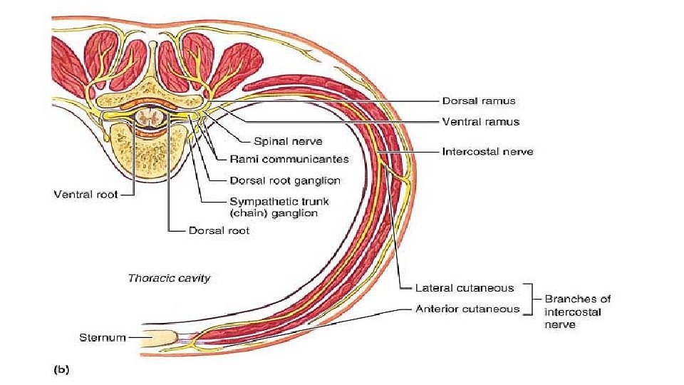
Spinal Nerves • Thirty-one pairs of spinal nerves, each containing thousands of nerve fibers, arise from the spinal cord and supply all parts of the body except the head and some areas of the neck.

ANS Divisions The two arms of the ANS, the parasympathetic and sympathetic divisions: • The parasympathetic division, sometimes called the “resting and digesting” system, keeps body energy use as low as possible, even as it directs vital “housekeeping” activities like digestion and elimination of feces and urine. • The sympathetic division is often referred to as the “fight-or-flight” system. Its activity is evident when we are excited or find ourselves in emergency or threatening situations, such as being frightened by street toughs late at night.

Comparison of the Somatic and Autonomic Nervous Systems
")
Overview of ANS Functional Differences Sympathetic • “Fight or flight” • Catabolic (expend energy) Parasympathetic • “Feed & breed”, “rest & digest” • Homeostasis » Dual innervation of many organs — having a brake and an accelerator provides more control
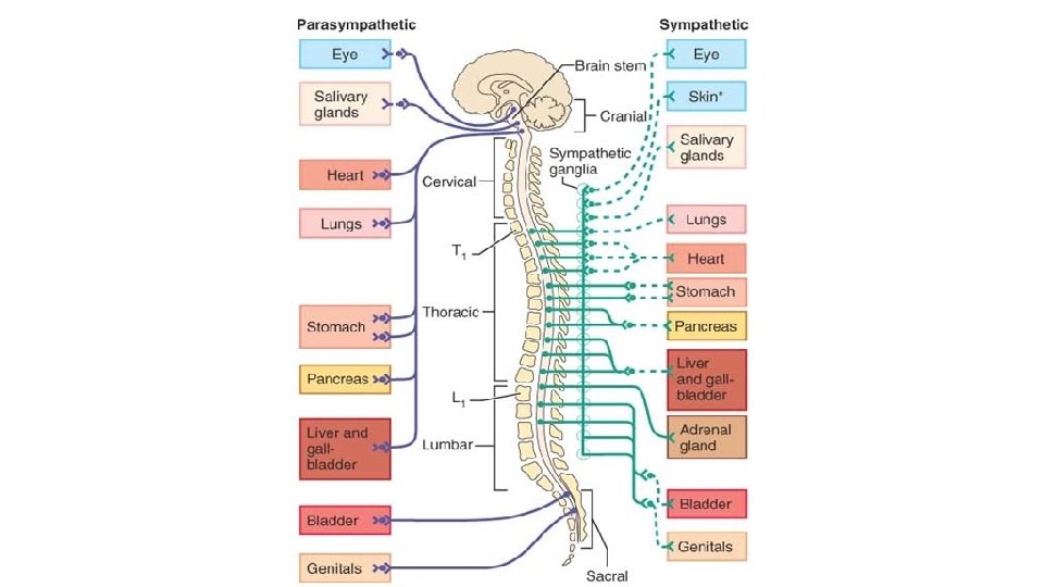

Overview of the Autonomic Nervous System Differences between Sympathetic & Parasympathetic Location of Preganglionic Cell Bodies Sympathetic Parasympathetic Thoracolumbar Craniosacral T 1 – L 2/L 3 levels of the spinal cord Brain: CN III, VII, IX, X Spinal cord: S 2 – S 4

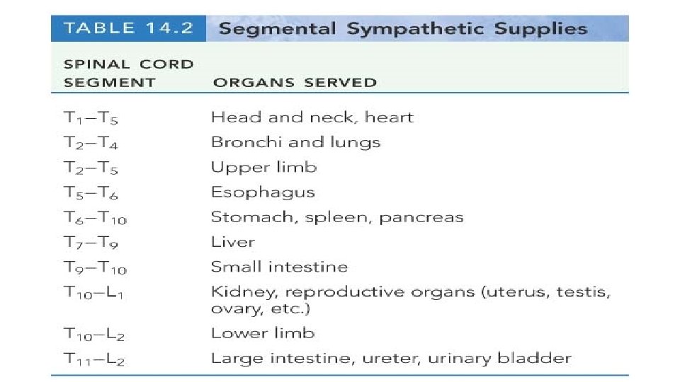
Sympathetic System: Preganglionic Cell Bodies • Preganglionic cell bodies in intermediolateral gray • T 1 – L 2/L 3 • Somatotopic organization somatic tissues (body wall, limbs) visceral tissues (organs) intermediolateral gray columns T 1 – L 2/L 3 lateral horn Clinical Relevance » dysfunction due to cord injury » spinal nerve impingement & OMM » referred pain Moore’s COA 6 2010

Sympathetic System: Postganglionic Cell Bodies 1. Paravertebral ganglia • Located along sides of vertebrae • United by preganglionics into Sympathetic Trunk • Preganglionic neurons are thoracolumbar (T 1–L 2/L 3) but postganglionic neurons are cervical to coccyx • Some preganglionics ascend or descend in trunk Paravertebral ganglia sympathetic trunk (chain) synapse at same level Prevertebral ganglia • celiac ganglion • sup. mesent. g. • inf. mesent. g. ascend to synapse at higher level descend to synapse at lower level aorta Moore’s COA 6 2010
 ganglia • Located anterior to abdominal")
Sympathetic System: Postganglionic Cell Bodies 2. Prevertebral (preaortic) ganglia • Located anterior to abdominal aorta, in plexuses surrounding its major branches • Preganglionics reach prevertebral ganglia via abdominopelvic splanchnic nerves Paravertebral ganglia sympathetic trunk (chain) Prevertebral ganglia abdominopelvic splanchnic nerve • celiac ganglion • sup. mesent. g. • inf. mesent. g. aorta Moore’s COA 6 2010
 division of the ANS. Solid lines indicate preganglionic fibers; dashed")
• Sympathetic (thoracolumbar) division of the ANS. Solid lines indicate preganglionic fibers; dashed lines indicate postganglionic fibers. Sympathetic innervation to peripheral structures (blood vessels, glands, and arrector pili muscles) is shown only in the cervical area, but occurs in all areas.

Parasympathetic Pathways Cranial outflow • CN III, VII, IX, X • Four ganglia in head • Vagus nerve (CN X) is major preganglionic parasymp. supply to thorax & abdomen • Synapse in ganglia within wall of the target organs (e. g. , enteric plexus Sacral outflow of GI tract) • S 2–S 4 via pelvic splanchnics • Hindgut, pelvic viscera, and external genitalia Clinical Relevance » Surgery for colorectal cancer puts pelvic splanchnics at risk » Damage causes bladder & sexual dysfunction Moore’s COA 6 2010
 division of the ANS. Solid lines indicate preganglionic nerve fibers.")
• Parasympathetic (craniosacral) division of the ANS. Solid lines indicate preganglionic nerve fibers. Dashed lines indicate postganglionic fibers. Terminal ganglia of the vagus and pelvic splanchnic nerve fibers are not shown; most of these ganglia are located in or on the target organ.
- Slides: 27