The Peripheral Nervous System PNS PART A Peripheral
 PART A")
 PNS – all neural structures outside the brain and spinal")


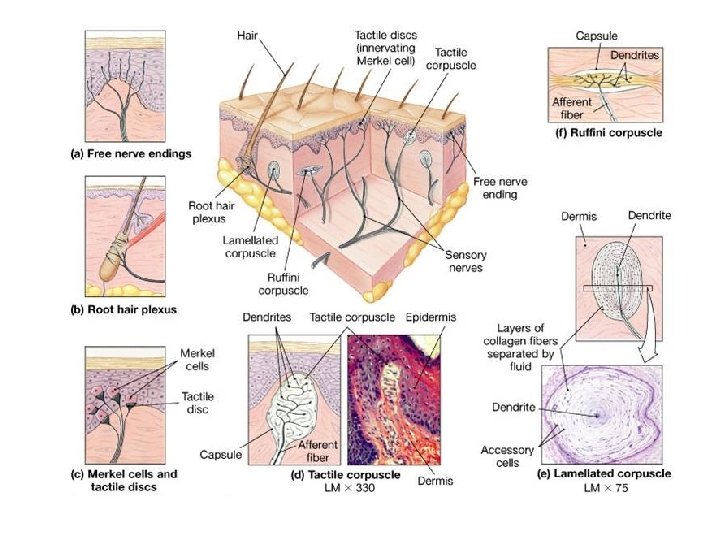
 ¡ Pacinian corpuscles (lamellated corpuscles) ¡ Muscle")


 – carry impulse to the CNS ¡ Motor (efferent)")


, maxillary (V 2), and mandibular")


 PART B")


")


 Reflexes ¡ The flexor reflex happens on the limb receiving the")


- Slides: 111
The Peripheral Nervous System (PNS) PART A
Peripheral Nervous System (PNS) PNS – all neural structures outside the brain and spinal cord ¡ Includes sensory receptors, peripheral nerves, associated ganglia, and motor endings ¡ Provides links to and from the external environment ¡
PNS in the Nervous System Figure 13. 1
Sensory Receptors Structures specialized to respond to stimuli ¡ Activation of sensory receptors results in depolarizations that trigger impulses to the CNS ¡ The realization of these stimuli, sensation and perception, occur in the brain ¡
Receptor Classification by Stimulus Type Mechanoreceptors – respond to touch, pressure, vibration, stretch, and itch ¡ Thermoreceptors – sensitive to changes in temperature ¡ Photoreceptors – respond to light energy (e. g. , retina) ¡ Chemoreceptors – respond to chemicals (e. g. , smell, taste, changes in blood chemistry) ¡ Nociceptors – sensitive to paincausing stimuli ¡
Receptor Class by Location: Exteroceptors Respond to stimuli arising outside the body ¡ Found near the body surface ¡ Sensitive to touch, pressure, pain, and temperature ¡ Include the special sense organs ¡
Receptor Class by Location: Interoceptors Respond to stimuli arising within the body ¡ Found in internal viscera and blood vessels ¡ Sensitive to chemical changes, stretch, and temperature changes ¡
Receptor Class by Location: Proprioceptors Respond to degree of stretch of the organs they occupy ¡ Found in skeletal muscles, tendons, joints, ligaments, and connective tissue coverings of bones and muscles ¡ Constantly “advise” the brain of one’s movements ¡
Receptor Classification by Structural Complexity Receptors are structurally classified as either simple or complex ¡ Most receptors are simple and include encapsulated and unencapsulated varieties ¡ Complex receptors are special sense organs ¡
Simple Receptors: Unencapsulated Free dendritic nerve endings l Respond chiefly to temperature and pain ¡ Merkel (tactile) discs ¡ Hair follicle receptors ¡
Simple Receptors: Encapsulated Meissner’s corpuscles (tactile corpuscles) ¡ Pacinian corpuscles (lamellated corpuscles) ¡ Muscle spindles, Golgi tendon organs, and Ruffini’s corpuscles ¡ Joint kinesthetic receptors ¡
Unencapsulated Receptors Table 13. 1. 1
Simple Receptors: Encapsulated Table 13. 1. 2
From Sensation to Perception Sensation is the awareness of changes in the internal and external environment ¡ Perception is the conscious interpretation of those stimuli ¡
Organization of the Somatosensory System Input comes from exteroceptors, proprioceptors, and interoceptors ¡ The three main levels of neural integration in the somatosensory system are: l Receptor level – the sensor receptors l Circuit level – ascending pathways l Perceptual level – neuronal circuits in the cerebral cortex ¡
Figure 13. 2
Processing at the Receptor Lever The receptor must have specificity for the stimulus energy ¡ The receptor’s receptive field must be stimulated ¡ Transduction l Conversion of the energy of a stimulus into the energy of a nerve signal ¡
Processing at the Receptor Lever Receptor potential l It is a graded potential happening on a receptor ¡ Depolarization or hyperpolarization ¡ Generator potential l It is a receptor potential strong enough to cause an action potential in an afferent fiber ¡
Adaptation of Sensory Receptors ¡ Adaptation is a reduction in sensitivity in the presence of a stimulus l Receptor membranes become less responsive l Receptor potentials decline in frequency or stop
Adaptation of Sensory Receptors ¡ Tonic receptors l Have little peripheral adaptation ¡ Chemical interoceptors ¡ Pain receptors ¡ Macula in the vestibular apparatus ¡ Proprioceptors
Adaptation of Sensory Receptors ¡ Phasic receptors l Are fast adapting receptors ¡ Pressure ¡ Touch ¡ Smell
Processing at the Circuit Level Chains of three neurons that conduct sensory impulses to the cerebral cortex ¡ First-order neurons – soma reside in dorsal root or cranial ganglia, and conduct impulses from the skin to the spinal cord or brain stem ¡
Processing at the Circuit Level Second-order neurons – soma reside in the dorsal horn of the spinal cord or medullary nuclei and transmit impulses to the thalamus or cerebellum ¡ Third-order neurons – located in the thalamus and conduct impulses to the somatosensory cortex of the cerebrum ¡
Processing at the Perceptual Level The thalamus projects fibers to: l The somatosensory cortex l Sensory association areas ¡ The exact point in the cortex that is activated will refer to where in the body the stimulus is happening ¡ The result is an internal, conscious image of the stimulus ¡
Main Aspects of Sensory Perception Perceptual detection – detecting that a stimulus has occurred and requires summation ¡ Magnitude estimation =intensity of the stimulus l Frequency of action potentials ¡
Main Aspects of Sensory Perception ¡ Spatial discrimination – identifies the location of the stimulus. It depends on the size of the receptor field. l Two-point discrimination test – smaller fields equals finer twopoint discrimination test
Main Aspects of Sensory Perception Feature abstraction – used to identify a specific feature of the stimulus (texture or shape) ¡ Quality discrimination – the ability to identify submodalities of a sensation (e. g. , sweet or sour tastes) ¡ Pattern recognition – ability to recognize patterns in stimuli (e. g. , melody, familiar face) ¡
Structure of a Nerve – peripheral axons enclosed by connective tissue ¡ Connective tissue coverings include: l Endoneurium – loose connective tissue that surrounds axons l Perineurium – coarse connective tissue that bundles fibers into fascicles l Epineurium – tough fibrous sheath around a nerve ¡
Structure of a Nerve Figure 13. 3 b
Classification of Nerves Sensory (afferent) – carry impulse to the CNS ¡ Motor (efferent) – carry impulses from CNS ¡ Mixed nerves – carry somatic and autonomic (visceral) impulses l Most common type ¡
Peripheral Nerves The four types of mixed nerves are: l Somatic ¡ Sensory ¡ Motor l Visceral ¡ Sensory ¡ Motor ¡ Peripheral nerves can be cranial or spinal ¡
Regeneration of Nerve Fibers Mature neurons are amitotic ¡ If the soma remains intact, damage can be repaired ¡ Steps l Separated ends seal themselves l Wallerian degeneration of the distal axon by macrophages l Formation of a regeneration tube by the Schwann cell ¡ Guide the axon growth distally ¡
Regeneration of Nerve Fibers Figure 13. 4
Regeneration of Nerve Fibers Figure 13. 4
Cranial Nerves Twelve pairs of cranial nerves arise from the brain ¡ They have sensory, motor, or both sensory and motor (mixed nerves) functions ¡ Each nerve is identified by a number (I through XII) and a name ¡
Cranial Nerves Figure 13. 5 a
Summary of Function of Cranial Nerves Figure 13. 5 b
Cranial Nerve I: Olfactory Arises from the olfactory epithelium ¡ Passes through the cribriform plate of the ethmoid bone ¡ Fibers run through the olfactory bulb and terminate in the primary olfactory cortex ¡ Function is the sense of smell ¡
Cranial Nerve I: Olfactory Figure I from Table 13. 2
Cranial Nerve II: Optic Arises from the retina of the eye ¡ Optic nerves pass through the optic canals and converge at the optic chiasm ¡ They continue to the thalamus where they synapse ¡ From there, the optic radiation fibers run to the visual cortex ¡ Functions carry impulses for vision ¡
Cranial Nerve II: Optic Figure II from Table 13. 2
Cranial Nerve III: Oculomotor Motor for movements of the eyes ¡ Parasympathetic fibers innervate the intrinsic muscles of the eye l Constricting the iris, and controlling lens shape ¡
Cranial Nerve III: Oculomotor Figure III from Table 13. 2
Cranial Nerve IV: Trochlear Figure IV from Table 13. 2
Cranial Nerve V: Trigeminal Three divisions: ophthalmic (V 1), maxillary (V 2), and mandibular (V 3) ¡ Conveys sensory impulses from various areas of the face (V 1) and (V 2), and supplies motor fibers (V 3) for mastication ¡
Cranial Nerve V: Trigeminal Figure V from Table 13. 2
Cranial Nerve VI: Abducens • Primarily a somatic motor nerve Figure VI from Table 13. 2
Cranial Nerve VII: Facial Somatic Motor to the muscles of facial expression, and the transmittal of ¡ Visceral motor to lacrimal and salivary glands ¡ Sensory function is taste from the anterior two-thirds of the tongue ¡
Cranial Nerve VII: Facial Figure VII from Table 13. 2
Cranial Nerve VIII: Vestibulocochlear Fibers arise from the hearing and equilibrium apparatus of the inner ear, ¡ Two divisions – cochlear (hearing) and vestibular (balance) ¡ A sensory nerve ¡
Cranial Nerve VIII: Vestibulocochlear Figure VIII from Table 13. 2
Cranial Nerve IX: Glossopharyngeal Nerve IX is a mixed nerve with motor and sensory functions ¡ Somatic Motor – innervates part of the tongue and pharynx, and ¡ Visceral Motor fibers to the parotid salivary gland ¡ Visceral Sensory –taste and general sensory impulses from the tongue and pharynx ¡
Cranial Nerve IX: Glossopharyngeal Figure IX from Table 13. 2
Cranial Nerve X: Vagus The only cranial nerve that extends beyond the head and neck ¡ The vagus is a mixed nerve ¡ Most visceral motor fibers are parasympathetic fibers to the heart, lungs, and visceral organs ¡ Its visceral sensory function is in taste ¡
Cranial Nerve X: Vagus Figure X from Table 13. 2
Cranial Nerve XI: Accessory ¡ Primarily a somatic motor nerve l Supplies fibers to the larynx, pharynx, and soft palate l Innervates the trapezius and sternocleidomastoid, which move the head and neck
Cranial Nerve XI: Accessory Figure XI from Table 13. 2
Cranial Nerve XII: Hypoglossal ¡ Somatic motor innervates the muscles of the tongue, which contribute to swallowing and speech
Cranial Nerve XII: Hypoglossal Figure XII from Table 13. 2
The Peripheral Nervous System (PNS) PART B
Spinal Nerves Thirty-one pairs of mixed nerves arise from the spinal cord and supply all parts of the body except the head ¡ They are named according to their point of issue l 8 cervical (C 1 -C 8) l 12 thoracic (T 1 -T 12) l 5 Lumbar (L 1 -L 5) l 5 Sacral (S 1 -S 5) l 1 Coccygeal (C 0) ¡
Spinal Nerves Figure 13. 6
Spinal Nerves: Roots Each spinal nerve connects to the spinal cord via two medial roots ¡ Each root forms a series of rootlets that attach to the spinal cord ¡ Ventral roots arise from the anterior horn and contain motor (efferent) fibers ¡ Dorsal roots arise from sensory neurons in the dorsal root ganglion and contain sensory (afferent) fibers ¡
Spinal Nerves: Roots Figure 13. 7 a
Spinal Nerves: Rami ¡ The short spinal nerves branch into three or four mixed, distal rami l Small dorsal ramus l Larger ventral ramus l Rami communicantes at the base of the ventral rami in the thoracic region ¡ visceral nerve fibers
Nerve Plexuses All ventral rami except T 2 -T 12 form interlacing nerve networks called plexuses ¡ Plexuses are found in the cervical, brachial, lumbar, and sacral regions ¡ Each resulting branch of a plexus contains fibers from several spinal nerves ¡
Nerve Plexuses Each muscle receives a nerve supply from more than one spinal nerve ¡ Damage to one spinal segment cannot completely paralyze a muscle ¡
Spinal Nerve Innervation: Back, Anterolateral Thorax, and Abdominal Wall The back is innervated by dorsal rami via several branches ¡ The thorax is innervated by ventral rami T 1 -T 12 as intercostal nerves ¡ Intercostal nerves supply muscles of the ribs, anterolateral thorax, and abdominal wall ¡
Spinal Nerve Innervation: Back, Anterolateral Thorax, and Abdominal Wall Figure 13. 7 b
Cervical Plexus Most branches are cutaneous nerves of the neck, ear, back of head, and shoulders ¡ The most important nerve of this plexus is the phrenic nerve l Motor and sensory nerve of the diaphragm ¡
Cervical Plexus Figure 13. 8
Brachial Plexus It gives rise to the nerves that innervate the upper limb ¡ There are four major branches of this plexus l Roots l Trunks l Divisions l Cords ¡
Brachial Plexus Figure 13. 9 a
Brachial Plexus: Nerves Axillary ¡ Musculocutaneous ¡ Median ¡ Ulnar ¡ Radial ¡
Brachial Plexus: Distribution of Nerves Figure 13. 9 c
Brachial Plexus: Nerves Figure 13. 9 b
Lumbar Plexus Innervates the thigh, abdominal wall, and psoas muscle ¡ The major nerves are the ¡ Femoral l For anterior thigh muscles ¡ Obturator l Adductors muscles ¡
Lumbar Plexus Figure 13. 10
Sacral Plexus Serves the buttock, lower limb, pelvic structures, and the perineum (pudendal nerve) ¡ The major nerve is the sciatic, the longest and thickest nerve of the body l Lower limb (except anteromedial thigh muscles) ¡ Branches into two nerves: the tibial and the common fibular (peroneus) ¡
Sacral Plexus Figure 13. 11
Dermatomes A dermatome is the area of skin innervated by the cutaneous branches of a single spinal nerve ¡ All spinal nerves except C 1 participate in dermatomes ¡
Dermatomes Figure 13. 12
Innervation of Joints ¡ Hilton’s law: any nerve serving a muscle that produces movement at a joint also innervates the joint itself and the skin over the joint
Motor Endings ¡ PNS elements that activate effectors by releasing neurotransmitters at: l Skeletal muscles l Smooth muscle and glands
Levels of Motor Control ¡ The three levels of motor control are l Segmental level ¡ Spinal cord circuit l Projection level ¡ Pyramidal and extrapyramidal systems l Precommand level ¡ Cerebellum and basal nuclei
Hierarchy of Motor Control Figure 13. 13
Segmental Level The segmental level is the lowest level of motor hierarchy ¡ It consists of segmental circuits of the spinal cord ¡ Its circuits control locomotion and specific, oft-repeated motor activity ¡
Projection Level Controls the spinal cord ¡ Consists of: l Cortical motor areas that produce the direct (pyramidal) system l Brain stem motor areas that oversee the indirect (multineuronal) system ¡ Send information to lower motor neurons and also to higher center ¡
Precommand Level ¡ Cerebellar and basal nuclei systems that: l Regulate motor activity l Precisely start or stop movements l Coordinate movements with posture l Block unwanted movements l Monitor muscle tone l Control the output of the cortex and brain stem motor centers
Reflexes A reflex is a rapid, predictable motor response to a stimulus ¡ Reflexes may: l Be inborn (intrinsic) or learned (acquired) l Involve, peripheral nerves, brain stem and spinal cord l Somatic and visceral reflexes ¡
Reflex Arc ¡ There are five components of a reflex arc l Receptor l Sensory neuron l Integration center l Motor neuron l Effector
Reflex Arc Figure 13. 14
Somatic Reflexes Spinal: ¡ Stretch reflex ¡ Golgi tendon reflex ¡ Withdrawal reflex ¡ Crossed-extensor reflex ¡ Superficial: ¡ Plantar l Babinski’s ¡ Abdominal ¡
Stretch and Deep Tendon Reflexes ¡ For skeletal muscles to perform normally: l The Golgi tendon organs (proprioceptors) must constantly inform the brain as to the state of the muscle l Stretch reflexes initiated by muscle spindles must maintain healthy muscle tone
Stretch reflex - monosynaptic Muscle Spindle ¡ Are composed of intrafusal muscle fibers that lack myofilaments in their central regions, are noncontractile, and serve as receptive surfaces ¡ Afferent fibers ¡ Motor fibers: l Extrafusal fibers l Intrafusal fibers ¡
Muscle Spindles Figure 13. 15
Operation of the Muscle Spindles Stretching the muscles activates the muscle spindle l There is an increased rate of action potential on sensory fibers ¡ Contracting the muscle reduces tension on the muscle spindle l There is a decreased rate of action potential on sensory fibers ¡
Operation of the Muscle Spindle Figure 13. 17
Stretch Reflex - monosynaptic Stretching the muscle activates the muscle spindle ¡ Excited motor neurons causes the muscle to contract ¡ Afferent impulses from the spindle result in inhibition of the antagonist ¡ Example: patellar reflex l Tapping the patellar tendon stretches the quadriceps and starts the reflex action l The quadriceps contract and the antagonistic hamstrings relax ¡
Stretch Reflex Figure 13. 16
Golgi Tendon Reflex - polysynaptic The opposite of the stretch reflex ¡ Contracting the muscle activates the Golgi tendon organs ¡ Afferent Golgi tendon neurons are stimulated, neurons inhibit the contracting muscle, and the antagonistic muscle is activated ¡ As a result, the contracting muscle relaxes and the antagonist contracts ¡ It moderates the muscle contraction ¡
Golgi Tendon Reflex Figure 13. 18
Flexor ( Withdrawal) Reflexes ¡ The flexor reflex happens on the limb receiving the painful stimulus l Withdrawal reflex by contraction of the flexor muscles l Reciprocal inhibition of the extensors l Polysynaptic reflex
Crossed Extensor Reflex ¡ The crossed extensor reflex l Happens on the opposite limb l Contraction of the extensor muscles l Relaxation of the flexor muscles l Polysynaptic
Crossed Extensor Reflex + Interneurons + – Afferent fiber + + – Efferent fibers Extensor inhibited Flexor stimulated s xe e l F Flexor inhibited Arm movements Extensor stimulated nds e Ext Key: + Excitatory synapse – Inhibitory synapse Right arm (site of stimulus) Left arm (site of reciprocal activation) 106 Figure 13. 19
Superficial Reflexes Initiated by gentle cutaneous stimulation ¡ Example: l Plantar reflex is initiated by stimulating the lateral aspect of the sole of the foot l The response is downward flexion of the toes ¡
Superficial Reflexes Indirectly tests for proper corticospinal tract functioning l Babinski’s sign: abnormal plantar reflex indicating corticospinal damage where the great toe dorsiflexes and the smaller toes fan laterally l
The Babinski Reflexes Figure 13. 23
Developmental Aspects of the PNS Spinal nerves branch from the developing spinal cord and neural crest cells l Supply motor and sensory function to developing muscles ¡ Cranial nerves innervate muscles of the head ¡
Developmental Aspects of the PNS Distribution and growth of spinal nerves correlate with the segmented body plan ¡ Sensory receptors atrophy with age and muscle tone lessens ¡ Peripheral nerves remain viable throughout life unless subjected to trauma ¡