The Peripheral Nervous System Honors Anatomy Physiology Chapter


 olfactory receptors")


 ¥ ¥")


 fibers muscle ¥ Dorsal ¥ sensory (afferent)fibers")
 ¥ ¥ ¥ 1. 2. 3. ramus = branch supply entire somatic")
")


- Slides: 53
The Peripheral Nervous System Honors Anatomy & Physiology Chapter 13
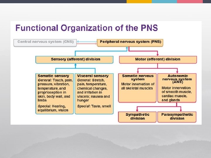
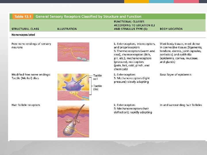
Classification of Sensory Receptors by Stimulus Type 1. Thermoreceptors: ¥ 2. Mechanoreceptors: ¥ 3. respond to light Chemoreceptors: ¥ 5. respond to mechanical force: touch, pressure, vibration, stretch Photoreceptors: ¥ 4. respond to temperature change respond to chemicals in solution Nociceptors: ¥ respond to pain
Pain Receptors ¥ activated 1. 2. by: extremes of pressure & temperature Chemicals ¥ ¥ ¥ histamine K+ ATP acids bradykinin
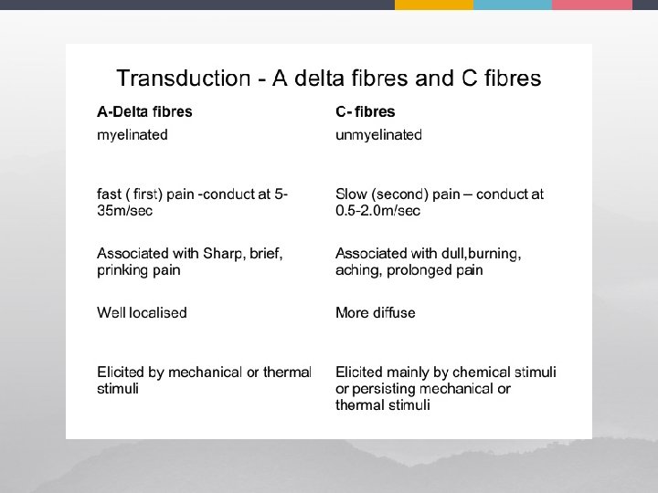
Types of Pain Sharp Pain ¥ myelinated A delta fibers Burning Pain ¥ unmyelinated C fibers
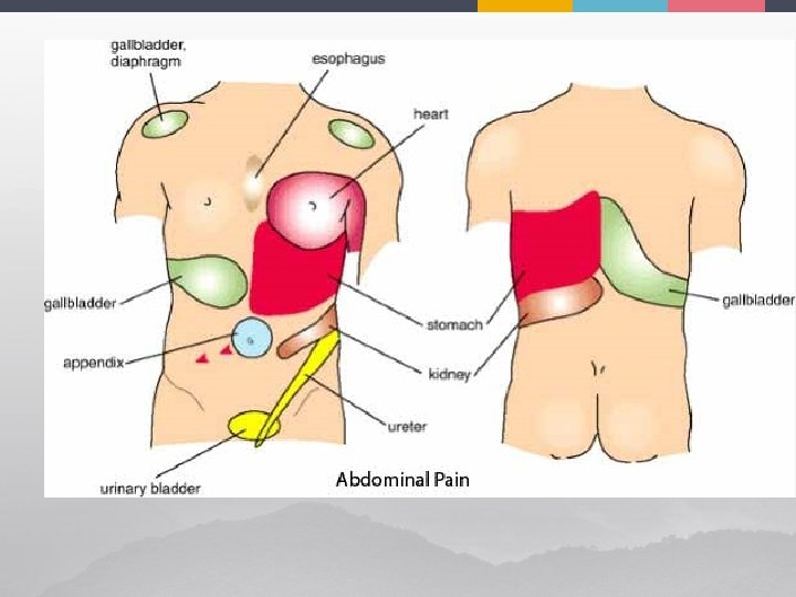
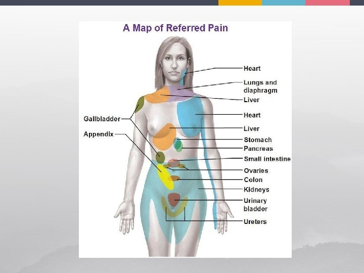
Referred Pain ¥ pain stimuli arising in one part perceived as pain from another part ¥ example: pain from heart attack can be felt as pain in medial aspect of left arm ¥ cause: T 1 – T 5 spinal segments innervate both
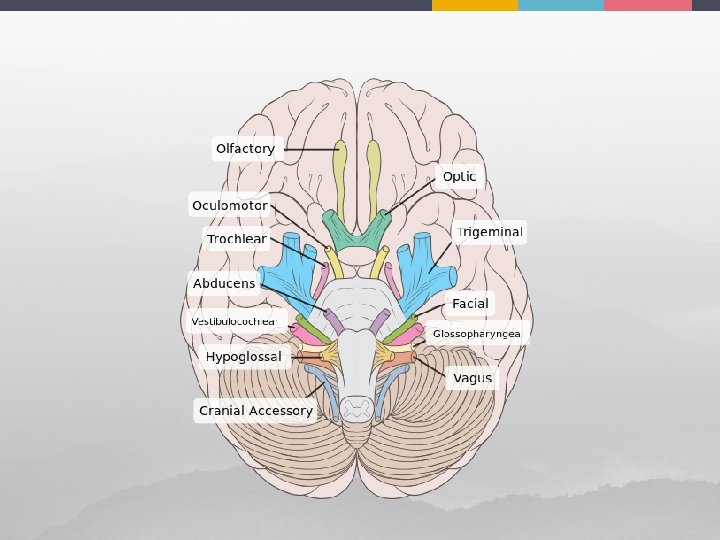
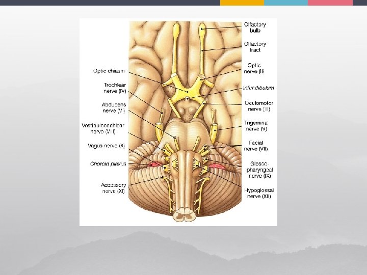
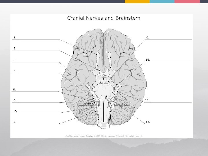
Cranial Nerves ¥ 12 paired on base of brain ¥ name refers to their function ¥ numbered by Roman numerals I and II attach to forebrain ¥ III – XII brain stem ¥
I - Olfactory Nerve ¥ sensory only ¥ nasal mucosa synapse in olfactory bulbs
¥ ¥ ¥ test: have patient smell ammonia damage: anosmia (total loss) olfactory receptors are bipolar neurons ¥ each: single odor-sensitive dendrite
II – Optic Nerve ¥ sensory only
Optic Nerve - II ¥ ¥ test: ¥ vision: eye chart ¥ visual fields: mark chart at point patient first sees an object ¥ view fundus with opthalmoscopeto check for swelling of optic disc (where optic n. leaves eyeball) & examine blood vessels *only place in body can directly visualize vessels damage: II: blindess in affected eye if beyond optic chiasma partial loss
III - Oculomotor ¥ ¥ “eye mover” motor mostly (only sensory proprioceptors) ¥ ¥ somatic 4 of 6 extrinsic eye muscles parasympathetic circular muscles of iris (constriction of pupil) & to ciliary muscle (controls shape of lens for focusing)
III - Oculomotor ¥ ¥ test: examine pupils for size, shape, symmetry damage: ¥ ¥ ¥ eye cannot be moved up, down, or inward; @ rest eye rotates laterally upper eyelid droops (ptosis) patient has double vision &trouble focusing on close objects
IV - Trochlear ¥ ¥ “pulley” motor ¥ ¥ sensory: proprioceptors supplies extrinsic eye muscle that loops through a pulley-shaped ligament (superior oblique muscle)
IV – Trochlear Nerve ¥ test: eye movement down & out ¥ damage: double vision & impairs ability to rotate eye inferolaterally
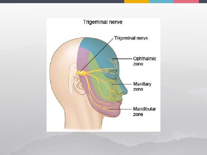
V - Trigeminal ¥ ¥ ¥ 3 branches largest cranial nerve sensory to face/ motor to chewing muscles
V – Trigeminal Nerve test: check blink reflex, touch to side of face, clench teeth, move jaw side to side ¥ damage: trigeminal neuralgia: worst pain known, inflammation of V, ? due to compression by a vessel – treatment: surgery ¥
VI - Abducens ¥ ¥ motor only controls eye muscle that abducts eyeball (lateral rectus)
VI – Abducens Nerve ¥ clinical test: check eye movements ¥ damage: @ rest eyeball rotates medially on affected side (internal strabismus)
VI – Abducens Nerve ¥ normal test ¥ abnormal test
VII - Facial ¥ mixed ¥ ¥ sensory: taste anterior 2/3 of tongue motor: muscles of facial expression; autonomic motor: lacrimal & salivary glands
VII – Facial Nerve ¥ ¥ clinical test: test taste in anterior 2/3 of tongue, check symmetry of face, assess tearing (ammonia) damage: Bell’s palsyparalysis of facial muscles on affected side, +/continuous tearing causing dry eye
Bell’s Palsy ¥? caused by herpes simplex which causes swelling & inflammation of facial nerve ¥ treatment: corticosteroids ¥ clinically: ask patient to smile
VIII - Vestibulocochlear ¥ mostly sensory: hearing & balance
VIII – Vestibulocochlear ¥ clinically: ¥ check hearing by air & bone conduction ¥ damage to vestibular division dizziness, nystagmus, loss of balance, nausea, vomiting ¥ cochlear division central deafness ¥
IX – Glossopharyngeal Nerve ¥ ¥ tongue & pharynx mixed: ¥ ¥ sensory: taste posterior 2/3 tongue, baroreceptors in carotid sinus, chemoreceptors in carotid bodies motor: upper pharynx, autonomic fibers to parotid glands
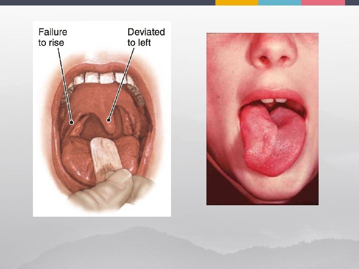
IX - Glossopharyngeal ¥ test: ¥ ¥ check position of uvula: will deviate away from affected side when patient says “ahh” check gag reflex ask patient to speak & cough taste check
X – Vagus Nerve ¥ ¥ *only cranial nerve to extend beyond head & neck Mixed: ¥ ¥ Motor: somatic to muscles of pharynx & larynx/ parasympathetic to heart (HR), lungs (RR), abd viscera (peristalsis) Sensory: from thoracic & abd viscera, chemoreceptors for respiration (carotid & aortic bodies) and taste bud in epiglottis, proprioceptors
X – Vagus Nerve ¥ ¥ ¥ “the wanderer” test: same as for IX damage: hoarseness or loss of voice, dysphagia (difficulty swallowing), impaired digestive motility
XI – Accessory Nerve ¥ ¥ motor ¥ trapezius & sternocleidomastoid ¥ only sensory is proprioception emerges from spinal cord (C 1 – C 5) up thru foramen magnum travels with X
XI – Accessory Nerve ¥ test: injury to 1 side causes head to turn to affected side (sternocleidomastoid); patient has weak shoulder shrug on affected side
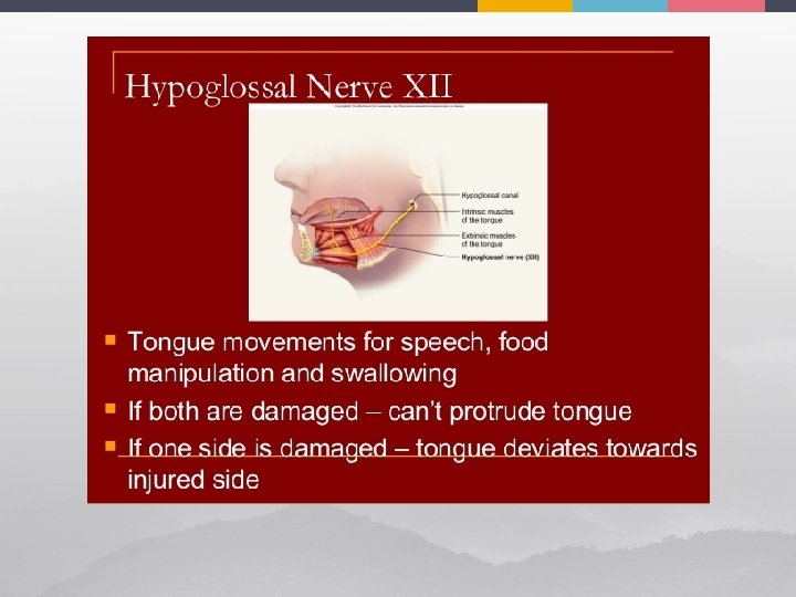
XII – Hypoglossal Nerve ¥ ¥ ¥ “below tongue” mostly motor: tongue: controls movements of tongue that mix & manipulate food when chewing, also contributes to speech & & swallowing test: protrude/ retract tongue, check for deviations
Spinal Nerves ¥ 31 pairs: ¥ ¥ ¥ 8 cevical C 1 – C 8 12 thoracic T 1 – T 12 5 lumbar L 1 – L 5 5 sacral S 1 – S 5 4 coccygeal Co 1 – Co 5
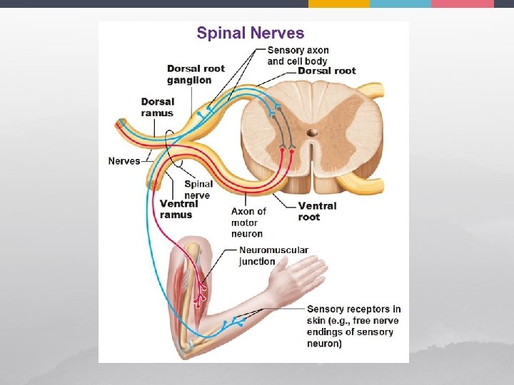
Spinal Roots ¥ Ventral ¥ motor (efferent) fibers muscle ¥ Dorsal ¥ sensory (afferent)fibers sensory receptors both pass laterally from cord & the 2 unite to form a spinal nerve (1 -2 cm) ¥
Rami (Ramus) ¥ ¥ ¥ 1. 2. 3. ramus = branch supply entire somatic region of body (skeletal muscle & skin) spinal nerve divides (1 -2 cm from vertebra) dorsal ramus: posterior body ventral ramus: anterior body + limbs meningeal branch: reenters vertebral canal to innervate the meninges
Nerve Plexuses ¥ ¥ ¥ all ventral rami (except T 2 - T 12) branch & join each other lateral to vertebrae forming complicated nerve networks called: nerve plexuses where fibers crisscross (so each muscle receives innervation from >1 spinal nerve) cervical plexus brachial plexus lumbar plexus sacral plexus
Nerve Plexuses
Reflex Arc