The Pancreatic Hormones and the blood Glucose regulation

The Pancreatic Hormones and the blood Glucose regulation Dr. Waleed R. Ezzat

Lecture Objectives: 1. Discuss principal hormones that affect blood glucose concentration. 2. Discuss metabolic effects of insulin. 3. Discuss the regulation of insulin secretion. 4. Discuss physiological effects of glucagon. 5. Describe the regulation of glucagon secretion.

Introduction n The pancreas is an exocrine and endocrine gland. Its endocrine portion secretes: 1. 2. 3. 4. 5. Insulin (from β cells, 60% of cells of Islet of Langerhans) Glucagon (from α cells, 25% of cells of Islet of Langerhans) Amylin (from β cells) Somatostatin (from delta cells (δ), 10% of cells of Islet of Langerhans) Pancreatic Polypeptide ( from PP cells, 5% of cells of Islet of Langerhans) Insulin, when secreted, inhibits neighbor α cells from releasing glucagon. Amylin inhibits insulin secretion, somatostatin inhibits both insulin and glucagon secretion n Insulin is a hormone that stores the excess energy. It is secreted in great quantities when there is great abundance of energy-giving foods in the diet. Insulin is a protein of a half-life of 6 minutes. The enzyme insulinase (from the liver) degrades insulin and clear it from circulation n Pancreatic polypeptide Inhibits gall bladder contraction and Inhibits secretion of pancreatic digestive enzymes n
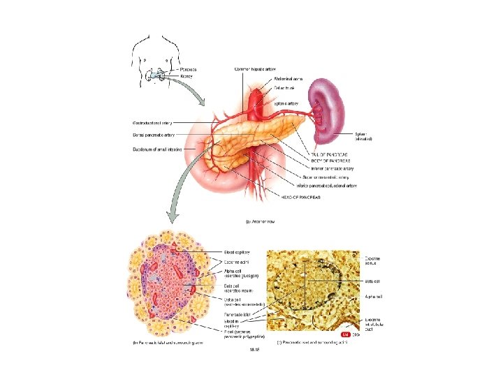
 n n n n In the pancreas the islets of Langerhans")
Introduction (cont. ) n n n n In the pancreas the islets of Langerhans represent only 1%– 2% of the mass of the pancreas, however, they receive approximately 10%– 15% of the pancreatic blood flow. Venous blood from the pancreas drains into the hepatic portal vein. Therefore, the liver, a principal target organ for the physiologic effects of pancreatic hormones, is exposed to the highest concentrations of pancreatic hormones. More than 50% of insulin is degraded during its first pass through the liver. Parasympathetic, and sensory nerves richly innervate the pancreatic islets. Acetylcholine, vasoactive intestinal polypeptide (VIP), pituitary adenylate cyclase-activating polypeptide, and gastrin-releasing peptide are released from the parasympathetic nerve terminals. Norepinephrine, galanin, and neuropeptide Y are released from sympathetic nerve terminals. Vagal nerve activation stimulates the secretion of insulin, glucagon, somatostatin, and pancreatic polypeptide. Sympathetic nerve stimulation inhibits basal and glucosestimulated insulin secretion (through α 2 -adrenergic mechanism) and somatostatin release and stimulates glucagon and pancreatic polypeptide secretion.

Insulin

Effect of Insulin on cells 1. Insulin (an anabolic polypeptide hormone with a half-life of 3 -8 minutes) increases the glucose uptake of 80% of body cells (except for brain neurons, renal epithelium, intestinal epithelium, erythrocytes, and liver) by increasing cellular glucose transport proteins. In the absence of insulin, these protein carriers will be removed from cell membrane within 3 -5 minutes. 2. Insulin makes cell membrane more permeable for many amino acids, K+, and phosphate ions to move internally. 3. Insulin remolds many cellular enzymatic machinery to achieve its metabolic goals. Note: The number of available insulin receptors is modulated by exercise, diet, insulin, and other hormones. Chronic exposure to high insulin levels, obesity, and excess growth hormone all lead to a downregulation of insulin receptors. In contrast, exercise and fasting upregulate the number of receptors, improving insulin responsiveness.

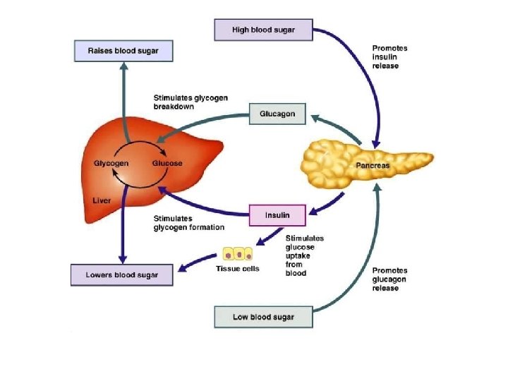
Effect of Insulin on carbohydrate metabolism A. Insulin promotes glucose uptake by all tissues and glycogen synthesis by muscles and liver. The storage of glycogen in the liver is 100 mg. During exercise the skeletal muscle become permeable to glucose even in the absence of insulin, this is because of the contraction process itself. In addition to glucose, exercising muscles consume fatty acids as well. The effect of insulin on glucose metabolism in the liver can be summarized in this diagram: Note: Unlike the liver, the muscle cannot convert glycogen back to glucose and release it to blood as it lacks the enzyme glucose phosphatase. Therefore, the liver is the organ that is responsible for the maintenance of stable serum glucose level (i. e. responsible for homeostasis).
 B. Insulin promotes the conversion of")
Effect of Insulin on carbohydrate metabolism (Cont. ) B. Insulin promotes the conversion of glucose into fatty acids by the liver, whereas gluconeogenesis is inhibited. C. Insulin has little effect on brain uptake or use of glucose. Brain cells are already permeable to glucose and cannot use fatty acids only with difficulty. Hypoglycemia down to 20 -50 mg/dl → hypoglycemic shock (irritability, fainting, and even coma).

Effect of insulin on fat metabolism A. Insulin promotes fat synthesis and storage in adipose tissue. This function is achieved by (1) sparing the use of fat since it increases glucose utilization and (2) converting extra glucose to acetyl-Co. A then → fatty acids → triglycerides in the liver. Triglycerides will be transported by blood lipoproteins to adipose cells. It should be noted: 1. Insulin activates capillary lipoprotein lipase in the adipose tissue. This enzyme converts triglycerides into fatty acids to be absorbed into the adipose cells, and then triglycerides will be reconstructed and stored inside the fat cells. 2. Insulin inhibits the enzyme lipase of fat cells. Insulin antagonizes catecholamine-induced lipolysis. B. Insulin deficiency causes: 1. Lipolysis of stored fat and release of free fatty acids due to activation of lipase in fat cells. This is evident in diabetes and after removal of pancreas. 2. ↑ in plasma cholesterol and phospholipids concentrations due to liver conversion of some fatty acids into these products and discharging them in blood in the lipoproteins → severe atherosclerosis (commonly seen in diabetics) 3. Ketosis and acidosis due to formation of acetoacetic acid by the liver due to excess acetyl-Co. A formation. Acetoacetic acid and substances that can be derived from it (hydroxyl-butyric acid and acetone) are called Ketone bodies.

Effect of insulin on protein metabolism and growth 1. Insulin promotes protein synthesis and storage by increasing the cellular uptake of amino acids (i. e. like growth hormone) and turns on the ribosomal machinery. 2. Insulin inhibits protein catabolism especially in muscle cells 3. Insulin inhibits hepatic gluconeogenesis. This conserves amino acids in body protein. Therefore, insulin lack → ↑ amino acids in circulation → ↑ urea excretion. 4. Combination of insulin and growth hormone are essential for growth. The two hormones function synergistically to promote growth.

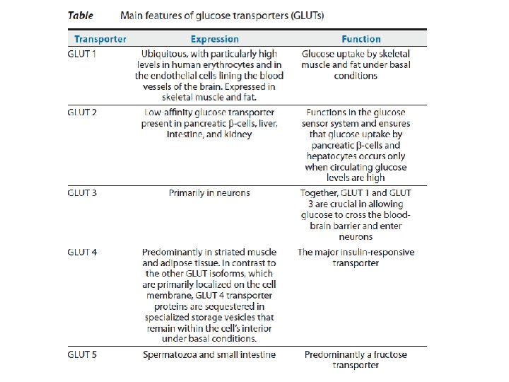
Control of insulin secretion 1. 2. 3. 4. 5. The pancreatic β-cell functions as a neuroendocrine integrator that responds to changes in plasma levels of energy substrates (glucose and amino acids), hormones (insulin, glucagon-like peptide I, somatostatin, and epinephrine), and neurotransmitters (norepinephrine and acetylcholine) by increasing or decreasing insulin release. Glucose is the principal stimulus for insulin release from the pancreatic β-cells. Glucose enters the β-cell through a membrane-bound glucose transporter 2 (GLUT 2) → ATP formation → closure of ATPsensitive K+ channels → opening of voltage-dependent Ca 2+ channels → exocytosis of insulin into the extracellular space. Some amino acids, such as arginine and lysine, cause a small rise in insulin secretion if glucose remains stable. However, secretion is enhanced with hyperglycemia even more than if glucose rises alone, i. e. amino acids potentiate the glucose stimulus for insulin secretion. Gastro-intestinal hormones; such as gastrin, secretin, CCK, and gastric inhibitory peptide; cause mild secretion of insulin.

Agents that Affect Insulin Secretion Primary Stimuli Secondary Stimuli Inhibitors Glucose Glucagon Somatostatin Mannose Secretin Epinephrine Leucine Cholecystokinin Norepinephrine Arginine Gastrin Starvation Lysine Gastric inhibitory peptide Exercise Short chain fatty acids Acetylcholine Long chain fatty acids Prostaglandin E 1 and E 2 Acetoacetate (Ketoacid) Obesity Physiologic β-Hydroxybutyrate Pharmacological N-Acetylglucosamine Theophylline Diazoxide Glyceraldehyde Caffeine Mannoheptulose Dihydroxyacetone Isobutyl-methylxanthine 2 -Deoxyglucose Glucosamine Sulfonylureas Iodoacetate Inosine β-adrenergic agonists α-adrenergic agonists

Glucagon, is a 29 -amino acid polypeptide hormone secreted by the α-cells of the islets of Langerhans. It is structurally related to the secretin family of peptide hormones. • Glucagon has a short half-life (5 -10 minutes) and is degraded mostly in the liver. • The principal target tissue for glucagon is the liver. However, glucagon receptor is expressed in liver, pancreatic β-cells, kidney, adipose tissue, heart, and vascular tissues, as well as in some regions of the brain, stomach, and adrenal glands. The role of glucagon receptors in many tissues other than the liver is still unclear. • In general, the function of this hormone is opposite to that of insulin; i. e. it is glycogenolytic; gluconeogenic; lipolytic; ketogenic; and stimulates secretion of GH, insulin, and pancreatic somatostatin. •

Mechanism of secretion 1. 2. 3. 4. Blood glucose is the principal control factor in exactly the opposite direction for the effect of glucose on insulin secretion. Hypoglycemia → ↑ plasma concentration of glucagon Hyperglycemia → ↓ plasma concentration of glucagon Increased levels of amino acids, especially alanine and arginine, in blood (after protein intake mainly by mouth) → stimulation of glucagon secretion. This is a similar effect to insulin secretion. This response aids gluconeogenesis. CCK and gastrin assists further to glucagon secretion by this mechanism. Exercise induces glucagon secretion. The mechanism is unknown. It could be due to sympathetic stimulation of the islets of Langerhans (via β 2 receptors), while stimulation of α receptors inhibits glucagon secretion. It should be noted that sympathetic β receptors predominate in the pancreas. Vagal (parasympathetic) stimulation increases glucagon release.

Factors that Regulate Glucagon Secretion Stimuli Inhibitors Hypoglycemia Hyperglycemia Low fatty acid levels High fatty acid levels Most amino acids Ketone bobies Epinephrine Secretin Norepinephrine Somatostatin Acetylcholine Serotonin Dopamine Gastrin Cholecystokinin Gastric inhibitory polypeptide Vasoactive intestinal polypeptide Exercise Starvation

The pancreatic Somatostatin • Pancreatic somatostatin inhibits glucagon and insulin secretion. This effect will prevent the rapid exhaustion of the food by insulin. • Somatostatin release is inhibited by insulin. • It is released in response to factors related to food ingestion: 1. 2. 3. 4. ↑ serum glucose level ↑ serum amino acids ↑ serum fatty acids ↑ level of many GI hormones

Functions of pancreatic somatostatin 1. 2. 3. 4. 5. ↓ secretion of both insulin and glucagon. ↓ stomach, duodenal, and gallbladder contractions. ↓ secretion and absorption in the GIT. ↓ the tone of mesenteric arteries and portalsystemic collateral veins (the mechanism is unknown). Therefore, somatostatin analogues are used in the treatment of portal hypertension. It may have a protective role against liver fibrosis.
 n Type I (Insulin-dependent diabetes mellitus or IDDM) - this type")
Diabetes Mellitus (DM) n Type I (Insulin-dependent diabetes mellitus or IDDM) - this type is due to lack of insulin secretion. Ø It could result from viral infection or autoimmune disorders that affect β cells, or due to hereditary tendency for β cell degeneration. Ø It can appear during childhood therefore, it is called juvenile diabetes mellitus. Ø Glucose spills into the urine when blood glucose concentration rises above 180 mg/dl (= the threshold). Glucose can reach up to 1200 mg/dl within few days or weeks. Ø High glucose → dehydration & osmotic diuresis → intracellular and extracellular dehydration + thirst sensation Ø Chronic high glucose → structural change in blood vessels and inadequate blood supply to the tissues → MI or CVA, kidney disease, retinopathy and blindness, and limb gangrene. Also it can lead to peripheral neuropathy & autonomic nervous system dysfunction (impaired CVS reflexes, impaired bladder control, decreased sensation in the extremities, etc. . ). DM is commonly associated with hypertension and atherosclerosis. Ø Diabetes mellitus → increased fat utilization and metabolic acidosis
 n Type II (non-insulin-dependent diabetes mellitus or NIDDM) - this type")
Diabetes Mellitus (DM) n Type II (non-insulin-dependent diabetes mellitus or NIDDM) - this type is due to decreased sensitivity of target tissues to the metabolic effect of insulin, i. e. insulin resistance. Ø Have the same metabolic abnormalities as Type I, but high levels of keto acids are usually not present in type II. Ø This type accounts for 80 -90% of all cases of diabetes. Ø The age onset is over 40 years of age. It develops gradually, therefore it called sometimes adult onset diabetes. Ø In contrast to type I, type II is associated increased plasma levels of insulin concentrations. Ø Late in the disease β cells become exhausted and are unable to produce enough insulin to prevent more severe hyperglycemia especially after starchy meal. Ø Insulin resistance is secondary to obesity. Mechanism of this is unknown, it could be due to down regulation of insulin receptors in skeletal muscle, liver, and adipose tissue. Ø How much pancreas can stand till it gets exhausted is genetically determined. Those who can sustain do not develop clinically significant DM for many years although they have increased insulin plasma level. Ø Drugs such as thiazolidinediones and metformin increase insulin sensitivity. Sulfonylurea causes additional insulin release Ø Glucose tolerance test: Is to give 1 mg of glucose/Kg body wt orally. Glucose should normally rise to 120 -140 mg/dl and back to normal within 2 hours.

Test Question: Q. Insulin regulates glucose transport into muscle and fat cells via which glucose transporter? A. GLUT-1. B. GLUT-2. C. GLUT-3. D. GLUT-4. E. GLUT-5.
- Slides: 23