
The Orthopedic Evaluation of The Child 06022007 The






- Slides: 98
The Orthopedic Evaluation of The Child 06/02/2007
The Orthopedic History n n n The chief complain. History of present illness. Birth history. Family history. Growth and development.
Growth and Development
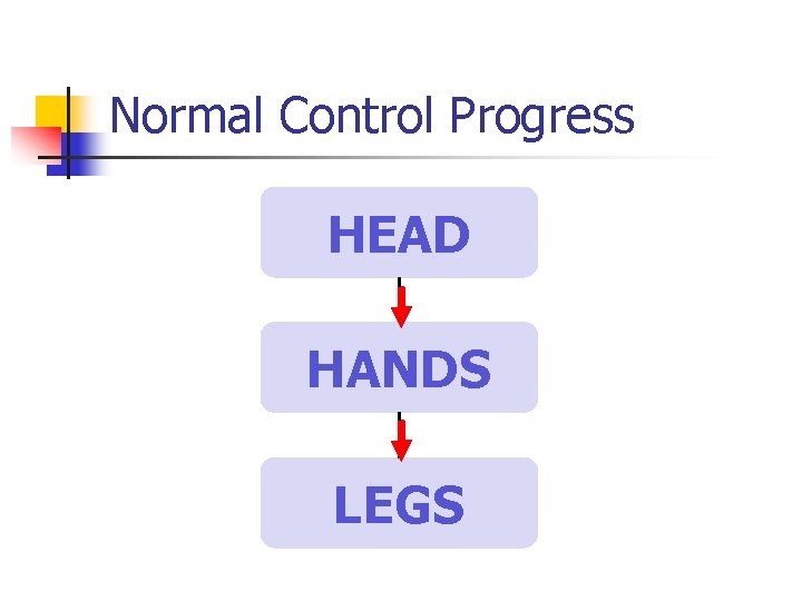
Normal Control Progress HEAD HANDS LEGS
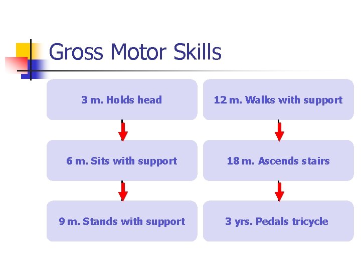
Gross Motor Skills 3 m. Holds head 12 m. Walks with support 6 m. Sits with support 18 m. Ascends stairs 9 m. Stands with support 3 yrs. Pedals tricycle
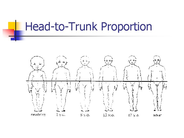
Head-to-Trunk Proportion
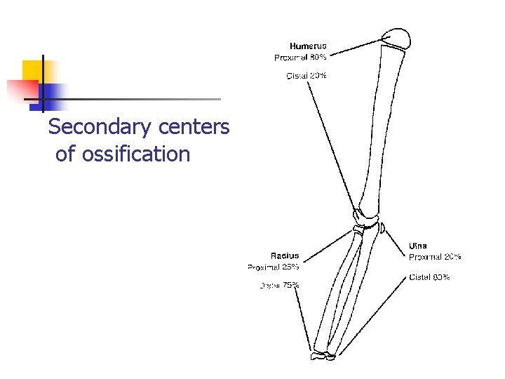
Secondary centers of ossification
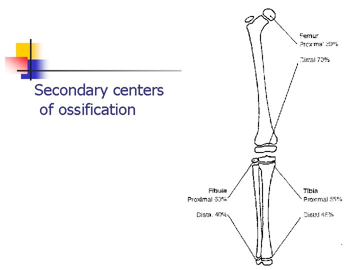
Secondary centers of ossification
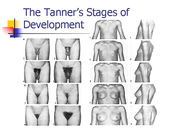
The Tanner’s Stages of Development
General Principles n Inspection (Angular deformities, Skin lesions…) n Joints Range of Motion. n Grading of Muscle Strength. n Developmental Reflexes.
The Neck
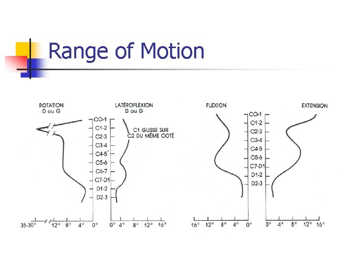
Range of Motion
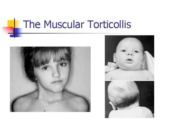
The Muscular Torticollis
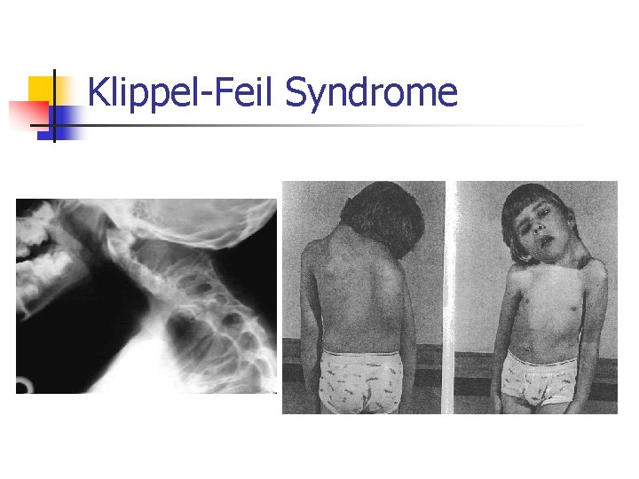
Klippel-Feil Syndrome
The Shoulder
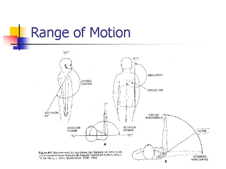
Range of Motion
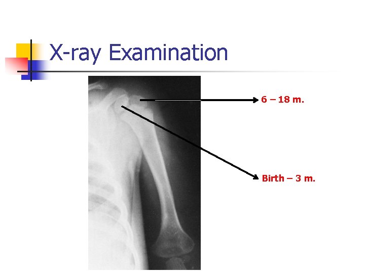
X-ray Examination 6 – 18 m. Birth – 3 m.
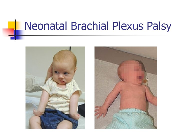
Neonatal Brachial Plexus Palsy
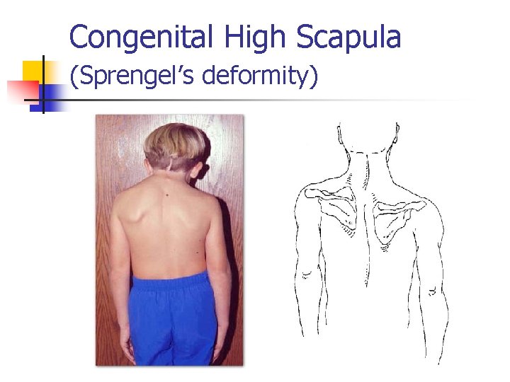
Congenital High Scapula (Sprengel’s deformity)
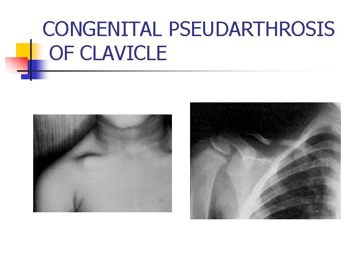
CONGENITAL PSEUDARTHROSIS OF CLAVICLE
The Elbow
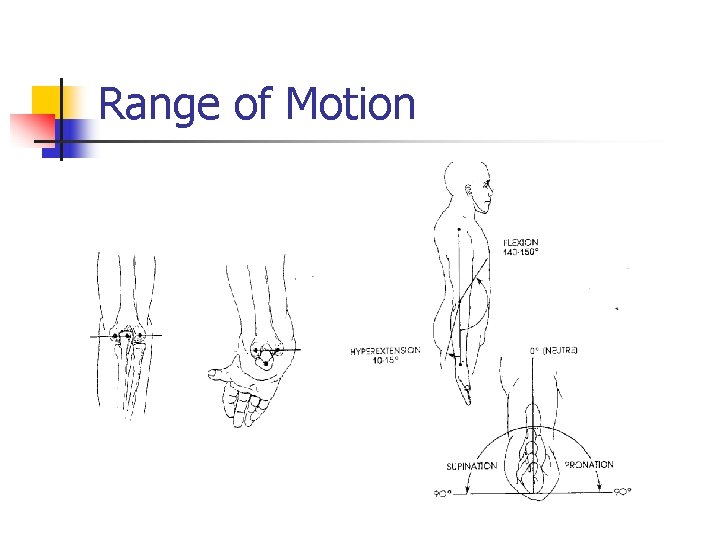
Range of Motion
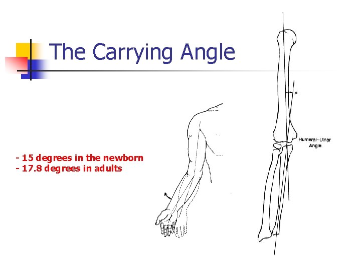
The Carrying Angle - 15 degrees in the newborn - 17. 8 degrees in adults
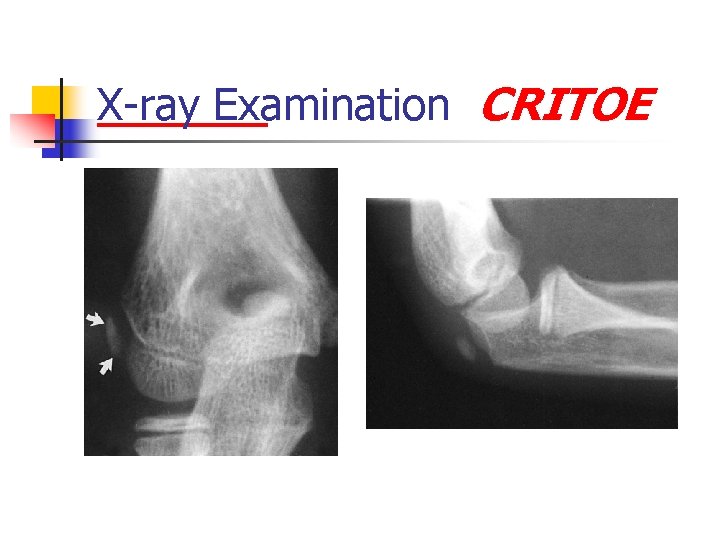
X-ray Examination CRITOE
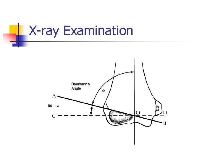
X-ray Examination
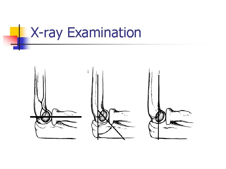
X-ray Examination
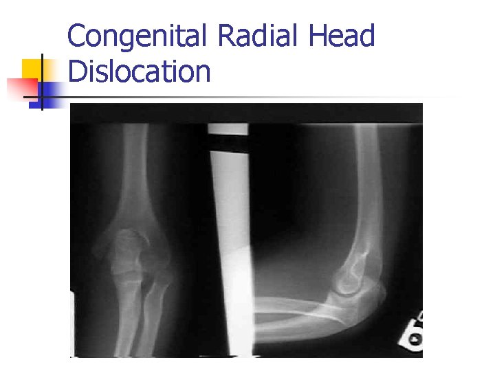
Congenital Radial Head Dislocation
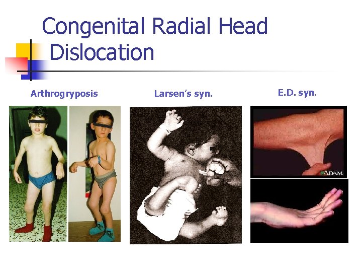
Congenital Radial Head Dislocation Arthrogryposis Larsen’s syn. E. D. syn.
The Hand
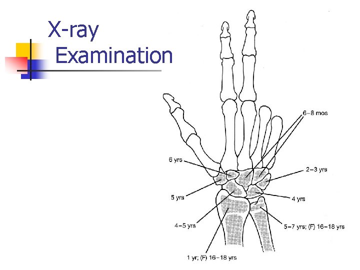
X-ray Examination
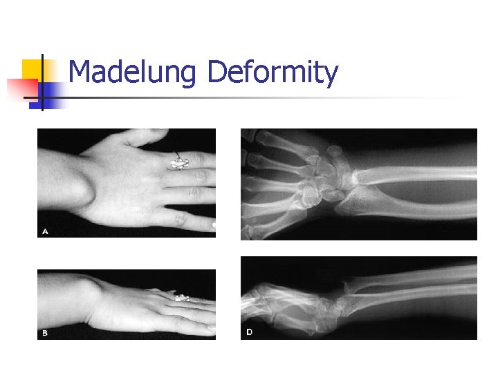
Madelung Deformity
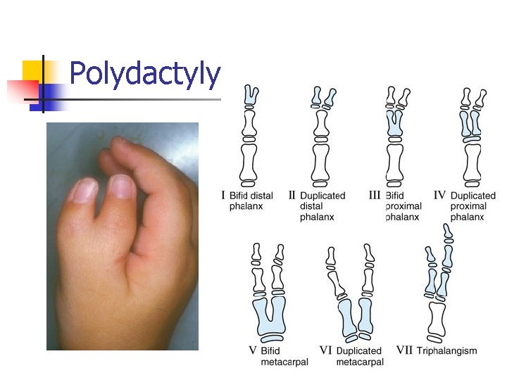
Polydactyly
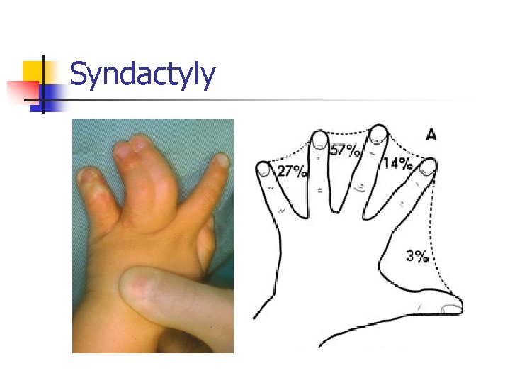
Syndactyly
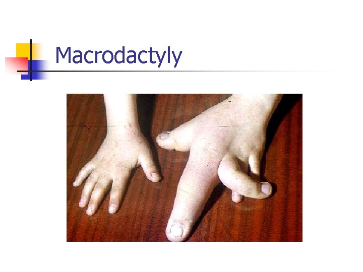
Macrodactyly
The Spine
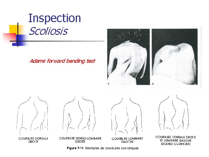
Inspection Scoliosis Adams forward bending test
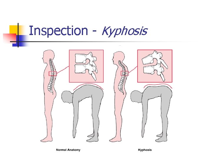
Inspection - Kyphosis
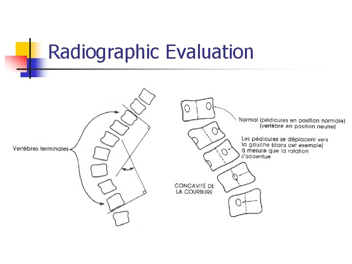
Radiographic Evaluation
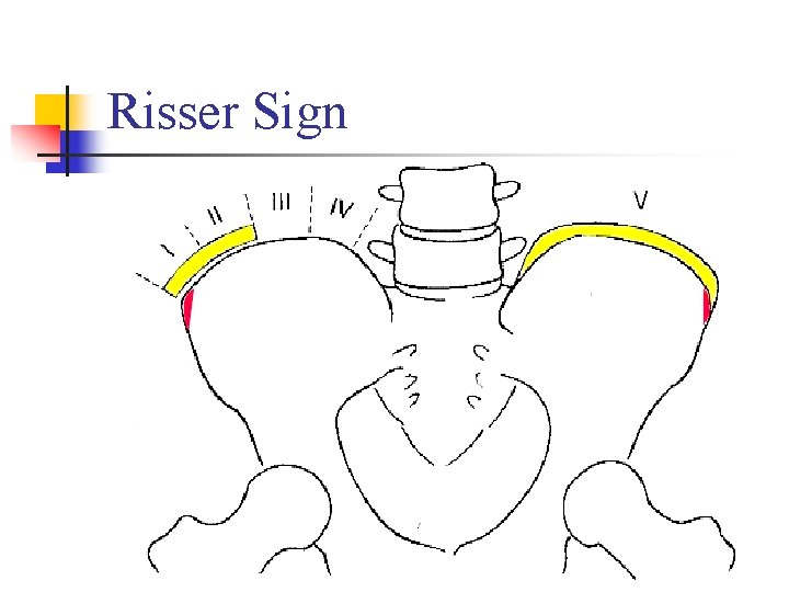
Risser Sign
The Hip
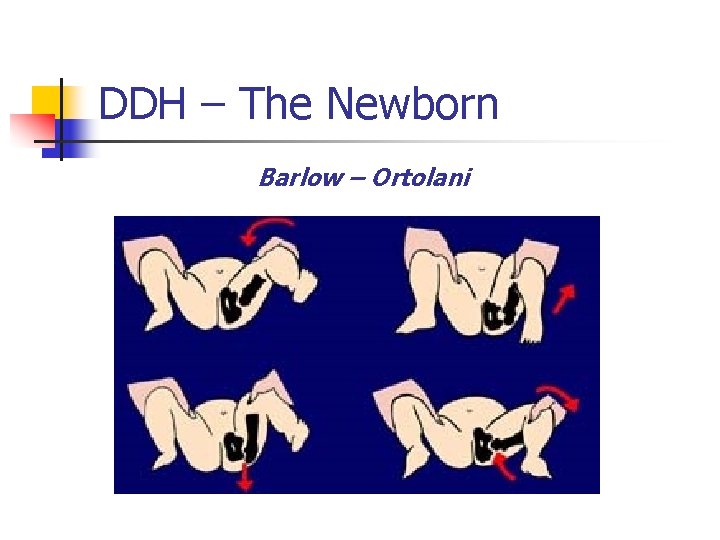
DDH – The Newborn Barlow – Ortolani
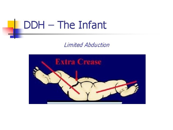
DDH – The Infant Limited Abduction
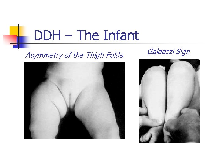
DDH – The Infant Asymmetry of the Thigh Folds Galeazzi Sign
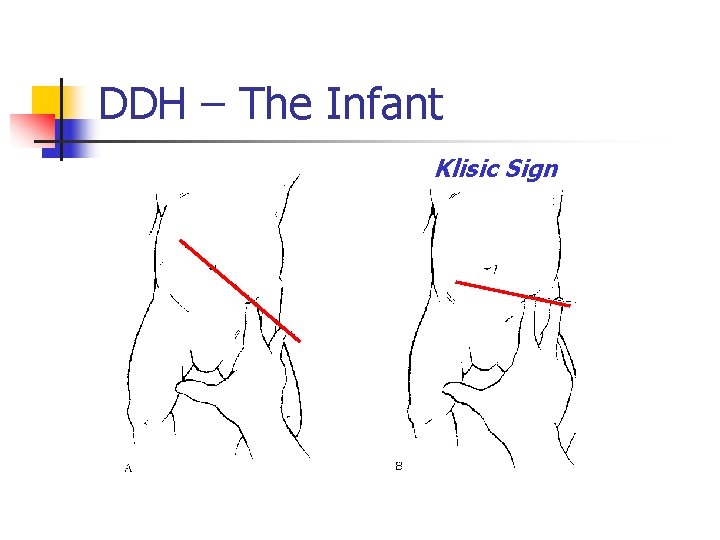
DDH – The Infant Klisic Sign
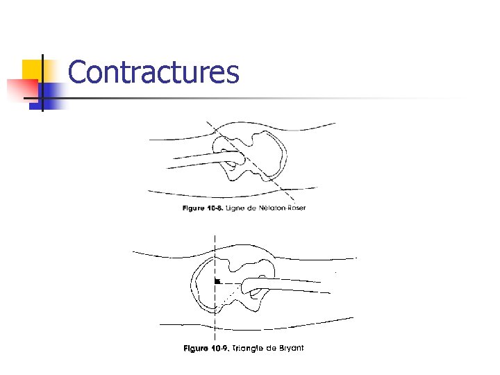
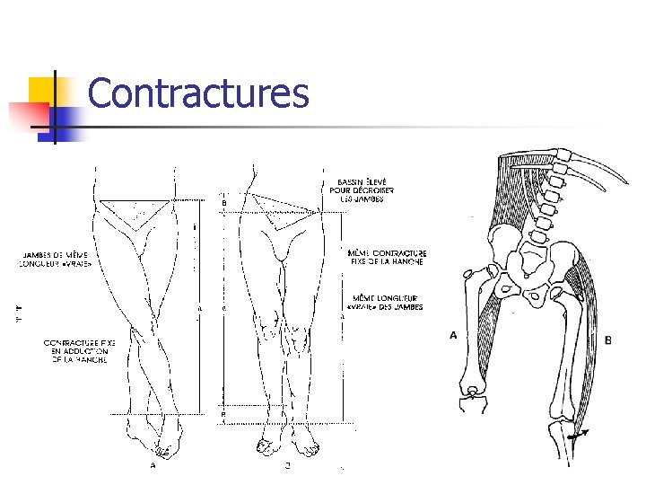
Contractures
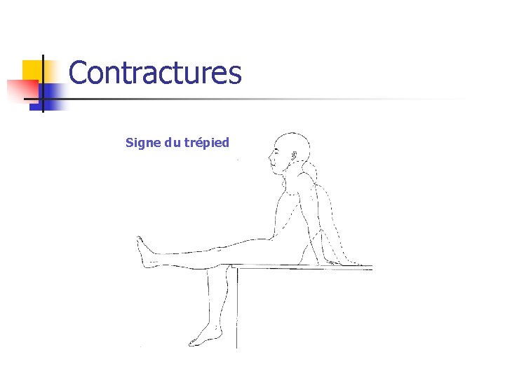
Contractures Signe du trépied
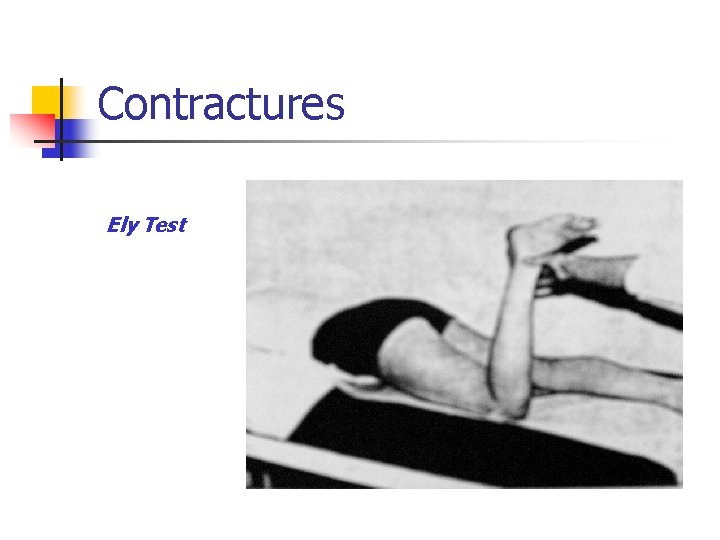
Contractures Ely Test
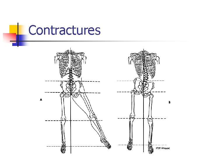
Contractures
Contractures
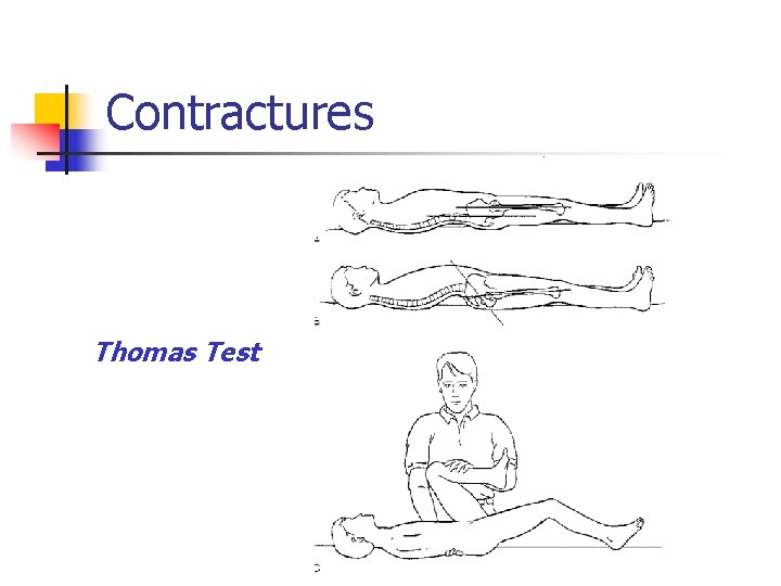
Contractures Thomas Test
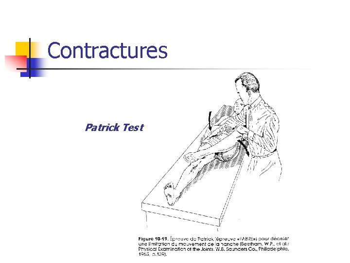
Contractures Patrick Test
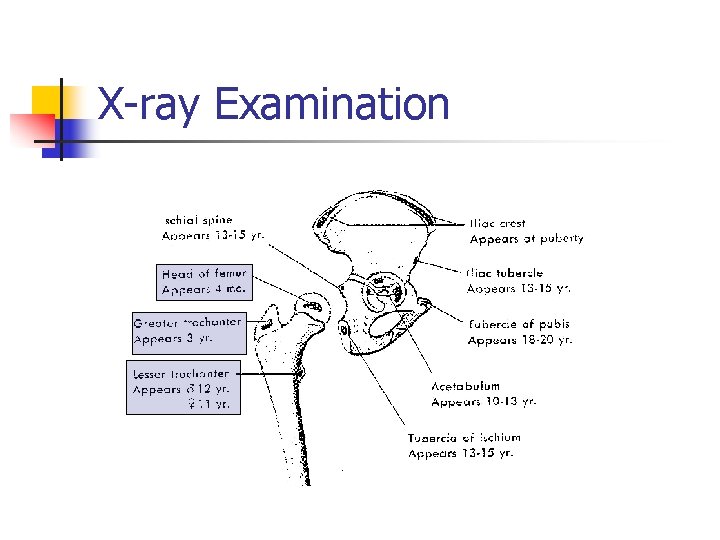
X-ray Examination
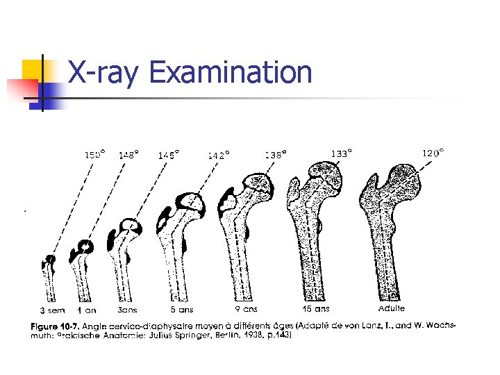
X-ray Examination
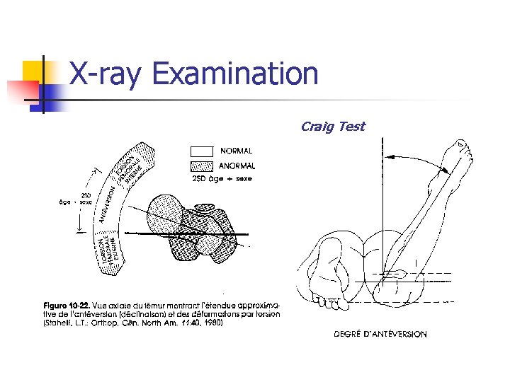
X-ray Examination Craig Test
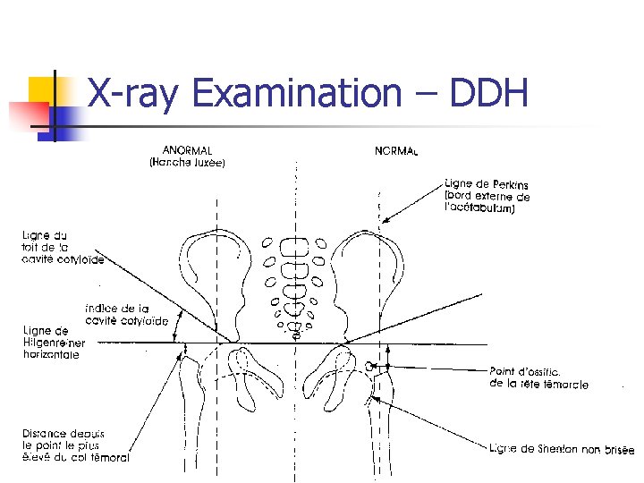
X-ray Examination – DDH
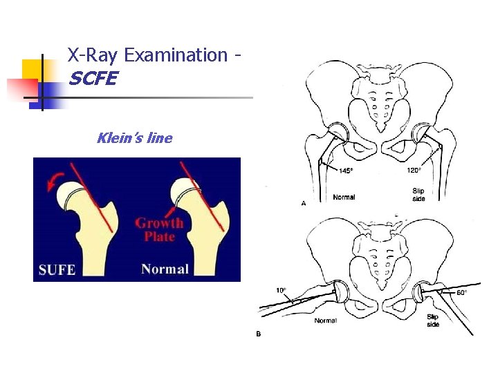
X-Ray Examination - SCFE Klein’s line
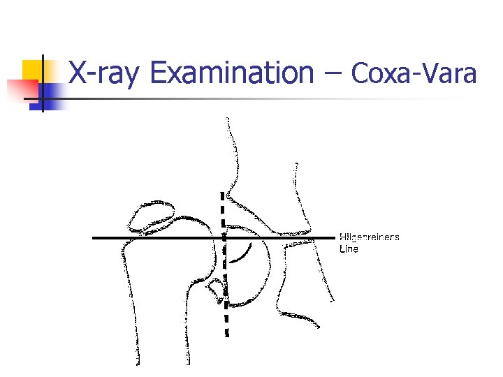
X-ray Examination – Coxa-Vara
The Knee and Leg
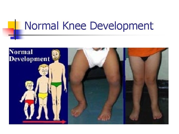
Normal Knee Development
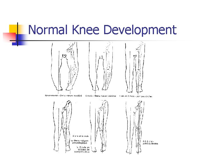
Normal Knee Development
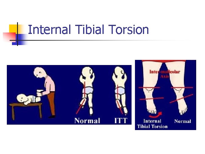
Internal Tibial Torsion
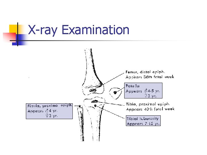
X-ray Examination
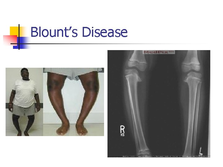
Blount’s Disease
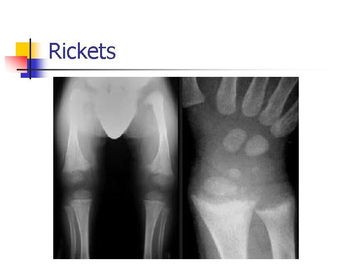
Rickets
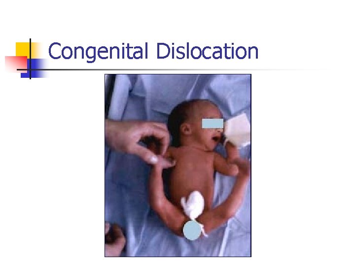
Congenital Dislocation
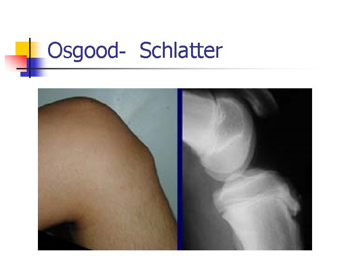
Osgood- Schlatter
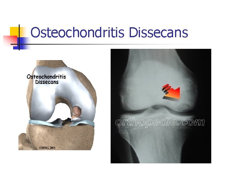
Osteochondritis Dissecans
The Foot
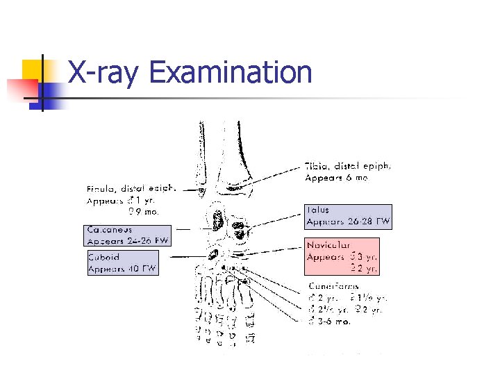
X-ray Examination
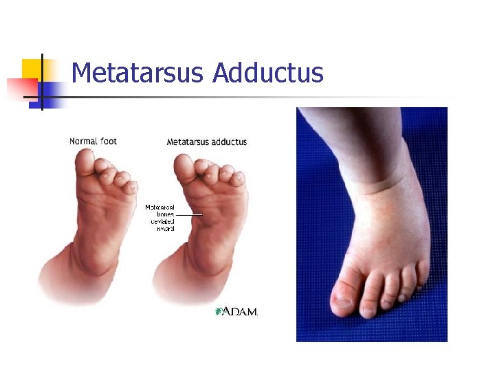
Metatarsus Adductus
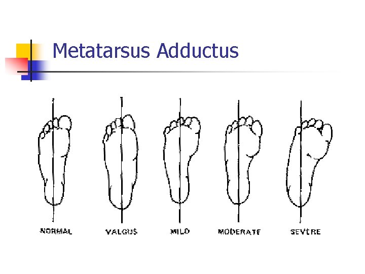
Metatarsus Adductus
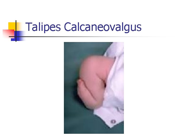
Talipes Calcaneovalgus
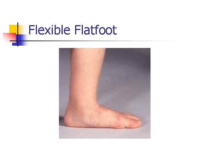
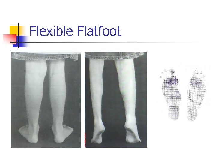
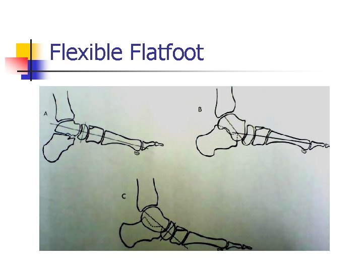
Flexible Flatfoot
Flexible Flatfoot
Flexible Flatfoot
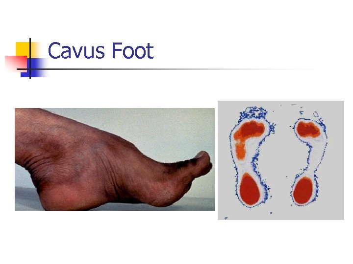
Cavus Foot
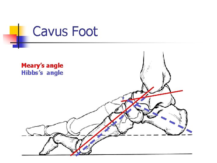
Cavus Foot Meary’s angle Hibbs’s angle
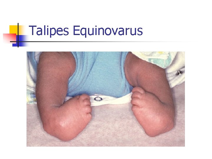
Talipes Equinovarus
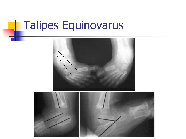
Talipes Equinovarus
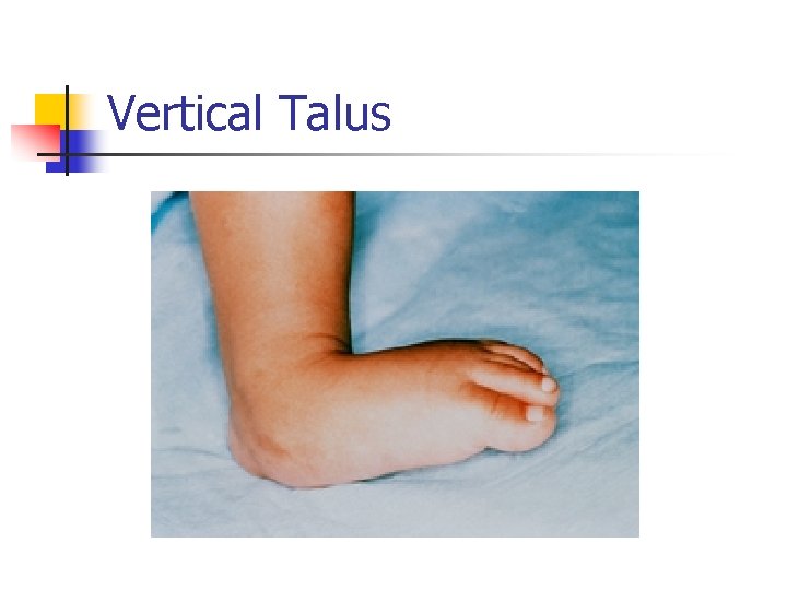
Vertical Talus
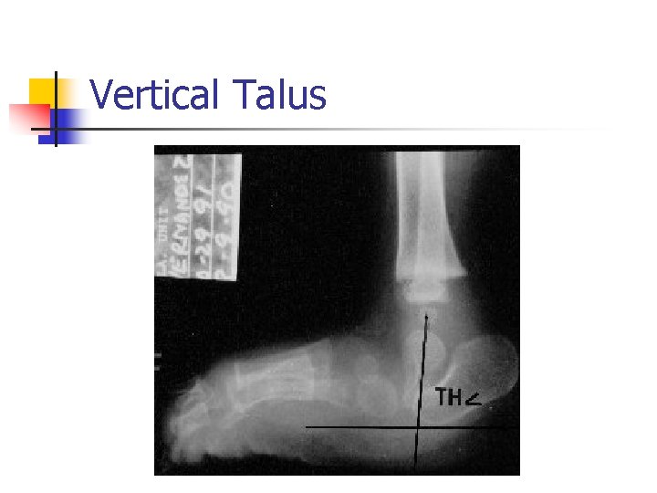
Vertical Talus
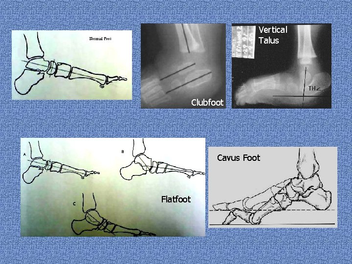
Vertical Talus Clubfoot Cavus Foot Flatfoot
The Gait Analysis
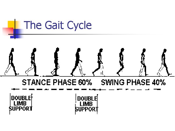
The Gait Cycle
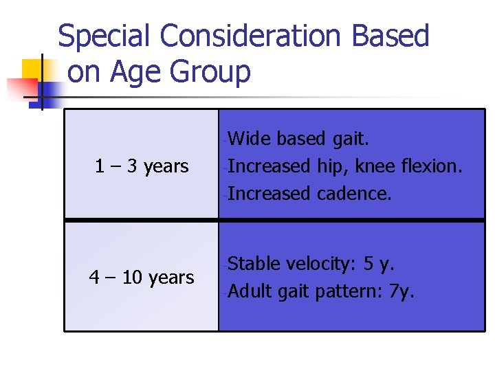
Special Consideration Based on Age Group Wide based gait. -Increased hip, knee flexion. -Increased cadence. - 1 – 3 years 4 – 10 years Stable velocity: 5 y. -Adult gait pattern: 7 y. -
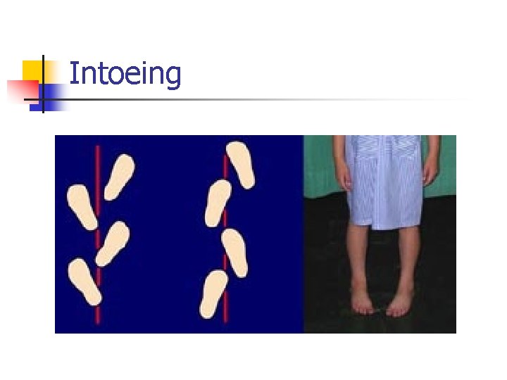
Intoeing
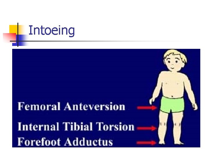
Intoeing
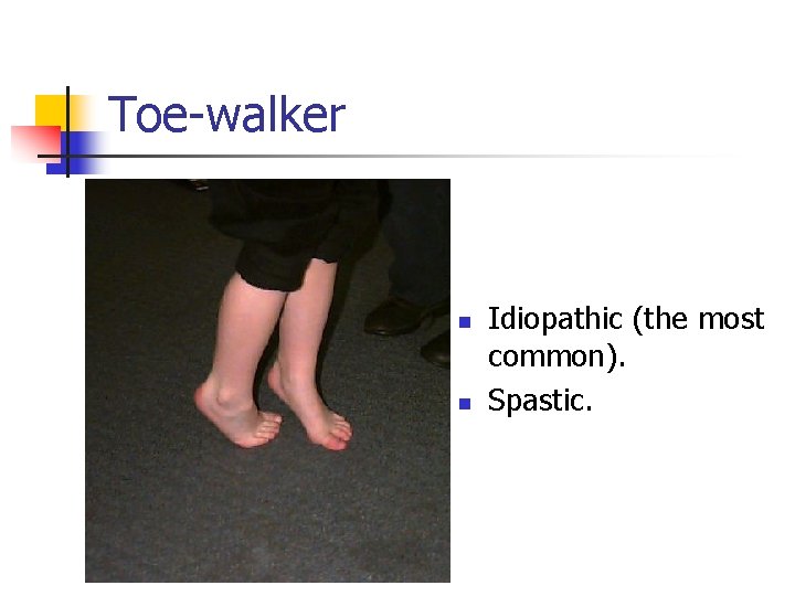
Toe-walker n n Idiopathic (the most common). Spastic.
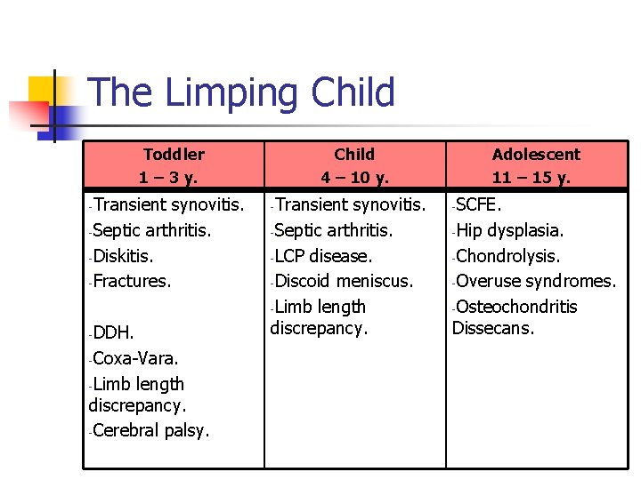
The Limping Child Toddler 1 – 3 y. Transient synovitis. -Septic arthritis. -Diskitis. -Fractures. - DDH. -Coxa-Vara. -Limb length discrepancy. -Cerebral palsy. - Child 4 – 10 y. Transient synovitis. -Septic arthritis. -LCP disease. -Discoid meniscus. -Limb length discrepancy. - Adolescent 11 – 15 y. SCFE. -Hip dysplasia. -Chondrolysis. -Overuse syndromes. -Osteochondritis Dissecans. -
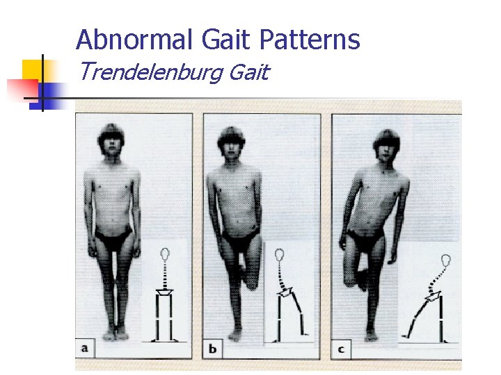
Abnormal Gait Patterns Trendelenburg Gait
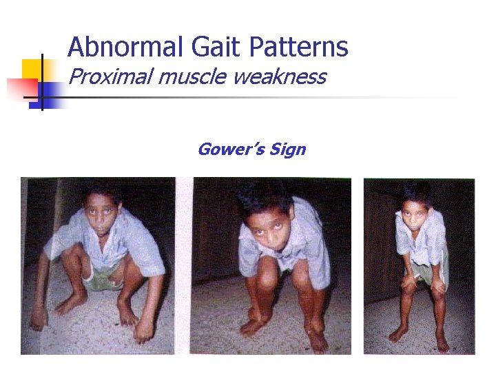
Abnormal Gait Patterns Proximal muscle weakness Gower’s Sign
Thank You