The Orbit Orbital Contents and Cranial Nerves III

The Orbit, Orbital Contents and Cranial Nerves III, IV and VI

Lecture Objectives • Describe the location of the orbit. • Make a list of structures making the orbit starting from orbital margin. • Define each component. • Describe openings into orbital cavity. • Define the orbital fascia. • Describe muscles of the orbit, their cone arrangement, origin, insertion, nerve supply and their function. • Describe the nerves of the orbit, their courses, important relations and their targets • Describe blood supply and lymph drainage of the orbit.

The Orbit: Orbital Margin

The Orbit: Orbital Cavity • Shape. . • Orientation. . • Walls. .
 Infraorbital groove, canal, &")
Openings into Orbital Cavity • • • Supraorbital notch (foramen) Infraorbital groove, canal, & foramen Nasolacrimal canal Inferior & superior orbital fissures Anterior & posterior ethmoidal foramina

Orbit: Content • • Eyelid Lacrimal apparatus Eyeball Fascia Extraocular muscles Nerves Blood vessels Fatty tissue

Eye: Fascia • Covers the eye • Separate the eye from surrounding orbital fat • Facilitates movement of eye • Pierced by orbital muscles – Tubular sheath • Attaches to orbital walls – Medial & lateral check ligaments • Suspensory ligament of the eye

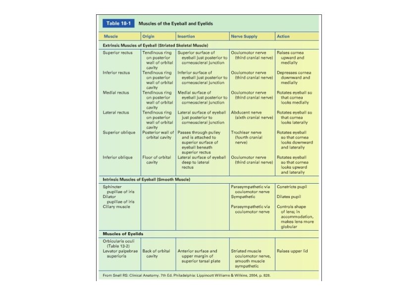
Eye: Muscles • Extrinsic muscles – – – Superior rectus Inferior rectus Medial rectus Lateral rectus Superior oblique • trochlea – Inferior oblique • Intrinsic muscles – Ciliary m. ‐ parasympathetic – Constrictor pupillae of the iris ‐ parasympathetic – Dilator pupillae of the iris ‐ sympathetic

Axes of Eyeball movements

Movements around transverse axis

Movements around vertical and A-P axes

Movements of Eyeball

Nerves of the Orbit
 • Trochlear (IV) • Abducens (VI)")
Nerves that Moves the Eyeball • Oculomotor (III) • Trochlear (IV) • Abducens (VI)
 • Mixed nerve; principally motor (GSE, GVE) (with proprioseptive) • Midbrain")
Oculomotor Nerve (III) • Mixed nerve; principally motor (GSE, GVE) (with proprioseptive) • Midbrain (anteriorly) • Cavernous sinus • Superior and inferior branches • Superior orbital fissure
 – Location – Relations • PAG,")
Oculomotor Nerve Nuclei • Main motor nucleus (GSE) – Location – Relations • PAG, superior colliculus – Connections • Cortex, superior colliculus – Fibers course • Accessory parasympathetic nucleus (Edinger‐Westphal nucleus)(GVE) – Location – Relations • Main motor nucleus – Connections • Pretectal nucleus – Fibers course

Pupillary Light Reflex
 • The superior branch – Superior rectus and levator palpebrae superioris")
Oculomotor Nerve (III) • The superior branch – Superior rectus and levator palpebrae superioris mm. • The inferior branch – Medial rectus, inferior rectus, and inferior oblique mm. – Parasympathetic innervation via the ciliary ganglion to the intrinsic eye muscles • Major functions – Regulating movements of upper eyelid and eyeball – Adjustment of lens for near vision, and constriction of pupil
: Lesion • Ptosis (denervation of levator palpebrae) • External ophthalmoplegia: Eye")
Oculomotor Nerve (III): Lesion • Ptosis (denervation of levator palpebrae) • External ophthalmoplegia: Eye look down & out (denervation of extraocular muscles) – Diplopia • Internal ophthalmoplegia: Dilated, fixed pupil & Paralysis of accommodation • Test – Asked to move the eye • Unable to move up, down, or medial – At rest looks down & lateral
 • Mixed nerve; primarily motor (GSE) (with proprioceptive) • Smallest of")
Trochlear Nerve (IV) • Mixed nerve; primarily motor (GSE) (with proprioceptive) • Smallest of the cranial nerves • Midbrain* • Cavernous sinus • Superior orbital fissure • Innervate the superior oblique muscle *Only one to arise from the posterior aspect of the brain stem ‐ Decussate and rotate around the brainstem
: Lesion • Difficulty to turn eye downward & laterally • Diplopia")
Trochlear Nerve (IV): Lesion • Difficulty to turn eye downward & laterally • Diplopia in looking downward
 – Location – Relations • PAG,")
Trochlear Nerve Nucleus • Trochlear nerve nucleus (GSE) – Location – Relations • PAG, inferior colliculus, main oculomotor nerve nucleus – Connections • Cortex, superior colliculus – Fibers course
 • Mixed nerve; primarily motor (GSE) (with proprioceptive) • Pons (anteriorly)")
Abducens Nerve (VI) • Mixed nerve; primarily motor (GSE) (with proprioceptive) • Pons (anteriorly) • Cavernous sinus • Superior orbital fissure • Called abducens because it causes abduction of the eyeball (lateral rotation) • Innervates the lateral rectus muscle
: Lesion • Nerve lesion causes internal strabismus & diplopia • Unable")
Abducens Nerve (VI): Lesion • Nerve lesion causes internal strabismus & diplopia • Unable to turn eyeball laterally
 – Location – Relations • 4")
Abducens Nerve Nucleus • Abducent nerve nucleus (GSE) – Location – Relations • 4 th ventricle • Colliculus facialis – Connections • Cortex, superior colliculus – Fibers course
 Superior orbital fissure •")
Nerves of the Orbit Branches of ophthalmic nerve (V 1) Superior orbital fissure • Frontal nerve Scalp – Branches: • Supraorbital & Supratochlear nn. • Lacrimal nerve Lateral part of upper eyelid – Carry parasympathetic fibers to lacrimal gland via zygomaticotemporal nerve
 • Nasociliary nerve‐ Branches:")
Nerves of the Orbit Branches of ophthalmic nerve (V 1) • Nasociliary nerve‐ Branches: – Comunicating branch to ciliary ganglion‐ sensory fibers from short ciliary nn. – Long ciliary nn. ‐ carry sympathetic fibers (dilator pupillae m. ) – Posterior ethmoidal n. (ethmoid & sphenoid sinuses) – Anterior ethmoidal n. • External nasal branch (tip of nose) – Infratrochlear n. (medial part of upper eyelid & part of nose)
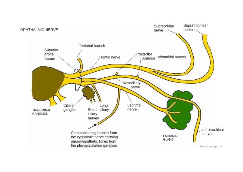

Nerves of the Orbit

Orbit: Ophthalmic Artery Optic canal • Branches of ophthalmic artery Supraorbital a. Supratrochlear a. Central retinal a. Ciliary aa. (short & long) Ethmoidal aa. (anterior & posterior) – Lacrimal a. – Dorsal nasal a. – – –

Orbit: Ophthalmic Veins ** No lymphatic vessels or nodes in orbit
- Slides: 32