The Nervous Tissue Contains two different varieties of


")


 endoneurium Connective tissue perineurium epineurium")


- Slides: 26
The Nervous Tissue Contains two different varieties of cells 1. Neurons 2. Neuroglia = supporting cells
NEURON Also called nerve cells Structural unit of nervous system Character: § Excitability § conductivity Conduct messages in the form of nerve impulses from one part of the body to another = synapse
Structure of neuron 1 - Cell body 2 -Cell processes = Soma around nucleus perikaryon axon dendrites
. Perikaryon = nerve cell body contains 1. Nucleus : large, central, large amount of euchromatin (intense synthetic activity) & prominent nucleolus 2. Rough endoplasmic reticulum (RER) - lots of r. ER for synthesis of structural and transport proteins= Nissl bodies seen with light microscope are condensations of this r. ER and free ribosomes. 3. Golgi apparatus : only found near nucleus in perikaryon. Expected, since intense synthetic activity of neurotransmitters that must be packaged in vesicles. 4. Mitochondria : abundant for high energy requirements 5. Neurofilaments : intermediate filaments 6. Microtubules : transport of materials (neurotransmitters) 7. Inclusions : pigment e. g. Lipofuscin pigment - residual bodies from autophagosome activity increase with age. H & E stains Silver stain
AXON The nerve cell processes: . Dendrite Origin From axon hillock From any part of cell Number Always single Usually multiple length long Short Thickness Thin Thick near cell then tapers away Branching Does not branch except at its termination Many branches. Organelles Few No Nissl bodies No golgi Contains most of organelles Contain golgi + Nissl Surrounding structures Surrounded with sheaths Not surrounded with sheaths Direction of impulse Away, centrifugal Towards, centripetal Axoplasm Axolemma
Classification of neurons Functionally Morphologically Sensory Size Motor 1. Golgi I (long axon ) Interneurons 2. Golgi II (short axon) Neurosecretory No of Processes 1. one Process 2. two Processes 3. more Processes
According to the number of processes
Multipolar Neurons According to the shape of the cell body Stellate Pyriform Pyramidal Granule
Function of neuroglia 1. 2. 3. 4. 5. 6. 7. Supportive Nutritive Electrical insulation= formation of myelin sheath Repair Formation of blood brain barrier Formation of CSF Phagocytosis Neuroglia small 1. Can not transmit nerve impulse 2. Able to divide 3. Not form synapse Neuron large 1. Can transmit nerve impulse 2. Not able to divide 3. Form synapse
Neuroglia Definition : Supporting cells of the nervous system Stain : Gold chloride Silver stain Immuno-histochemical Site : Inside CNS macroglia =astrocytes, microglia, ependymal cell , oligodendroglia Outside CNS Schwann cell, satellite cells in ganglia, pituicytes in posterior lobe of pituitary gland. Shape : Branching cells …. Origin : All ectodermal in origin except microglia derived from blood monocytes = mesodermal in origin
Types of neuroglia Name of the cell Location Function v Astrocytes protoplasmic & Fibrous CNS Grey & white matter Regulate microenvironment B. B. B v Oligodendrocytes CNS White & grey matter Myelination of axon in CNS v Microglia CNS Grey & white matter Mesodermal in origin Phagocytic v Ependymal cell CNS Ventricle & central canal of spinal cord------with a ciliated simple columnar shape Assist in producing & controlling composition of CSF v Schwann cell PNS Myelination of axon Structural support v Satellite cell PNS In ganglia Regulate microenvironment of neurons
Protoplasmic Fibrous Site Gray matter White matter Processes No, length, thickness, course Numerous, short thick & wavy Fewer, longer, thinner & straight GFAP Few bundles Rich in it
Myelin Sheath Segmented protein-lipoid sheath around most long or large-diameter axons Functions : 1. Protect and electrically insulate the axon 2. Increase speed of nerve impulse transmission Myelin Sheaths in the PNS Schwann cells wraps many times around the axon Myelin sheath —concentric layers of Schwann cell membrane Nodes of Ranvier ---? ? Myelin sheath gaps between adjacent Schwann cells Myelinated nerve are separated by nodes of Ranvier, at these points , the axons are bare. Impulses jump from one node to the next ---Saltatory Conduction Unmyelinated Axons. Thin nerve fibers are unmyelinated. One Schwann cell may incompletely enclose 15 or more unmyelinated axons. Conduction in Myelin Sheaths in the CNS Formed by processes of oligodendrocytes, not the whole cells Nodes of Ranvier are present No neurilemma Thinnest fibers are unmyelinated
v Schwann cell plasma membrane v Schwann cell Cytoplasm A Schwann cell envelopes an axon. Schwann cell nucleus v Axon Neurilemma The Schwann cell then rotates around the axon, wrapping its plasma membrane loosely around it in successive layers. Myelin sheath Myelination of a nerve fiber (axon)------? ? Unmyelinated nerve fiber (axon)
SYNAPSE Def : Synapse is a specialized area of functional contact. Through synapses nerve impulses are transmitted from a presynaptic neuron to a postsynaptic cell (another neuron, muscle or gland).
Classification of synapse: 1 -According to the components: Axodendritic: between axon and dendrite Axosomatic: between axon and cell body. Axo-axonic: between two axons. Dendrodendritic: between two dendrites. Axodendritic and axosomatic synapses are common axoaxonic and dendrodendritic synapses are rare. 2 - According to the effect on the target neuron, synapse could be: Excitatory Inhibitory
3 - According to mode of transmission of nerve impulse: Chemical synapse: • Number: The most common mode of communication between neurons. • Structure : With EM, chemical synapse is formed of: • Terminal bouton: It is a bulbous expansion "synaptic knob" found at the terminations of the axon. It contains numerous mitochondria and many synaptic vesicles assembled around the presynaptic membrane. These membrane- bound synaptic vesicles are filled with the neurotransmitter e. g. acetylcholine and biogenic amines. • Presynaptic membrane: it is the electron dense membrane of the axon terminal end at the site of synapse. • Synaptic cleft: it is a gap of 20 nm between the pre and postsynaptic membranes. Into this gap the neurotransmitter is released by exocytosis. • Postsynaptic membrane: it is the electron dense membrane of the target neuron at the site of synapse. It contains the specific receptors for the neurotransmitter.
postsynaptic membrane presynpatic membrane vesicles mitochondria Synaptic cleft
Electrical synapse: • Number: Few in the nervous system. • Structure: It is formed of gap junctions. • Function: It allows movement of ions between neurons "electrical coupling" thus permits the direct spread of electrical current from one cell to another. • Transmission of impulse: Direct, bidirectional and do not require release of neurotransmitters.
The Axon+ sheath = Nerve fiber • Cytoplasm is called axoplasm • Plasma membrane is called axolemma • One axon per cell arising from the axon hillock • Long , thin • No Nissl bodies • Knoblike axon terminals (synaptic knobs) • Numerous terminal branches • Occasional branches (axon collaterals) • Mostly myelinated some axons not myelinated • Generates and transmits nerve impulses (action potentials) away from the cell body
Peripheral nerves are made up of axon +sheath (NF) endoneurium Connective tissue perineurium epineurium • Nerves can be injured by: ischaemia , compression, traction, laceration or burning. v. Damage varies in severity from: transient and quickly recoverable loss of function vto complete interruption and degeneration. • There may be a mixture of types of damage in the various fascicles of a single nerve trunk.
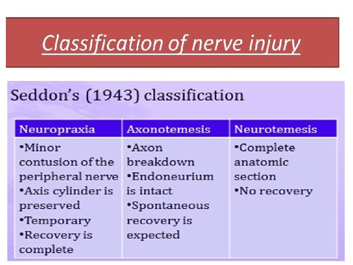
CLASSIFICATION OF NERVE INJURIES Seddon's Sunderland Three different types v. Neurapraxia v. Axonotmesis v. Neurotmesis First degree injury served as a useful classification for many years. • Increasingly, however, it has been recognized that many cases fall into an area somewhere between axonotmesis and neurotmesis. axonotmesis. Axonal degeneration takes place but, because the endoneurium is preserved, regeneration can, lead to complete, or near complete, recovery without the need for intervention. This embraces transient ischaemia and neurapraxia, reversible. Second degree injury= Seddon's Third degree injury worse than axonotmesis. The endoneurium is disrupted but the perineurial sheaths are intact and internal damage is limited. The chances of the axons reaching their targets are good, but fibrosis will limit recovery.
Types of nerve injuries
Nerve injury and repair Cause: damage of cell body, axon, myelin sheath, connective tissue, blood supply Normal axon and target organ (striated muscle). (b) Following nerve injury the distal part of the axon disintegrates and the myelin sheath breaks up. The nerve cell nucleus becomes eccentric and Nissl bodies are sparse. (c) New axonal tendrills grow into the mass of proliferating Schwann cells. One of the tendrill will find its way into the old endoneurial tube and (d) the axon will slowly regenerate. Ø Three basic processes Ø Wallerian degeneration Distal axon degeneration, following section or severe injury, with degeneration of the myelin. The process occurs within 7 -10 days of injury and this portion of the nerve is inexcitable electrically. Ø Axon degeneration Distal degenerated nerve is inexcitable electrically. Regeneration can occur since the basement membrane of the Schwann cell survives and act as a skeleton along which the axon regrows up to a rate of about 1 mm per day. Ø Demyelination Segmental destruction of the myelin sheath occurs without axonal damage. The primary lesion affects the Schwann cell and causes marked slowing of conduction or conduction block. Local demyelination is caused by inflammation, e. g : Guillain-Barre syndrome.
Thank You