
The nervous system v CNS v Brain Spinal
The nervous system v. CNS: v. Brain Spinal cord v. Peripheral NS: v Somatic nerves : voluntary v Autonomic nervous system (ANS) : involuntary; Controls functions of Heart, smooth muscle of blood vessels and viscera, exocrine gland. 1
The ANS Consists of: v 1. Sympathetic nervous system (Thoraco-lumbar) : - ergotropic ( spends energy). v 2. Parasympathetic nervous system (Cranio-sacral) - trophotropic (conserves energy) & thus promotes growth. v 3. Enteric nervous system (ENS) is included as 3 rd division of ANS 2
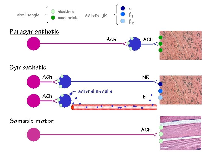
Neurotransmitter v. A chemical substance released from nerve endings & carries impulses across the synapses. It combines & stimulates its specific postsynaptic receptors v. Main neurotransmitters in ANS are : Acetylcholine ( in parasympathetic NS) Noradrenaline ( in sympathetic NS) and 3

General arrangement of ANS Each of the major 2 divisions of ANS consists of : v Pregangiolinic fibers : arise from cell bodies of neurons in CNS (preganglionic neurons) v Autonomic Ganglia : site of synapses between preganglonic fibers and cell bodies of postganglionic neurons v Postganglionic fibers : arise from cell bodies of postganglionic neurons in ganglia to innervate various effector tissues Central regulation is done by hypothalamus and centers in medulla oblongata in CNS. 4
5

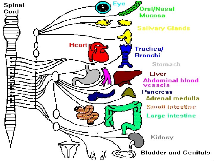
Sympathetic nervous system v. Preganglionic myelinated fibers leave CNS through anterior roots of thoracic & lumbar spinal nerves v. Sympathetic Ganglia in the paravertebral sympathetic chains. - connection is by myelinated white rami communicantes. v. From ganglia, post-ganglionic unmyelinated fibers run via grey rami communicantes back to spinal nerves, and then along blood vessels to innervate effector tissues (neuro-effector junctions) 6
7

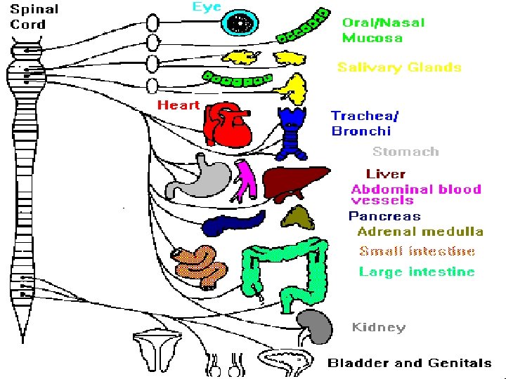
Parasympathetic system v. Preganglionic myelinated fibers leave CNS with cranial nerves (3 rd oculomotor, 7 th facial, 9 th glossopharyngeal, 10 th vagus nerves) & S 2, S 3 and S 4 spinal nerves Ganglia associated with above cranial nerves are ciliary ganglia with III nerve, pterygopalatine and submandibular ganglia with VII nerve, and otic ganglia with IX nerve. They are located close to innervated organs. In case of vagus nerve X, ganglion cells are located in wall of viscera in thorax, abdomen, and pelvis. From ganglion cells, short unmyelinated 8 postganglionic fibers run to innervate target tissues
9
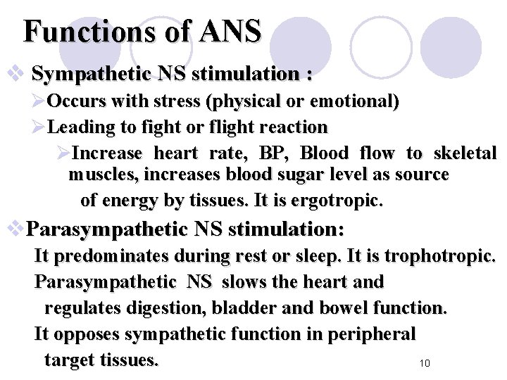
Functions of ANS v Sympathetic NS stimulation : ØOccurs with stress (physical or emotional) ØLeading to fight or flight reaction ØIncrease heart rate, BP, Blood flow to skeletal muscles, increases blood sugar level as source of energy by tissues. It is ergotropic. v. Parasympathetic NS stimulation: It predominates during rest or sleep. It is trophotropic. Parasympathetic NS slows the heart and regulates digestion, bladder and bowel function. It opposes sympathetic function in peripheral target tissues. 10
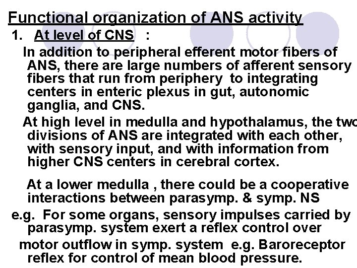
Functional organization of ANS activity 1. At level of CNS : In addition to peripheral efferent motor fibers of ANS, there are large numbers of afferent sensory fibers that run from periphery to integrating centers in enteric plexus in gut, autonomic ganglia, and CNS. At high level in medulla and hypothalamus, the two divisions of ANS are integrated with each other, with sensory input, and with information from higher CNS centers in cerebral cortex. At a lower medulla , there could be a cooperative interactions between parasymp. & symp. NS e. g. For some organs, sensory impulses carried by parasymp. system exert a reflex control over motor outflow in symp. system e. g. Baroreceptor reflex for control of mean blood pressure.

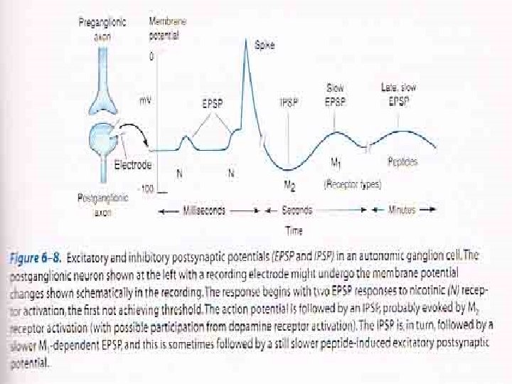
2. At level of receptors : Presynaptic regulation : feedback inhibition by presynaptic receptors : these can be autoreceptors or heteroceptors Postsynaptic regulation : upregulation or downregulation of postsynaptic receptors 3. Ganglionic modulation : Modulation of effect of Ach in autonomic ganglia by secondary events evoked by same transmitter (Ach) or different transmitter acting on different postsynaptic receptors
3. Enteric Nervous System (ENS) v. Collections of neurons in wall of the GIT vconsidered as 3 rd division of the ANS v. Includes: ØA. Myenteric plexus of Auerbach ØB. Submucosal plexus of Meissner v. Regulates motor & secretory functions of GIT 14
ENS contains: ØCholinergic fibers ØAdrenergic fibers ØNonadrenergic noncholinergic (NANC) neurons Note : Sympathetic and parasympathetic fibers in ENS play a modulatory role; removal of both fibers does not completely stop activity in either the enteric plexus neurons or in the smooth muscle and gland innervated by them 15

NANC neurons v. Motor & sensory neurons in autonomic target organs (gut, bronchi & bladder) v. Neurotransmitters: ØPeptides e. g opioid peptides, substance P, VIP ØATP Ø NO ØSerotonin (5 -Hydroxytryptamine) 16
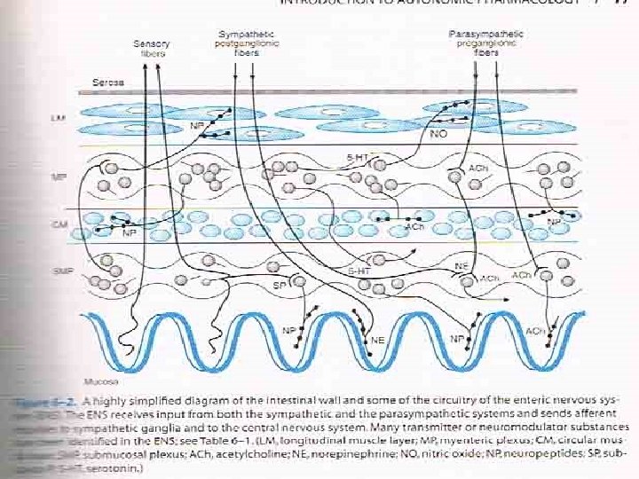
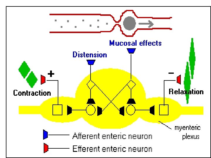
Peristalsis is a distinctive pattern of smooth muscle contractions that propels food distally through the esophagus, stomach, and intestines. Peristalsis is a manifestation of two major reflexes within the enteric nervous system that are stimulated by a bolus of foodstuff in the lumen. Mechanical distension and perhaps mucosal irritation stimulate afferent sensory enteric neurons. These sensory neurons synapse with two sets of cholinergic interneurons, which lead to two distinct effects :
1. One group of interneurons activates excitatory motor neurons above the bolus - these neurons, which contain acetylcholine and substance P, stimulate contraction of smooth muscle above the bolus. 2. Another group of interneurons activates inhibitory motor neurons that stimulate relaxation of smooth muscle below the bolus. These inhibitor neurons appear to use nitric oxide, vasoactive intestinal peptide and ATP as inhibitory neurotransmitters. Peristalsis is not affected to any degree by vagotomy or sympathetectomy, indicating its mediation by the intestine's local, intrinsic nervous system.
Cholinergic Transmission v. Acetylcholine ( Ach ) is the neurotransmitter v. Acts on cholinoceptors present in: ØAutonomic ganglia (sympathetic & parasympathetic) ØNeuro-effector junction of parasympathetic post-ganglionic fibers ØAdrenal medulla ØNMJ (Neuro-muscular junction) endplates ØCNS 21
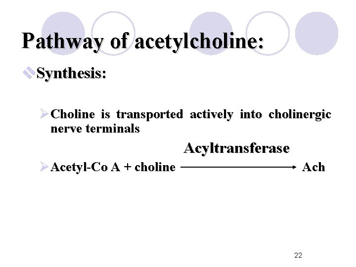
Pathway of acetylcholine: v. Synthesis: ØCholine is transported actively into cholinergic nerve terminals Acyltransferase ØAcetyl-Co A + choline Ach 22
Pathway of Acetylcholine Storage: Ø Ach is actively transported from cytoplasm into storage vesicles where it is protected from degradation Release : by exocytosis Binding : Ach binds to & activates cholinoceptors on post-synaptic tissues leading to various actions 23
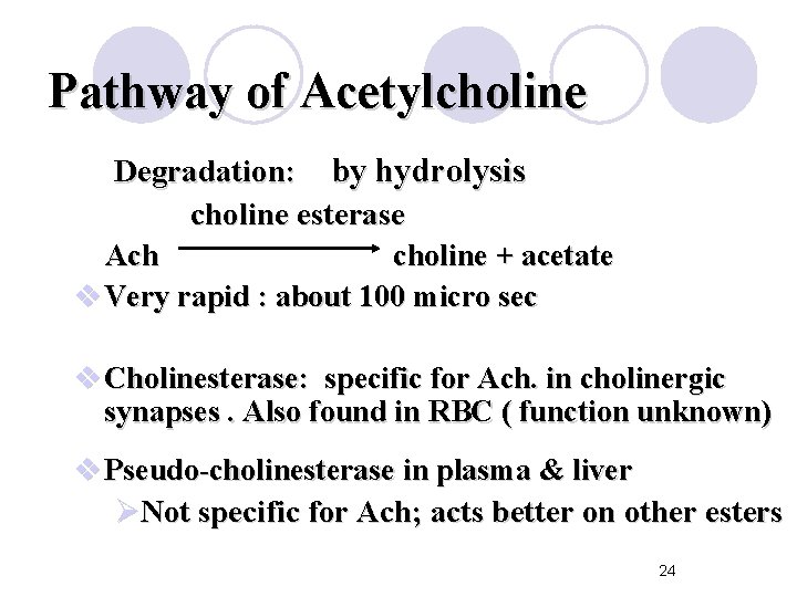
Pathway of Acetylcholine Degradation: by hydrolysis choline esterase Ach choline + acetate v Very rapid : about 100 micro sec v Cholinesterase: specific for Ach. in cholinergic synapses. Also found in RBC ( function unknown) v Pseudo-cholinesterase in plasma & liver ØNot specific for Ach; acts better on other esters 24
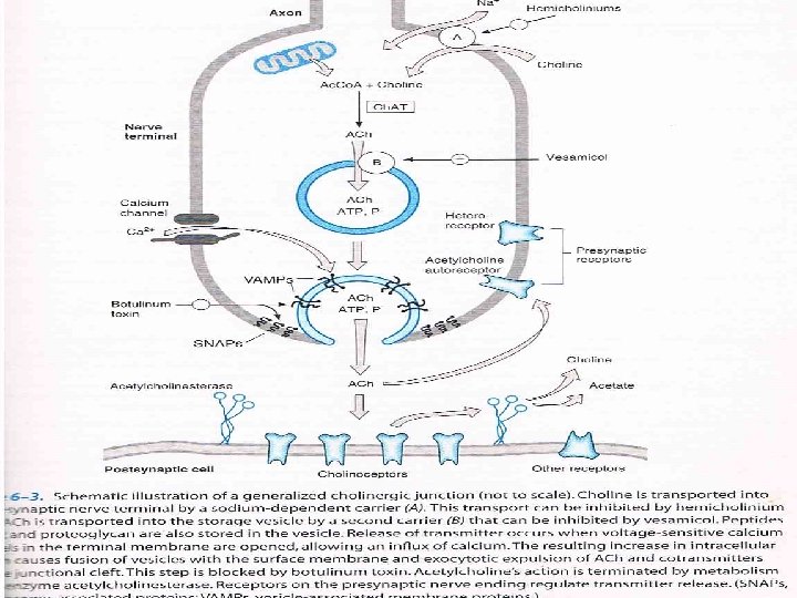
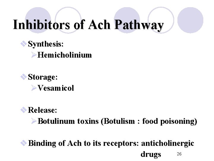
Inhibitors of Ach Pathway v Synthesis: ØHemicholinium v Storage: ØVesamicol v Release: ØBotulinum toxins (Botulism : food poisoning) v Binding of Ach to its receptors: anticholinergic 26 drugs
Cholinoceptors v. Muscarinic receptors v. Nicotinic receptors 27
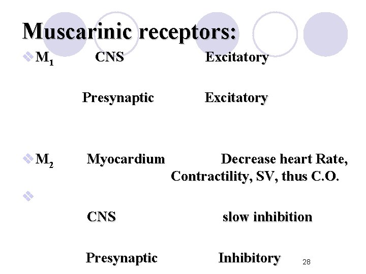
Muscarinic receptors: v M 1 CNS Presynaptic v M 2 Myocardium Excitatory Decrease heart Rate, Contractility, SV, thus C. O. v CNS Presynaptic slow inhibition Inhibitory 28
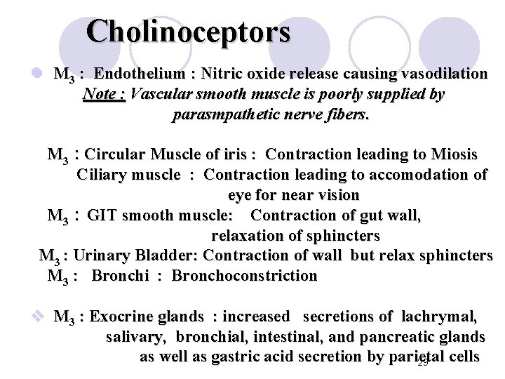
Cholinoceptors l M 3 : Endothelium : Nitric oxide release causing vasodilation Note : Vascular smooth muscle is poorly supplied by parasmpathetic nerve fibers. M 3 : Circular Muscle of iris : Contraction leading to Miosis Ciliary muscle : Contraction leading to accomodation of eye for near vision M 3 : GIT smooth muscle: Contraction of gut wall, relaxation of sphincters M 3 : Urinary Bladder: Contraction of wall but relax sphincters M 3 : Bronchi : Bronchoconstriction v M 3 : Exocrine glands : increased secretions of lachrymal, salivary, bronchial, intestinal, and pancreatic glands as well as gastric acid secretion by parietal cells 29
Nicotinic receptors v Nicotinic NN : Autonomic ganglia (Stimulation) : Adrenal medulla : considered as large target sympathetic ganglion (Noradrenaline & adrenaline release) v Nicotinic NM : Stimulation of NMJ endplate leading to membrane depolarization and skeletal muscle contraction NM receptors are composed of 5 subunits while NN are receptors are composed of 4 subunits. Note : If Ach persists at nicotinic receptors, stimulation is followed by inhibition due to persistent depolarization 30 of post-synaptic cells
Receptor transduction mechanisms : Muscarinic receptors : GPCRs. metabotropic M 1 and M 3 stimulate Gq and leads to breakdown of PIP 2 in cell membrane to IP 3 and DAG M 2 stimulates Gi leading to inhibition of adenyl cyclase and a decrease in cytoplasmic c. AMP; also increases K+ permeability in resting pacemaker cells in SA node; it also decreases the slow calcium current during phase 2 of cardiac action potential Nicotinic receptors : ionotropic receptors in autonomic ganglia or NMJ that increases cationic (sodium and potassium) permeability and lead to depolarization
- Slides: 31