The Nervous System The human nervous system can


 • A physiological mechanism that alters the permeability of brain")


 is composed of endothelial cells")


, while best remembered for the disease state named")


 is a")


 DESCRIPTION SINEMET* (Carbidopa-Levodopa) is a combination of carbidopa and levodopa for the")
 are endogenous opioid biochemical compounds. They are")


 • http: //www. epilepsy. com/web/animation. php? swf=what_is")

 “Fight or flight”")


, also known as noradrenaline")


 is a bicyclic antidepressant morpholine derivative that")


.")
 • Attention-Deficit/Hyperactivity Disorder (ADHD) (sometimes referred to as ADD")

- Slides: 94
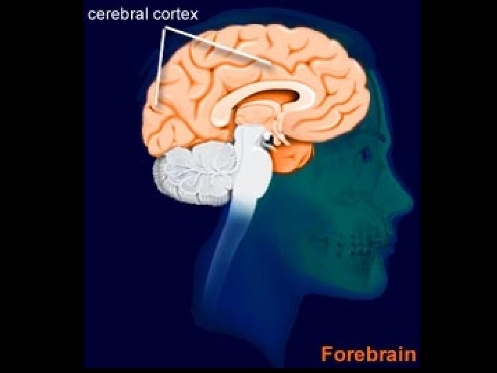
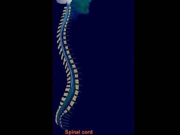
The Nervous System • The human nervous system can be divided into two parts: the central nervous system (CNS) and the peripheral nervous system (PNS) • http: //pennhealth. com/health_info/anima tionplayer/nerve_conduction. html
Central Nervous System Drugs that affect the CNS can: • Selectively relieve pain • Reduce fever • Suppress disordered movement • Induce sleep or arousal • Reduce appetite • Allay the tendency to vomit • Be used to treat anxiety, depression, schizophrenia, Parkinson’s Disease, Alzheimer’s Disease, epilepsy, migraine, etc.
How do drugs work in the CNS? • “A central underlying concept of neuropharmacology is that drugs that influence behavior and improve the functional status of patients with neurological or psychiatric diseases act by enhancing or blunting the effectiveness of specific combinations of synaptic transmitter actions. ”
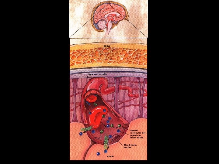
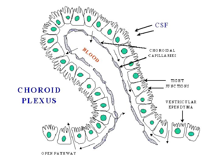
Blood Brain Barrier (BBB) • A physiological mechanism that alters the permeability of brain capillaries, so that some substances, such as certain drugs, are prevented from entering brain tissue, while other substances are allowed to enter freely. • The separation of the brain, which is bathed in a clear cerebrospinal fluid, from the bloodstream. The cells near the capillary beds external to the brain selectively filter the molecules that are allowed to enter the brain, creating a more stable, nearly pathogen-free environment.
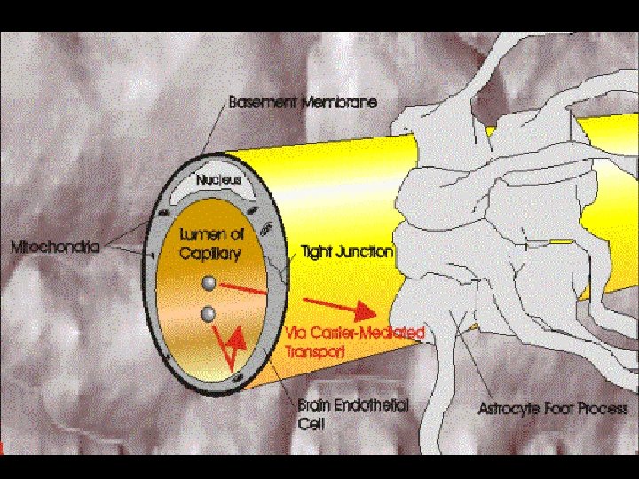
Diagram of a cerebral capillary enclosed in astrocyte end-feet. Characteristics of the blood-brain barrier are indicated: (1) tight junctions that seal the pathway between the capillary (endothelial) cells; (2) the lipid nature of the cell membranes of the capillary wall which makes it a barrier towater -soluble molecules; (3), (4), and (5) represent some of the carriers and ion channels; (6) the 'enzymatic barrier'that removes molecules from the blood; (7) the efflux pumps which extrude fatsoluble molecules that have crossed into the cells
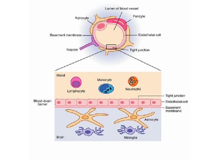
Blood-Brain-Barrier • Oxygen, glucose, and white blood cells are molecules that are able to pass through this barrier. Red blood cells cannot.
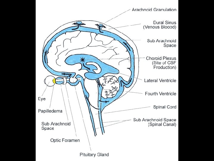
Blood Brain Barrier • The blood-brain barrier (abbreviated BBB) is composed of endothelial cells packed tightly in brain capillaries that more greatly restrict passage of substances from the bloodstream than do endothelial cells in capillaries elsewhere in the body. • Processes from astrocytes surround the epithelial cells of the BBB providing biochemical support to the epithelial cells. • The BBB should not be confused with the bloodcerebrospinal fluid barrier (BCB), a function of the choroid plexus.
History of the BBB • The existence of such a barrier was first noticed in experiments by Paul Ehrlich in the late-19 th century. Ehrlich was a bacteriologist who was studying staining, used for many studies to make fine structures visible. Some of these dyes, notably the aniline dyes that were then popular, would stain all of the organs of an animal except the brain when injected. At the time, Ehrlich attributed this to the brain simply not picking up as much of the dye.
• However, in a later experiment in 1913, Edwin Goldmann (one of Ehrlich's students) injected the dye into the spinal fluid of the brain directly. • He found that in this case the brain would become dyed, but the rest of the body remained dye-free. This clearly demonstrated the existence of some sort of barrier between the two sections of the body.
History of the BBB • At the time, it was thought that the blood vessels themselves were responsible for the barrier, as there was no obvious membrane that could be found. • It was not until the introduction of the scanning electron microscope to the medical research fields in the 1960 s that this could be demonstrated. The concept of the blood-brain (then termed hematoencephalic) barrier was proposed by Lina Stern in 1921.
What is the purpose of the BBB? • The blood-brain barrier protects the brain from the many chemicals flowing around the body. • For example, many bodily functions are controlled by hormones, which are detected by receptors on the plasma membranes of targeted cells throughout the body. • The secretion of many hormones are controlled by the brain, but these hormones generally do not penetrate the brain from the blood, so in order to control the rate of hormone secretion effectively, there are specialized sites where neurons can "sample" the composition of the circulating blood.
• At these sites, the blood-brain barrier is 'leaky'; these sites include three important 'circumventricular organs', the subfornical organ, the area postrema and the organum vasculosum of the lamina terminalis (OVLT). • The blood-brain barrier is also an effective way to protect the brain from common infections. Thus infections of the brain are very rare; however, as antibodies are too large to cross the blood-brain barrier, when infections of the brain do occur they can be very serious and difficult to treat.
How does the BBB affect the design of therapeutic agents? • Mechanisms for drug targeting in the brain involve going either "through" or "behind" the BBB. • Modalities for drug delivery through the BBB entail disruption of the BBB by osmotic means, biochemically by the use of vasoactive substances such as bradykinin, or even by localized exposure to high intensity focused ultrasound (HIFU). • The potential for using BBB opening to target specific agents to brain tumors has just begun to be explored.
The Blood Brain Barrier • http: //www. clinicaloptions. com/HIV/Man agement%20 Series/Neuro. AIDS/Animati on/Blood%20 Brain%20 Barrier. aspx
Introduction to the CNS • http: //biosingularity. wordpress. com/200 7/05/07/neurons-and-how-they-workanimation • http: //www. healthscout. com/animation/6 8/10/main. html
Neurotransmitters found in the CNS
It’s a balancing act!! • Current models of CNS diseases often attribute the physiological cause of the disease to an imbalance of neurotransmitters.
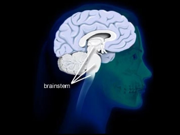
Acetylcholine • All ACh receptors in the CNS are nicotinergic. The stimulating effect of nicotine is due to the influence of these receptors. • Acetylcholine is transmitted within cholinergic pathways that are concentrated mainly in specific regions of the brainstem and are thought to be involved in cognitive functions, especially memory. Severe damage to these pathways is the probable cause of Alzheimer’s disease.
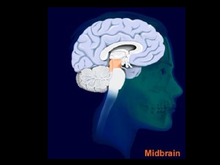
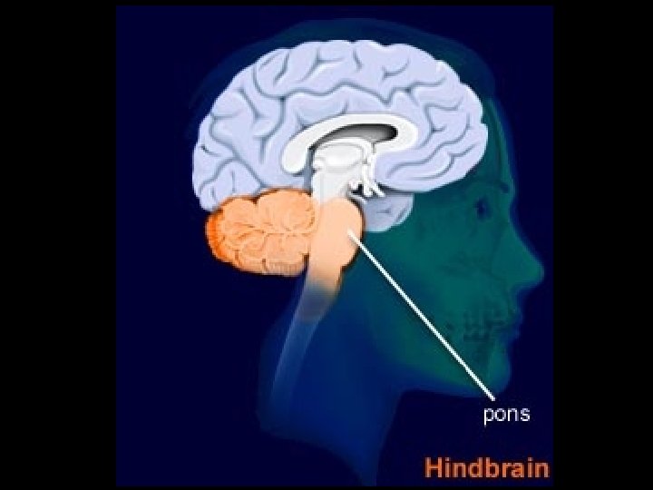
Norepinephrine • Most cell bodies of noradrenergic neurons are in the locus coeruleus, a center in the brain stem. These neurons send their axons to the limbic system (appetite inhibition), the subcortical centers and the cerebral cortex (arousal). • Noradrenaline is classed as a monoamine neurotransmitter and noradrenergic neuronsハ are found in the locus coeruleusハ, the pons and the reticular formation in the brain. These neurons provide projections to the cortex, hippocampusハ, thalamusハ and midbrain. The release of noradrenaline tends to increase the level of excitatory activity within the brain, and noradrenergic pathways are thought to be particularly involved in the control of functions such as attention and arousal.
Locus ceruleus • The Locus ceruleus, also spelled locus caeruleus or locus coeruleus (Latin for 'the blue spot'), is a nucleus in the brain stem responsible for physiological responses to stress and panic. The locus ceruleus (or "LC") is located within the dorsal wall of the upper pons, under the cerebellum in the caudal midbrain, surrounded by the fourth ventricle. This nucleus is one of the main sources of norepinephrine in the brain, and is composed of mostly medium-sized neurons. Melanin granules inside the LC contribute to its blue color; it is thereby also known as the nucleus pigmentosus pontis, meaning "heavily pigmented nucleus of the pons".
Locus ceruleus
hippocampus
Thalamus
• Dopamine is also classed as a monoamine neurotransmitter and is concentrated in very specific groups of neurons collectively called the basal ganglia. Dopaminergic neurons are widely distributed throughout the brain in three important dopamine systems (pathways): the nigrostriatal, mesocorticolimbic, and the tuberohypophyseal pathways. A decreased brain dopamine concentration is a contributing factor in Parkinsonユs disease, while an increase in dopamine concentration has a role in the development of schizophrenia.
Biosynthesis of Epinephrine
• Although dopamine is synthesized by only several hundred thousand cells, it fulfils an exceedingly important role in the higher parts of the CNS. These dopaminergic neurons can be divided into three subgroups with different functions. The first group regulates movements: a deficit of dopamine in this (nigrostriatal) system causes Parkinson's disease which is characterized by trembling, stiffness and other motor disorders, while in the later phases dementia can also set in. The second group, the mesolimbic, has a function in regulating emotional behavior. The third group, the mesocortical, projects only to the prefrontal cortex. This area of cortex is involved with various cognitive functions, memory, behavioral planning and abstract thinking, as well as in emotional aspects, especially in relation to stress. The earlier mentioned reward system is part of this last system. The nucleus accumbens is an important intermediate station here. Disorders in the latter two systems are associated with schizophrenia.
Dopamine and Parkinson’s Disease • In patients with Parkinson’s disease, there is disease or degeneration of the so-called basal ganglia in the deeper grey matter of the brain, particularly of that part known as the substantia nigra.
Parkinson’s Disease • The substantia nigra, which connects with the striatum (caudate nucleus and globus pallidus), contains black pigmented cells and, in normal individuals, produces a number of chemical transmitters, the most important of which is dopamine. Transmitters are chemicals that transmit, that is, pass on, a message from one cell to the next, either stimulating or inhibiting the function concerned; it is like electricity being the transmitter of sound waves in the radio. Other transmitters include serotonin, somatostatin and noradrenaline. In Parkinsonユs disease, the basal ganglia cells produce less dopamine, which is needed to transmit vital messages to other parts of the brain, and to the spinal cord, nerves and muscles.
In Parkinson’s disease, there is degeneration of the substantia nigra which produces the chemical dopamine deep inside the brain
Parkinson’s Disease • The basal ganglia, through the action of dopamine, are responsible for planning and controlling automatic movements of the body, such as pointing with a finger, pulling on a sock, writing or walking. If the basal ganglia are not working properly, as in Parkinson’s disease patients, all aspects of movement are impaired, resulting in the characteristic features of the disease ミ slowness of movement, stiffness and effort required to move a limb and, often, tremor. • Dopamine levels in the brain’s substantia nigra do normally fall with ageing. However, they have to fall to one-fifth of normal values for the symptoms and signs of parkinsonism to emerge.
Parkinson’s Disease • http: //www. parkinsonshealth. com/About PD/Section. aspx? Section. Id=798 d 598 e 2 a 1 c-4747 -ac 81 -cd 92 f 475744 b • http: //www. medindia. net/animation/parki nsons_disease. asp
History • James Parkinson (1755 -1824), while best remembered for the disease state named after him by Charcot, was a man of many talents and interests. Publishing on chemistry, paleontology and other diverse topics, he was, early in his career, a social activist championing the rights of the disenfranchised and poor. His efforts in this area were enough to result in his arrest and appearance before The Privy Council in London on at least one occasion. In collaboration with his son, who was a surgeon, he also offered the first description, in the English language, of a ruptured appendix.
History of Parkinson’s Disease • His small but famous publication, "Essay on the Shaking Palsy", appeared in 1817, 7 years before his death in 1824. The clinical description of 6 patients was a remarkable masterpiece testifying to his prodigious powers of observation for most of the 6 were never actually examined by Parkinson himself; rather, they were simply observed walking on the streets of London.
Treatment of Parkinson’s Disease • Since PD is related to a deficiency of dopamine, it would be appropriate to administer dopamine • Problem: Dopamine does not cross BBB, since it is too polar
History of Treatment of PD • Arvid Carlsson (b. January 25, 1923) is a Swedish scientist who is best known for his work with the neurotransmitter dopamine and its effects in Parkinson's disease. Carlsson won the Nobel Prize in Physiology or Medicine in 2000 along with corecipients Eric Kandel and Paul Greengard. Carlsson was born in Uppsala, Sweden, son of Gottfrid Carlsson, historian and later professor of history at the Lund University, where he began his medical education in 1941. Although Sweden was neutral during World War II, Carlsson's education was interrupted by several years of service in the Swedish Armed Forces. In 1951, he received his M. L. degree (the equivalent of the American M. D. ) and his M. D. (the equivalent of the American Ph. D. ). He then became a professor at the University of Lund. In 1959 he became a professor at the G嗾eborg University. In the 1950 s, Carlsson demonstrated that dopamine was a neurotransmitter in the brain and not just a precursor for norepinephrine, as had been previously believed. He developed a method for measuring the amount of dopamine in brain tissues and found that dopamine levels in the basal ganglia, a brain area important for movement, were particularly high. Carlsson then showed that giving animals the drug reserpine caused a decrease in dopamine levels and a loss of movement control. These effects were similar to the symptoms of Parkinson's disease. By administering to these animals L-Dopa, a precursor to dopamine, he could alleviate the symptoms. These findings led other doctors try L-Dopa with human Parkinson's patients and found it to alleviate some of the symptoms in the early stages of Parkinson's. LDopa is still today the cornerstone of Parkinson therapy.
Biosynthesis of Epinephrine
Wait a minute! • If dopamine is too polar to cross the BBB, how can L-DOPA cross it?
Answer! • L-DOPA is transported across the BBB by an amino acid transport system (same one used for tyrosine and phenylalanine) • Once across, L-DOPA is decarboxylated to dopamine by Dopa Decarboxylase (DDC).
• This is an example of a “prodrug”, that is, a molecule that is a precursor to the drug and is converted to the actual drug at an appropriate place in the body.
• In actual practice, L-DOPA is almost always coadminstered together with an inhibitor of aromatic L-amino acid decarboxylase, so it doesn’t get converted to dopamine before it crosses the BBB. • The inhibitor commonly used is carbidopa, which does not cross the BBB itself. • The inhibitor also prevents undesirable side effects of dopamine release into the PNS, including nausea.
SINEMET (CARBIDOPA-LEVODOPA) DESCRIPTION SINEMET* (Carbidopa-Levodopa) is a combination of carbidopa and levodopa for the treatment of Parkinson's disease and syndrome. • http: //www. learningcommons. umn. edu/ neuro/mod 6/carb. html
Endorphin • Endorphins (or more correctly Endomorphines) are endogenous opioid biochemical compounds. They are peptides produced by the pituitary gland the hypothalamus in vertebrates, and they resemble the opiates in their abilities to produce analgesia and a sense of well-being. In other words, they might work as "natural pain killers. " Using drugs may increase the effects of the endorphins.
Serotonin • Although the CNS contains less than 2% of the total serotonin in the body, serotonin plays a very important role in a range of brain functions. It is synthesised from the amino acid tryptophan. Within the brain, serotonin is localised mainly in nerve pathways emerging from the raphe nuclei, a group of nuclei at the centre of the reticular formation in the Midbrainハ, ponsハ and medulla. These serotonergic pathways spread extensively throughout the brainstemハ, the cerebral cortexハ and the spinal cordハ. In addition to mood control, serotonin has been linked with a wide variety of functions, including the regulation of sleep, pain perception, body temperature, blood pressure and hormonal activity. Outside the brain, serotonin exerts a number of important effects, particularly involving the gastrointestinal and cardiovascular systems.
What is serotonin? In the central nervous system, serotonin is believed to play an important role in the regulation of body temperature, mood, sleep, vomiting, sexuality, and appetite. Low levels of serotonin have been associated with several disorders, namely clinical depression, obsessivecompulsive disorder (OCD), migraine, irritable bowel syndrome, tinnitus, fibromyalgia, bipolar disorder, and anxiety disorders. [citation needed] If neurons of the brainstem that make serotonin—serotonergic neurons—are abnormal, there is a risk of sudden infant death syndrome (SIDS) in an infant. [1]
Understanding Serotonin • The pharmacology of 5 -HT is extremely complex, with its actions being mediated by a large and diverse range of 5 -HT receptors. At least seven different receptor "families" are known to exist, each located in different parts of the body and triggering different responses. As with all neurotransmitters, the effects of 5 -HT on the human mood and state of mind, and its role in consciousness, are very difficult to ascertain.
Understanding Serotonin • Serotonergic action is terminated primarily via uptake of 5 -HT from the synapse. This is through the specific monoamine transporter for 5 -HT, 5 -HT reuptake transporter, on the presynaptic neuron. Various agents can inhibit 5 -HT reuptake including MDMA (ecstasy), cocaine, tricyclic antidepressants (TCAs) and selective serotonin reuptake inhibitors (SSRIs). Recent research suggests that serotonin plays an important role in liver regeneration and acts as a mitogen (induces cell division) throughout the body.
Anatomy of the Brain (seizures) • http: //www. epilepsy. com/web/animation. php? swf=what_is
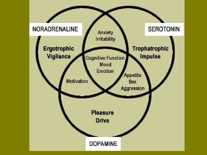
The action of drugs to treat mental illness • Serotonin, noradrenaline and dopamine are involved in the control of many of our mental states, sometimes acting on their own and at other times acting together (illustrated in the following diagram). These and other neurotransmitters are likely to play a pivotal role in the pathological basis of mental illness and diseases of the brain. Much of the evidence for this stems from the fact that most of the effective antidepressant drugs are thought to work by changing either serotonin and/or noradrenaline metabolism, or receptor sensitivity to these neurotransmitters
Definitions • Ergotropic: Energy expending systems (sympathetic division of the PNS) “Fight or flight” • Trophotropic: Nutrient accumulating systems (parasympathetic division of the PNS) “Rest and digest”
Schizophrenia • http: //www. healthscout. com/animation/6 8/49/main. html • http: //www. abilify. com/abilify/channels/s ch_content. jsp? BV_Use. BVCookie=Yes &channel. Name=Schizophrenia%2 f. Sch_ Brain_Sch_Abilify&referrer=null
Historical: Drugs to treat schizophrenia • http: //www. pbs. org/wgbh/aso/databank/ entries/dh 52 dr. html
• Histamine is a biogenic amine chemical involved in local immune responses as well as regulating physiological function in the gut and acting as a neurotransmitter (Marieb, 2001, p. 414). New evidence also indicates that histamine plays a role in chemotaxis of white blood cells.
• Histamine is released as a neurotransmitter. The cell bodies of neurons which release histamine are found in the posterior hypothalamus, in various tuberomammillary nuclei. From here, these histaminergic neurons project throughout the brain, to the cortex through the medial forebrain bundle. Histaminergic action is known to modulate sleep. Classically, antihistamines (H 1 histamine receptor antagonists) produce sleep. Likewise, destruction of histamine releasing neurons, or inhibition of histamine synthesis leads to an inability to maintain vigilance. Finally, H 3 receptor antagonists (which stimulate histamine release) increase wakefulness. It has been shown that histaminergic cells have the most wakefulness-related firing pattern of any neuronal type thus far recorded. They fire rapidly during waking, fire more slowly during periods of relaxation/tiredness and completely stop firing during REM and non-REM sleep. Histaminergic cells can be recorded firing just before an animal shows signs of waking.
• Sexual response: • Research has shown that histamine is released as part of the human orgasm from mast cells in the genitals, and the histamine release has been connected to the sex flush among women. If this response is lacking while a woman also has trouble achieving orgasm, this may be a sign of histapenia. In such cases, a doctor may prescribe diet supplements with folic acid and niacin (which used in conjunction can increase blood histamine levels and histamine release), or L-histidine. Conversely, men with high histamine levels may suffer from premature ejaculations.
Antibodies and the Immune Response • Antibodies are manufactured by the lymph system. Antibodies are specialized proteins that the body produces in response to invasion by a foreign substance. The process of antibody formation begins when an antigen stimulates specialized lymphocytes, called B cells, into action. Antibodies then counteract invading antigens by combining with the antigen to render it harmless to the body. • Production of white blood cells and antibodies in reaction to an invading disease organism is called an immune response. This response is one of the body's primary and most efficient lines of defense. In most cases, once antibodies have been produced to fight a certain organism, it no longer poses a great threat to the body. That is why one attack of a disease often prevents that same disease from infecting the body again -- the first attack causes production of antibodies that protect the body against subsequent attacks. With measles, for example, antibodies are produced as a result of having the disease or of being immunized with the measles vaccine. These antibodies are able to resist a second attack of the disease.
Antibodies and the Immune Response • Antibodies are not always beneficial. For example, when tissue from another body, such as a transplanted heart, is introduced, antibodies are produced to destroy the "invader. " Transplants usually are made possible only by means of drugs that act against the body's natural immune response. Also, when blood is transfused from one person to another, it must be of a matching type; otherwise, the recipient's immune system will manufacture antibodies to destroy the transfused blood. • Sometimes, the immune system causes reactions that make the body unusually sensitive to foreign material. When the immune response is disruptive to the body in this way, it is called an allergic reaction. Let's look at this important mechanism, and the types of allergens, in the next section.
Allergic Reaction • An allergy is a state of special sensitivity to a particular environmental substance, or allergen. An allergic reaction is the body's response to exposure to an allergen. • Although an allergy can be present almost immediately after exposure to an allergen, it usually develops over time, as the immune system forms antibodies against the foreign substance. Under normal conditions, such antibodies work to protect the body from further attack. In the case of an allergy, however, the antibodies and other specialized cells involved in this protective function trigger an unusual sensitivity, or overreaction, to the foreign substance. • The antibodies stimulate specialized cells to produce histamine, a powerful chemical. Histamine causes the small blood vessels to enlarge and the smooth muscles (such as those in the airways and the digestive tract) to constrict. Histamine release can also cause other reactions, such as hives.
Allergic Reaction • No one knows why allergies develop, but it is known that an allergy can appear, disappear, or reappear at any time and at any age. Allergic reactions rarely occur during the first encounter with the troublesome allergen because the body needs time to accumulate the antibodies. Also, an individual's sensitivity to certain allergens seems to be related to a family history of allergies. People who have a tendency to develop allergies are referred to as atopic. • An allergic reaction can be so mild that it is barely noticeable or so severe that it is life-threatening. An extremely severe allergic reaction, called anaphylactic shock, is marked by breathing difficulties (from swelling of the throat and larynx and narrowing of the bronchial tubes), itching skin, hives, and collapse of the blood vessels, as well as by vomiting, diarrhea, and cramps. This condition can be fatal if not treated immediately.
Allergic reaction: Histamine and Antihistamines • http: //www. healthscout. com/animation/6 8/20/main. html • http: //pennhealth. com/health_info/anima tionplayer/allergies. html
Antihistamines to Antipsychotics? • In the late 1930 s, such dicyclic antihistamines as phenbenzamine, diphenhydramine, and mepyramine were in wide clinical use. The antihistamines' most striking clinical side-effect was CNS depression -- drowsiness.
Antihistamines to Antipsychotics? • In common use, the term antihistamine refers only to H 1 -receptor antagonists, also known as H 1 -antihistamines. It has been discovered that these H 1 -antihistamines are actually inverse agonists at the histamine H 1 receptor, rather than antagonists per se.
Antihistamines to Antipsychotics? • In the late 1930 s, Paul Charpentier had synthesized the first tricyclic antihistamine, promethazine, which had a strong sedative effect. He then synthesized a variety of promethazine analogues, including chiorpromazine.
Antihistamines to Antipsychotics? • http: //ajp. psychiatryonline. org/cgi/conte nt/full/160/10/1895? etoc
Antihistamines to Antipsychotics? • Chlorpromazine was the first antipsychotic drug, used during the 1950 s and 1960 s. Used as chlorpromazine hydrochloride and sold under the tradenames Largactilィ and Thorazineィ, it has sedative, hypotensive and antiemetic properties as well as anticholinergic and antidopaminergic effects. It also has anxiolytic (alleviation of anxiety) properties. Today, chlorpromazine is considered a typical antipsychotic.
Antihistamines to Antipsychotics? • The drug had been developed by Laboratoires Rh冢e-Poulenc in 1950 but they sold the rights in 1952 to Smith-Kline & French (today's Glaxo. Smith. Kline). The drug was being sold as an antiemetic when its other use was noted. Smith-Kline was quick to encourage clinical trials and in 1954 the drug was approved in the US for psychiatric treatment. The effect of this drug in emptying psychiatric hospitals has been compared to that of penicillin and infectious diseases. [1] Over 100 million people were treated but the popularity of the drug fell from the late 1960 s as the severe extrapyramidal side effects and tardive dyskinesia became more of a concern. From chlorpromazine a number of other similar neuroleptics were developed (e. g. triflupromazine, trifluoperazine).
Antihistamines to Antipsychotics? • • Previously used as an antihistamine and antiemetic its effects on mental state were first reported by the French doctor Henri Laborit in 1951 or 1952 (different sources) as sedation without narcosis. It became possible to cause 'artificial hibernation' in patients, if used as a cocktail together with pethidine and hydergine. Patients with shock, severe trauma or burns, become, if treated so, sedated, without anxiety and unresponsive/indifferent to painful external stimuli like minor surgical interventions. The first published clinical trial was that of Jean Delay and Pierre Deniker at Ste. Anne H冱pital in Paris in 1952, in which they treated 38 psychotic patients with daily injections of chlorpromazine. [1] Drug treatment with chlorpromazine went beyond simple sedation with patients showing improvements in thinking and emotional behaviour. Ironically, the antipsychotic properties of chlorpromazine appear to be unrelated to its sedative properties. During long term therapy some tolerance to the sedative effect develops. Chlorpromazine substituted and eclipsed the old therapies of electro and insulin shocks and other methods such as psychosurgical means (lobotomy) causing permanent brain injury. Before the era of neuroleptics, starting with chlorpromazine, positive long-term results for psychotic patients were only 20%.
Definitions • Neuroleptic: A term that refers to the effects of antipsychotic drugs on a patient, especially on his or her cognition and behavior. • Neuroleptic drugs may produce a state of apathy, lack of initiative and limited range of emotion. In psychotic patients, neuroleptic drugs cause a reduction in confusion and agitation and tend to normalize psychomotor activity. The term comes from the Greek "lepsis" meaning a taking hold.
Definitions • Extrapyramidal side effects: Physical symptoms, including tremor, slurred speech, akathesia, dystonia, anxiety, distress, paranoia, and bradyphrenia, that are primarily associated with improper dosing of or unusual reactions to neuroleptic (anti-psychotic) medications.
Reward pathways in the CNS • The most important reward pathway in brain is the mesolimbic dopamine system. This circuit (VTA-NAc) is a key detector of a rewarding stimulus. Under normal conditions, the circuit controls an individualユs responses to natural rewards, such as food, sex, and social interactions, and is therefore an important determinant of motivation and incentive drive. In simplistic terms, activation of the pathway tells the individual to repeat what it just did to get that reward. It also tells the memory centers in the brain to pay particular attention to all features of that rewarding experience, so it can be repeated in the future. Not surprisingly, it is a very old pathway from an evolutionary point of view. The use of dopamine neurons to mediate behavioral responses to natural rewards is seen in worms and flies, which evolved 1 -2 billion years ago. • http: //www 3. utsouthwestern. edu/molpsych/pa ths_b 02. htm
Norepinephrine Reuptake Inhibitors as Antidepressants • Norepinephrine reuptake inhibitors (NRIs), also known as noradrenaline reuptake inhibitors (NARIs), are compounds that elevate the extracellular level of the neurotransmitter norepinephrine in the central nervous system by inhibiting its reuptake from the synaptic cleft into the presynaptic neuronal terminal. The drugs inhibit the class of neurotransmitter transporters known as norepinephrine transporters. They have virtually no action at other monoamine transporters.
Depression • http: //www. healthcentral. com/depression/intro duction-5003 -109. html • http: //www. healthscout. com/animation/68/10/ main. html • http: //www. insidecymbalta. com/patient_resou rces/neuro_animation. jsp
Norepinephrin Reuptake Inhibitors for Depression • Atomoxetine is classified as a norepinephrine reuptake inhibitor, and is approved for use in children, adolescents, and adults. • Atomoxetine is the first non-stimulant drug approved for the treatment of attention -deficit hyperactivity disorder (ADHD). It is sold in the form of the hydrochloride salt of atomoxetine. It is manufactured and marketed under the brand name Stratteraィ by Eli Lilly and Company as a generic Attentin by Torrent Pharmaceuticals. There is currently no generic available within the United States due to patent restrictions.
Atomoxetine • Strattera was originally intended to be a new antidepressant drug; however, in clinical trials, no such benefits could be proven. Since norepinephrine is believed to play a role in ADHD, Strattera was tested and subsequently approved as an ADHD treatment.
• Reboxetine is an antidepressant drug used in the treatment of clinical depression, panic disorder and ADD/ADHD. Its mesilate (i. e. methanesulfonate) salt is sold under tradenames including Edronaxィ, Noreboxィ, Proliftィ, Solvexィ or Vestraィ. • Unlike most antidepressants on the market, reboxetine is a noradrenaline reuptake inhibitor (NARI); it does not inhibit the reuptake of serotonin, therefore it can be safely combined with an SSRI.
• Viloxazine (Emovit, Vivalan, Vivarint, Vicilan) is a bicyclic antidepressant morpholine derivative that inhibits the reuptake of norepinephrine. • In 1976, Lippman and Pugsley reported that viloxazine, like imipramine, inhibited norepinephrine reuptake in the hearts of rats and mice; unlike imipramine, (or desipramine or amitriptyline, for that matter) it did not block reuptake of norepinephrine in neither the medullae nor the hypothalami of rats.
Further ‘tinkering’ with the structure of the antipsychotic drugs led to a drug which was useful in treating depression
Historical • Imipramine was, in the late 1950 s, the first tricyclic antidepressant to be developed (by Ciba-Geigy). Initially, it was tried against psychotic disorders (e. g. schizophrenia), but proved insufficient. • During the clinical studies its antidepressant qualities, unsurpassed until the advent of SSRIs, became evident. Subsequently it was extensively used as standard antidepressant and later served as a prototypical drug for the development of the later released tricyclics. • It is not as commonly used today but sometimes used to treat major depression as a second-line treatment.
“Tricyclic” Antidepressants • The ‘tricyclic’ antidepressants share the common structural feature of fused 6 -7 -6 membered rings, as shown below.
Tricyclic Antidepressants
Tricyclic antidepressants • Tricyclic antidepressants are a class of antidepressant drugs first used in the 1950 s. They are named after the drugs' molecular structure, which contains three rings of atoms (compare tetracyclic antidepressant). The term 'tricyclic antidepressant' is sometimes abbreviated to TCA. • The exact mechanism of action is not well understood, however it is generally thought that tricylic antidepressants work by inhibiting the reuptake of the neurotransmitters norepinephrine, dopamine, or serotonin by nerve cells. Tricyclics may also possess an affinity for muscarinic and histamine H 1 receptors to varying degrees. Although the pharmacologic effect occurs immediately, often the patient's symptoms do not respond for 2 to 4 weeks. [1] • Tricyclic antidepressants are used in numerous applications; mainly indicated for the treatment of clinical depression, pain, nocturnal enuresis, and ADHD, but they have also been used successfully for headache, bulimia nervosa, interstitial cystitis, irritable bowel syndrome, narcolepsy, persistent hiccups, pathological crying or laughing, smoking cessation, as an adjunct in schizophrenia, and in ciguatera poisoning. [1]
Definitions • Narcolepsy is a neurological condition most characterized by Excessive Daytime Sleepiness (EDS). A narcoleptic will most likely experience disturbed nocturnal sleep, confused with insomnia, and disorder of REM or rapid eye movement sleep. It is a type of dyssomnia. A person with narcolepsy is likely to become drowsy or to fall asleep, often at inappropriate times and places. • While the cause of narcolepsy has not yet been determined, scientists have discovered conditions that may increase an individual's risk of having the disorder. Specifically, there appears to be a strong link between narcoleptic individuals and certain genetic conditions. One factor that may predispose an individual to narcolepsy involves an area of Chromosome 6 known as the HLA (human leukocyte antigen) complex. • Certain variations in the HLA complex are thought to increase the risk of an auto-immune response to protein producing neurons in the brain. The protein produced, called hypocretin or orexin, is responsible for controlling appetite and sleep patterns. Individuals with narcolepsy often have reduced numbers of these protein-producing neurons in their brains.
Attention Deficit Hyperactivity Disorder (ADHD) • Attention-Deficit/Hyperactivity Disorder (ADHD) (sometimes referred to as ADD when only inattentiveness and distractibility are problematic) is a neurological disorder initially appearing in childhood which manifests itself with symptoms such as hyperactivity, forgetfulness, poor impulse control, and distractibility. • Research suggests that ADHD arises from a combination of various genes, many of which affect dopamine transporters. [27] Suspect genes include the 10 -repeat allele of the DAT 1 gene, [28] the 7 -repeat allele of the DRD 4 gene, [28] and the dopamine beta hydroxylase gene (DBH Taq. I). [29] Additionally, SPECT scans found people with ADHD to have reduced blood circulation, [30] and a significantly higher concentration of dopamine transporters in the striatum which is in charge of planning ahead.