The Nervous System Overview sensory input monitors changes


 - brain and spinal cord integrating")

 • supporting cells •")
")

")


 1. dendrites - short,")

")


 toward CNS 2. motor nerves - (efferent)")


 • supporting neuroglial")
- Slides: 48
The Nervous System Overview
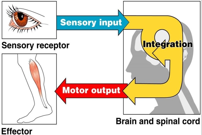
sensory input - monitors changes outside and inside the body
integration - processes and interprets input and makes decision
motor output - effects a response from either a muscle or a gland
Basic Functions: • sensory input - monitors changes outside and inside the body • integration - processes and interprets input and makes decision • motor output - effects a response from either a muscle or a gland
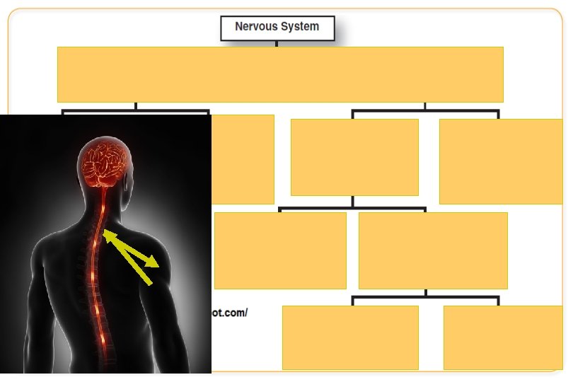
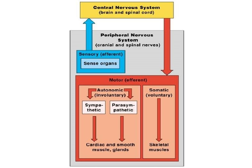
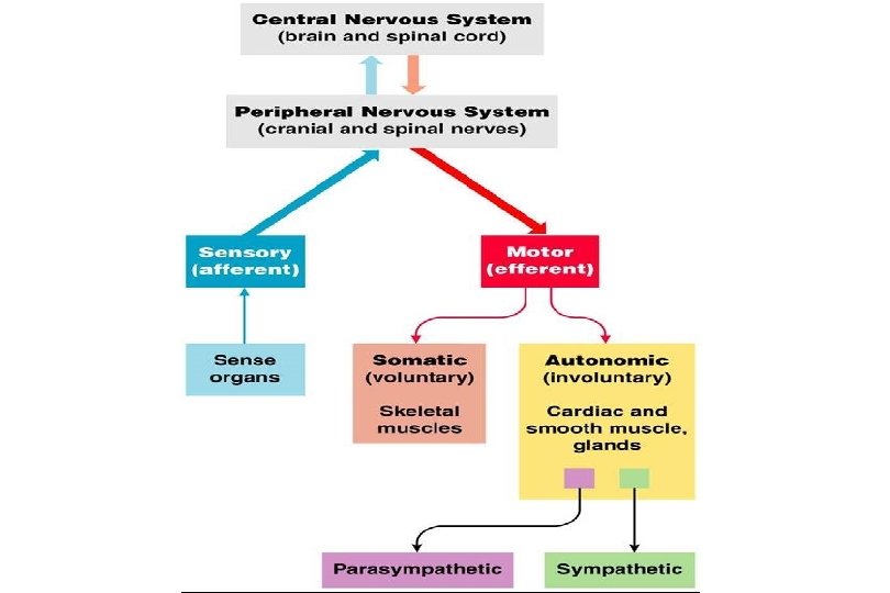
Divisions of the Nervous System • Central (CNS) - brain and spinal cord integrating and command centers • Peripheral (PNS) nerves outside of CNS
Histology of Nervous Tissue
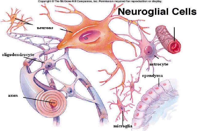
Nervous Tissue: Support Cells • collectively called neuroglia (nerve glue) • supporting cells • assist, segregate, and insulate neurons • 9 times more numerous than neurons
1. astrocytes • control ionic environment • attach neurons to capillaries (nutrients)
2. microglia • type of macrophage • engulf microorganisms
3. ependymal • form cerebrospinal fluid (CSF)
4. oligodendrocytes • form myelin sheath • insulates nerve fibers
5. Schwann cells • form myelin sheath • act as phagocytes • insulates
6. satellite cells • control chemical environment
Types of Glia Cells 1. astrocytes - control ionic environment, attach neurons to caps. (nutrients) 2. microglia - type of macrophage, engulf microorganisms 3. ependymal - form cerebrospinal fluid (CSF) 4. oligodendrocytes - form myelin sheath, insulates nerve fibers 5. Schwann cells - form myelin sheath, act as phagocytes, insulates 6. satellite cells - controlling chemical environment
The Neuron
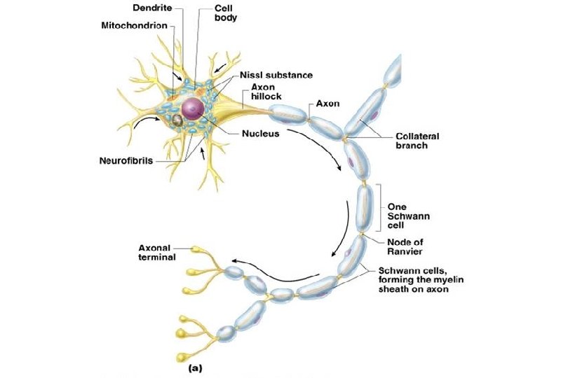
Neuron Structure • able to generate and conduct nerve impulse • can live and function for a lifetime amitotic • cell body - large nucleus and nucleolus – rough ER in form of Nissl bodies
• processes - cytoplasmic extensions (extend from cell body) 1. dendrites - short, branched – conduct impulses toward cell body
2. axon - only 1 may have side branches ends in terminals or synaptic bulbs which release neurotransmitters conduct impulses away from cell body
• insulated by myelin sheath • made of Schwann cells (wrap around)
• adjacent Schwann cells don’t touch – gaps are Nodes of Ranvier • increase speed of transmission Node of Ranvier Myelin sheath Schwann Cell
Formation of Myelin Sheath • As the Schwann cells wrap around the axon, the myelin sheath forms. • The neurilemma is the outer most part of the myelin sheath with the majority of the cytoplasm and nuclei. – innermost is called the myelin sheath
• White matter is a collection of myelinated fibers. • Gray matter is unmyelinated with only a single layer of Schwann cells. Gray matter is slow and found where distances are short.
Neuron Classification According To Structure number of processes extending from the cell body
1. Multipolar • • many dendrites 1 axon 1 cell body most common
2. Bipolar • • • 1 axon 1 dendrite 1 cell body rare ex: olfactory and retina
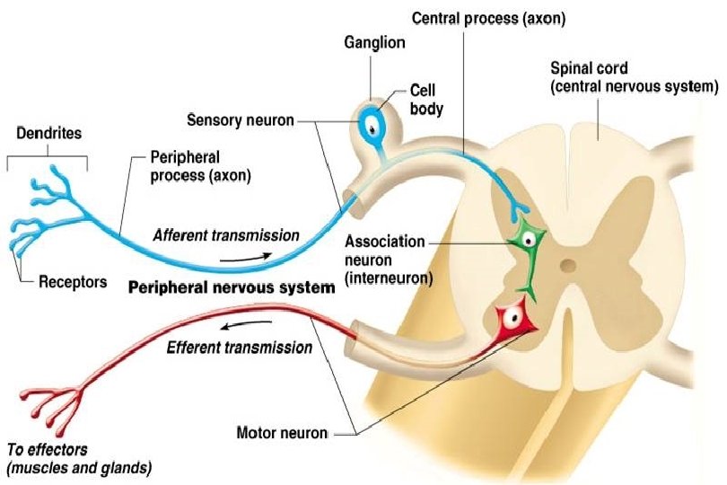
3. Unipolar • single process that merges with cell body • sensory neurons • ex: skin
Neuron Classification According To Function direction of impulse conduction
1. sensory / afferent – toward CNS, from skin or internal organs 2. motor / efferent – away from CNS, to muscle or glands, most multipolar 3. association neurons / interneurons – lie between sensory and motor neurons, shuttle signals
Nerves • Nerves are cord like bundles of nerve fibers wrapped by connective tissue. • Blood and lymph vessels are also found inside. • Nerves are only found in the PNS.
Types of Nerves 1. sensory nerves (afferent) toward CNS 2. motor nerves - (efferent) away from CNS 3. mixed nerves – both sensory (afferent) and motor (efferent) nerves, to and from CNS ** most nerves
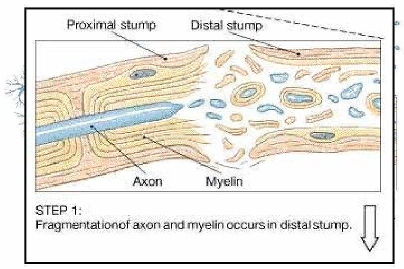
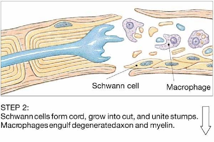
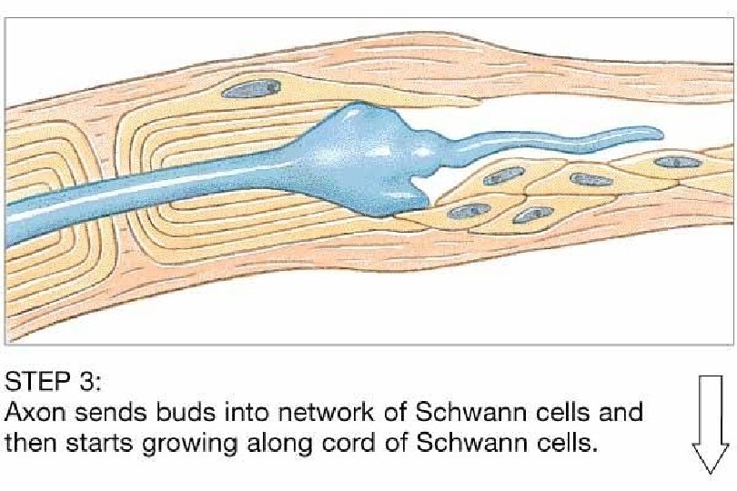
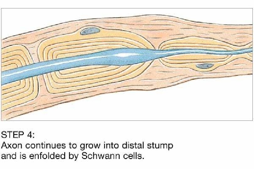
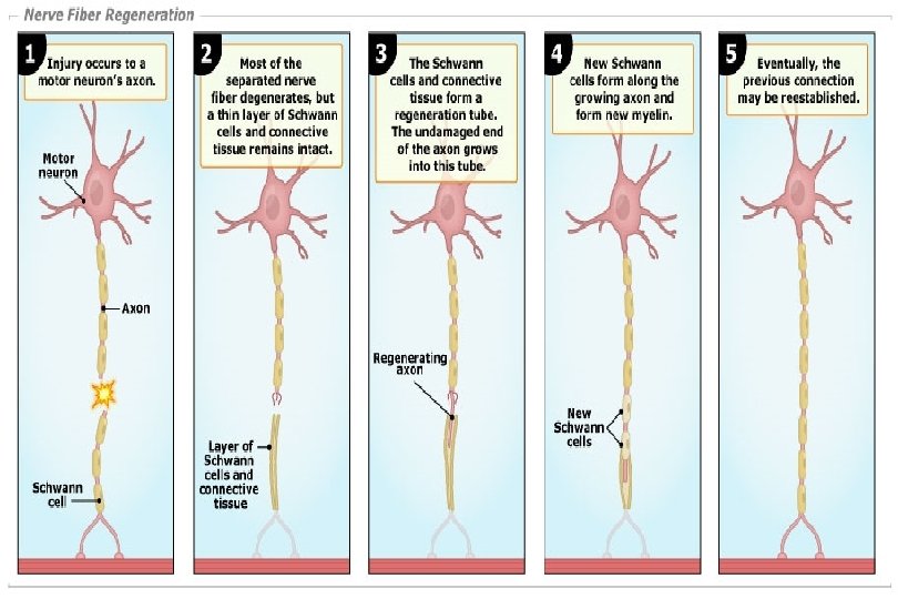
Regeneration of Nervous Tissue • mature neurons do not undergo mitosis • if damage is severe or close to cell body, neuron may die • however, cut or compressed axons can regenerate in the PNS
Regeneration of Nervous Tissue 1. Damage area is reabsorbed 2. Neurilemma of Schwann cells form a tunnel to guide axonal “sprouts” to their original contacts. Schwann cells also releases growth factor. – – – The grater the distance, the less chance of nerve recovery. Neurosurgeons align cut nerve endings surgically to enhance regeneration. Post trauma regrowth is never exactly the same.
Regeneration of Nervous Tissue • CNS cells never regenerate (lack neurilemma) • supporting neuroglial cells provide no guiding tunnel and scaring blocks axon sprouts