The Nervous System CNS Central Nervous System CNS

 • CNS develops from the embryonic neural tube ▫ The")
 Figure 7. 12 a")
 Diencephalon Brain stem Cerebellum")
 ▫ Paired (left and right)")
 • Shallow grooves =")
 divide the cerebrum into lobes •")


 — outer layer in the cerebral")


 • • Similar to blood plasma composition Formed by the choroid")


 • Commonly called a stroke • The result of a ruptured")


")


 • Nerves and ganglia outside the central nervous system •")


- Slides: 56
The Nervous System CNS
Central Nervous System (CNS) • CNS develops from the embryonic neural tube ▫ The neural tube becomes the brain and spinal cord ▫ The opening of the neural tube becomes the ventricles Four chambers within the brain Filled with cerebrospinal fluid
Central Nervous System (CNS) Figure 7. 12 a
Regions of the Brain • • Cerebral hemispheres (cerebrum) Diencephalon Brain stem Cerebellum
Regions of the Brain: Cerebrum • Cerebral Hemispheres (Cerebrum) ▫ Paired (left and right) superior parts of the brain ▫ Includes more than half of the brain mass ▫ The surface is made of ridges (gyri) and grooves (sulci)
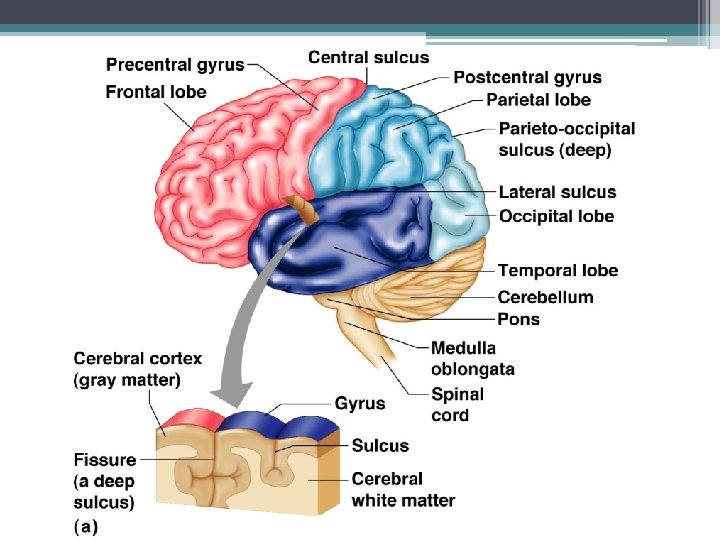
Cerebral Hemisphere • Marked by elevated ridges = gyri (gyrus) • Shallow grooves = sulci (sulcus) ▫ Sulci separate hemispheres into five lobes Frontal, parietal, occipital, temporal ▫ Central sulcus separates frontal lobe from parietal lobe • Deeper grooves = fissures ▫ Longitudinal fissure separates cerebral hemispheres ▫ Transverse cerebral fissure separates cerebral hemispheres from cerebellum
Lobes of the Cerebrum • Fissures (deep grooves) divide the cerebrum into lobes • Surface lobes of the cerebrum ▫ ▫ Frontal lobe Parietal lobe Occipital lobe Temporal lobe
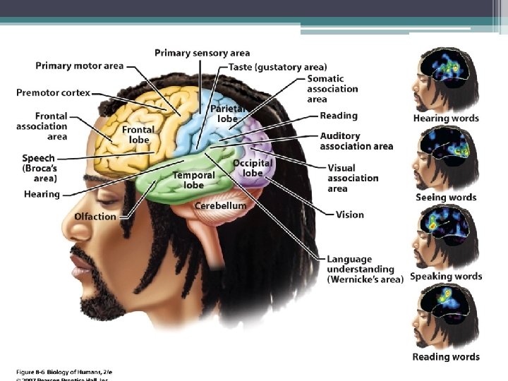
Specialized areas of the cerebrum • Primary somatic sensory area ▫ Receives impulses from the body’s sensory receptors ▫ Located in parietal lobe • Broca’s area ▫ Involved in our ability to speak • Primary motor area ▫ Sends impulses to skeletal muscles ▫ Located in frontal lobe
Regions of the Brain: Cerebrum Figure 7. 13 c
Figure 7. 14
Regions of the Brain: Cerebrum • Cerebral areas involved in special senses ▫ ▫ Gustatory area (taste) Visual area Auditory area Olfactory area • Interpretation areas of the cerebrum ▫ Speech/language region ▫ Language comprehension region ▫ General interpretation area
Regions of the Brain: Cerebrum Figure 7. 13 c
Layers of the cerebrum • Gray matter (cortex) — outer layer in the cerebral cortex composed mostly of neuron cell bodies • White matter —fiber tracts deep to the gray matter ▫ Corpus callosum connects hemispheres • Basal nuclei —islands of gray matter buried within the white matter
Regions of the Brain: Diencephalon Figure 7. 16
Regions of the Brain: Diencephalon • Sits on top of the brain stem • Enclosed by the cerebral hemispheres • Made of three parts ▫ Thalamus ▫ Hypothalamus ▫ Epithalamus
Regions of the Brain: Diencephalon Figure 7. 16 a
Diencephalon: Thalamus • Surrounds the third ventricle • The relay station for sensory impulses • Transfers impulses to the correct part of the cortex for localization and interpretation
Regions of the Brain: Diencephalon Figure 7. 16 a
Diencephalon: Hypothalamus • Under the thalamus • Important autonomic nervous system center ▫ Helps regulate body temperature ▫ Controls water balance ▫ Regulates metabolism • An important part of the limbic system (emotions) • The pituitary gland is attached to the hypothalamus
Diencephalon: Epithalamus • Forms the roof of the third ventricle • Houses the pineal body (an endocrine gland) • Includes the choroid plexus—forms cerebrospinal fluid
Regions of the Brain: Brain Stem • Attaches to the spinal cord • Parts of the brain stem ▫ Midbrain ▫ Pons ▫ Medulla oblongata faculty. ksu. edu. sa -
Brain Stem: Mid Brain • Mostly composed of tracts of nerve fibers • Has two bulging fiber tracts— cerebral peduncles • Has four rounded protrusions— corpora quadrigemina ▫ Reflex centers for vision and hearing faculty. ksu. edu. sa -
Brain Stem: Pons • The bulging center part of the brain stem • Mostly composed of fiber tracts • Includes nuclei involved in the control of breathing faculty. ksu. edu. sa -
Brain Stem: Medulla Oblongata • • The lowest part of the brain stem Merges into the spinal cord Includes important fiber tracts Contains important control centers ▫ ▫ ▫ Heart rate control Blood pressure regulation Breathing Swallowing Vomiting
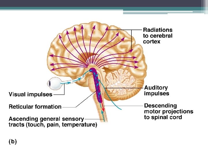
Brain Stem: Reticular formation • Diffuse mass of gray matter along the brain stem • Involved in motor control of visceral organs • Reticular activating system (RAS) plays a role in awake/sleep cycles and consciousness
Regions of the Brain: Cerebellum • Two hemispheres with convoluted surfaces • Provides involuntary coordination of body movements • The cerebellum integrates information from the motor cortex and sensory pathways to produce well-timed voluntary movements (automatic pilot) and controls equilibrium and posture
Regions of the Brain: Cerebellum Figure 7. 16 a
Protection of the Central Nervous System • • • Scalp and skin Skull and vertebral column Meninges Cerebrospinal fluid (CSF) Blood-brain barrier
Protection of the Central Nervous System Figure 7. 17 a
Meninges • Dura mater ▫ Double-layered external covering Periosteum—attached to inner surface of the skull Meningeal layer—outer covering of the brain ▫ Folds inward in several areas
Meninges • Arachnoid layer ▫ Middle layer ▫ Web-like § Pia mater § Internal layer § Clings to the surface of the brain
Cerebrospinal Fluid (CSF) • • Similar to blood plasma composition Formed by the choroid plexus Forms a watery cushion to protect the brain Circulated in arachnoid space, ventricles, and central canal of the spinal cord
Ventricles and Location of the Cerebrospinal Fluid Figure 7. 18 a–b
Ventricles and Location of the Cerebrospinal Fluid Figure 7. 18 c
Hydrocephalus in a Newborn • Hydrocephalus ▫ CSF accumulates and exerts pressure on the brain if not allowed to drain Figure 7. 19
Blood-Brain Barrier • Includes the least permeable capillaries of the body • Excludes many potentially harmful substances • Useless as a barrier against some substances ▫ ▫ ▫ Fats and fat soluble molecules Respiratory gases Alcohol Nicotine Anesthesia
Traumatic Brain Injuries • Concussion ▫ Slight brain injury ▫ No permanent brain damage • Contusion ▫ Nervous tissue destruction occurs ▫ Nervous tissue does not regenerate • Cerebral edema ▫ Swelling from the inflammatory response ▫ May compress and kill brain tissue
Cerebrovascular Accident (CVA) • Commonly called a stroke • The result of a ruptured blood vessel supplying a region of the brain • Brain tissue supplied with oxygen from that blood source dies • Loss of some functions or death may result
Alzheimer’s Disease • Progressive degenerative brain disease • Mostly seen in the elderly, but may begin in middle age • Structural changes in the brain include abnormal protein deposits and twisted fibers within neurons • Victims experience memory loss, irritability, confusion, and ultimately, hallucinations and death
Spinal Cord • Extends from the foramen magnum of the skull to the first or second lumbar vertebra • 31 pairs of spinal nerves arise from the spinal cord • Cauda equina is a collection of spinal nerves at the inferior end
Spinal Cord Anatomy
Spinal Cord Anatomy • Internal gray matter is mostly cell bodies ▫ Dorsal (posterior) horns ▫ Anterior (ventral) horns ▫ Gray matter surrounds the central canal Central canal is filled with cerebrospinal fluid • Exterior white mater—conduction tracts ▫ Dorsal, lateral, ventral columns
Spinal Cord Anatomy Figure 7. 21
Spinal Cord Anatomy • Meninges cover the spinal cord • Spinal nerves leave at the level of each vertebrae ▫ Dorsal root Associated with the dorsal root ganglia— collections of cell bodies outside the central nervous system ▫ Ventral root Contains axons
Pathways Between Brain and Spinal Cord Figure 7. 22
Peripheral Nervous System (PNS) • Nerves and ganglia outside the central nervous system • Nerve = bundle of neuron fibers • Neuron fibers are bundled by connective tissue
PNS: Structure of a Nerve • Endoneurium surrounds each fiber • Groups of fibers are bound into fascicles by perineurium • Fascicles are bound together by epineurium
PNS: Classification of Nerves • Mixed nerves ▫ Both sensory and motor fibers • Sensory (afferent) nerves ▫ Carry impulses toward the CNS • Motor (efferent) nerves ▫ Carry impulses away from the CNS
PNS: Cranial Nerves • 12 pairs of nerves that mostly serve the head and neck • Only the pair of vagus nerves extend to thoracic and abdominal cavities • Most are mixed nerves, but three are sensory only
PNS: Distribution of Cranial Nerves
PNS: Cranial Nerves • I Olfactory nerve—sensory for smell • II Optic nerve—sensory for vision • III Oculomotor nerve—motor fibers to eye muscles • IV Trochlear—motor fiber to eye muscles
PNS: Cranial Nerves • V Trigeminal nerve—sensory for the face; motor fibers to chewing muscles • VI Abducens nerve—motor fibers to eye muscles • VII Facial nerve—sensory for taste; motor fibers to the face • VIII Vestibulocochlear nerve— sensory for balance and hearing
PNS: Cranial Nerves • IX Glossopharyngeal nerve—sensory for taste; motor fibers to the pharynx • X Vagus nerves—sensory and motor fibers for pharynx, larynx, and viscera • XI Accessory nerve—motor fibers to neck and upper back • XII Hypoglossal nerve—motor fibers to tongue