The Nervous System Chapter 48 Although the Nervous


 = brain & spinal")
")
")


 How,")


 Neurotransmitter opens Na+ & K+ gates – generates action potential")
 Neurotransmitter causes K+ to rush out (or Clto enter) –")

")


- Slides: 37
The Nervous System Chapter 48
Although the Nervous System and the Endocrine System coordinate responses, there are 3 main differences. The nervous system: is much faster has pinpoint control has more structural complexity
Nervous system = 2 main parts: Central Nervous System (CNS) = brain & spinal cord Peripheral Nervous System (PNS) = nerves (throughout body) including: Afferent nerves: sensory Efferent nerves: motor
Efferent PNS has 2 main divisions Somatic PNS: responds to external environment (mostly voluntary) Ex. Skeletal muscle Autonomic PNS: regulate internal env (mostly involuntary) Ex. Invol. Muscle or glands
Autonomic PNS is further subdivided into: Sympathetic: stimulates organs & energy use (for action) Parasympathetic: causes energy intake and conservation – antagonistic
3 Overlapping functions of the Nervous System: sensory input: afferent neuron integration: info is interpreted and associated w/ responses by CNS motor output: efferent neuron sends signal to effector cells (muscle or gland)
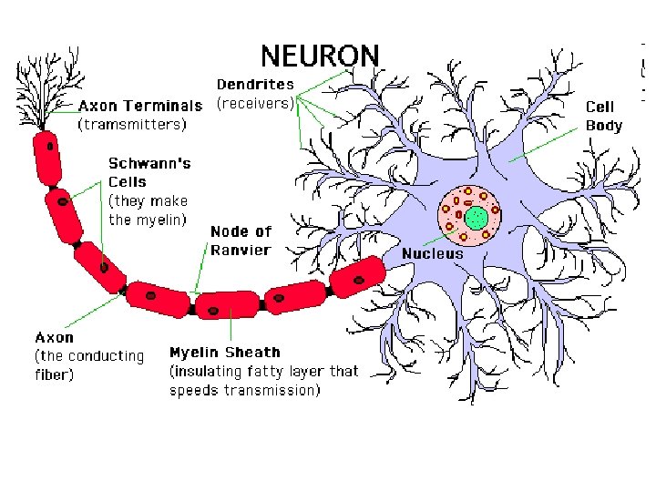
Neuron Structure: Dendrites: short, highly branched fibers. Receive stimuli and send electrical impulses to cell body. Cell Body: cytoplasm, nucleus, most organelles Axon: long, branched extension of cell body – conducts nerve impulse to another neuron or effector Axon (synaptic) terminals: release neurotransmitters Synapse = gap between synaptic node and next neuron or effector
Nerve impulse: Travels in one direction ONLY! Receptor dendrite axon synapse cell body
PNS Axon Structure: Most of the axons in the PNS have 2 coverings made of supportive glia cells : Both called Schwann cells Cellular sheath: aids in nerve regeneration Myelin (inner) sheath: membrane contains myelin which provides electrical insulation Nodes of Ranvier: gaps in myelin sheath – axon not insulated (allows impulse to jump)
Multiple Sclerosis: neurological disease – deterioration of myelin sheath Causes disruptions in nerve impulse transmission, leads to – loss of coordination &/or paralysis
CNS Axon Structure: The axons in the CNS are supported and insulated by glia cells Two types: Oligodendrocytes: insulate axons (w/ myelin) Astrocytes: encircle capillaries to form a blood-brain barrier
Note: Not all neurons are exactly alike. We’re focusing on a typical PNS neuron. When a PNS neuron is wrapped in Schwann cells (like we discussed), the axons are called “white fibers. ” (“white matter” in the brain is made of glial cells & myelinated axons) If, instead, several axons are held together by a single Schwann cell that does NOT wrap around them, the axons are called “gray fibers. ” (“grey matter” in brain is cell bodies and is unmyelinated)
Nerve = Bundle of hundreds of axons wrapped together in connective tissue.
Action Potentials in Neurons Do you Remember any of this? ? ? All cells have a resting membrane potential: net difference in charge across plasma membrane Inside of cells is negatively charged due to: large neg. charged organic molecules trapped inside, Limited permeability to + ions, and the action of the Na+/K+ pump Channel Proteins (in membrane): “gates” for Na+ or K+, voltage regulated
In Neurons An electrical or chemical disturbance near the dendrite causes a local depolarization which opens the Na+ gates, in and Na+ rushes ______. The depolarization causes an opening of the K+ gates and -- K+ moves out to cause repolarization The flow of ions and resulting change in membrane potential = action potential
All-or-None Law: once depolarization starts, the entire sequence occurs (regardless of stimulus size) How, then does the nervous system distinguish between strong and weak stimuli? Strong Stimuli = higher frequency of action potentials (neuron “fires” repeatedly)
Depolarization Speed is increased with: thicker axons Saltatory conduction: myelin sheath insulation prevents movement of ions through membrane. Therefore, action potentials only occur at gaps (nodes of Ranvier) which are spaced about 1 mm apart. Action potential: leaps from node to node.
Presynaptic cell sends to the postsynaptic cell through electrical or chemical synapses. Electrical Synapses: Cells must be connected by – gap junctions to allow ion currents to flow Responsible for certain rapid movements in some vertebrates (CNS): escape from predator… Much less common than chemical synapses in vertebrates (& many invertebrates)
Chemical Synapses: Cells are not connected: action potential cannot cross synapse Electrical signal of presynaptic cell must be converted to a chemical signal which crosses the synapse and is converted back to an electrical signal in the postsynaptic cell. Synaptic terminal (axon end): contains many synaptic vesicles full of neurotransmitters When the action potential reaches the terminal, it causes Ca+ to rush into cell through voltage-sensitive channels. The sudden influx of Ca+ causes synaptic vesicles to fuse with the presynaptic membrane and release neurotransmitters via: exocytosis.
Neurotransmitters binding to postsynaptic membrane: alter the membrane potential Example: Acetylcholine binds to gate channels in postsynaptic membrane to allow Na+ & K+ diffusion. The enzyme, cholinesterase, breaks it down for recycling. (restores resting potential)
Neurotransmitters Can excite membranes by: bringing the voltage closer to the threshold potential Can inhibit membranes by: hyperpolarizing the membrane
Excitatory Postsynaptic Potentials (EPSP’s) Neurotransmitter opens Na+ & K+ gates – generates action potential Summation: cumulative effect of postsynaptic potentials. 2 types: temporal: chemical transmissions occurring in quick succession (no time to return to resting potential) spatial: several different presynaptic neurons have an additive effect on the postsynaptic neuron AT THE SAME TIME
Inhibitory Postsynaptic Potentials (IPSP’s) Neurotransmitter causes K+ to rush out (or Clto enter) – hyperpolarizes the membrane Makes it much harder to generate action potential Example Neurotransmitters: dopamine, norepinephrin, serotonin, glutamic acid…
Invertebrate Nervous Systems Many invertebrates have ___ ventral ___ nerve cords and segmental ganglia: Clusters of nerve cell bodies. Invertebrate neurons: A. A dye-filled antennal neuron, within a Drosophila ganglion B. Giant neuron in a cricket ganglion C Leech ganglion from an embryo (with fluorescent dyes) D. Two auditory neurons in the crickets ganglion Source: http: //www. scholarpedia. org/article/Neuron
Organization of some Nervous Systems (textbook p. 1012)
Vertebrate Nervous Systems Vertebrates have ___ dorsal ___ nerve cords without segmental ganglia: Segmented ganglia lie just outside the cord. Invertebrate neurons: A. A dye-filled antennal neuron, within a Drosophila ganglion B. Giant neuron in a cricket ganglion C Leech ganglion from an embryo (with fluorescent dyes) D. Two auditory neurons in the crickets ganglion Source: http: //www. scholarpedia. org/article/Neuron
Embryonic Development of the Brain All vertebrates have three bilaterally symmetrical anterior bulges of the neural tube: Forebrain, midbrain, hindbrain During vertebrate evolution, the brain divided further. Birds and mammals have a much larger: forebrain See Figure 48. 23, p. 1028
The Brainstem • “Lower brain” • 3 parts: medulla oblongata, pons, midbrain • Structure: stalk at anterior end of spinal cord with caplike swellings
The Brainstem: medulla and pons Functions: • Release neurotransmitters which cause changes in: attention, alertness, appetite, and motivation • Control visceral (automatic, homeostatic) functions such as: breathing, heart / vessel activity, swallowing, vomiting and digestion
The Brainstem: medulla and pons • Controls information transmission: input from sensory neurons and motor instruction output travel through brainstem • Coordinates large-scale body movements -most axons carrying info. between spinal cord and mid- or forebrain cross over here: causing left side of brain to control movements on right side (and vice versa)
The Brainstem: midbrain • Contains centers for receipt and integration of sensory information: hearing (both) and vision (receipt & reflexes) The Brainstem: reticular formation Neuron network throughout the brainstem which filters sensory input to regulate: Sleep and arousal
The Cerebellum Functions: • ___ Coordination ___ during motor, perceptual and cognitive functions. • Learning and remembering ___ motor ___ skills such as bike riding. • Coordinates movements and balance such as in hand eye coordination.
The Diencephalon Found in Embryo: Develops into epithalamus, thalamus and hypothalamus. • Epithalamus includes: pineal gland & choroid plexus (capillary cluster) • Thalamus: processes input / output of info between senses, cerebrum & motor neurons • Hypothalamus: homeostatic regulation via hormones, basic survival mechanisms, mating behaviors, and pleasure
The Cerebrum Structure: • Divided into left and right ___ hemispheres___. • Each side has: gray matter -- (outer covering) cerebral cortex internal white matter basal nuclei – (groups of neurons involved in movement)
The Cerebrum: cerebral cortex • Largest and most complex part of human brain. • Functions: sensory info is analyzed, motor commands are issued, language is generated • Neocortex (outermost part of mammalian cerebrum) is highly convoluted in humans to provide: Huge surface area and about 80% of brain’s total mass.
The Cerebrum: corpus callosum • Thick band of ___ axons ___. • Function: enables communication between right and left cerebral cortex • Damage to one area of the cerebrum early in development can cause redirection of its functions to other areas. Even in adults, cerebral cortex damage can lead to: new brain circuits.