The Nervous System A Central Nervous System CNS
: • • • brain and spinal")


 • It is the outermost layer, made of dense fibrous")

 is an extracranial part of the central nervous")


 Connected by")

= ventral horn (Motor nuclei) • it is broad")


 Ts.")
 1 - Anterior &posterior")


- Slides: 29
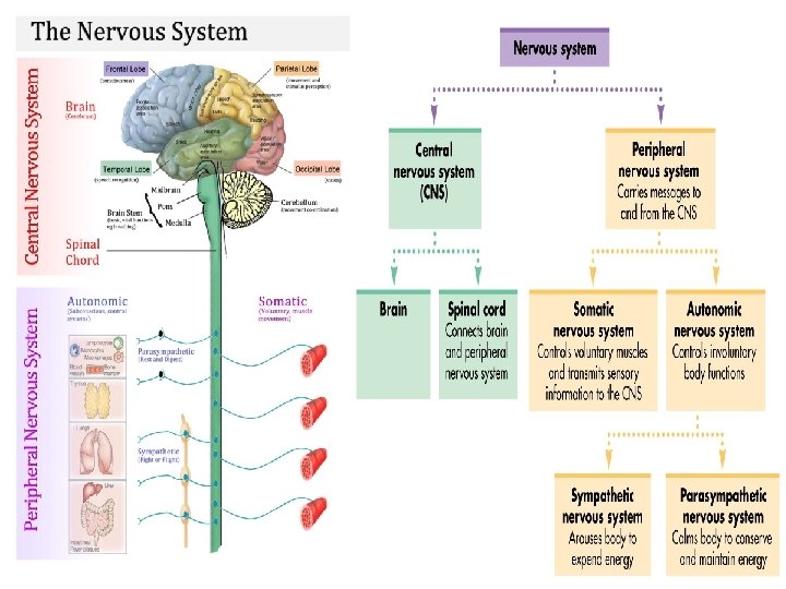
The Nervous System A- Central Nervous System (CNS): • • • brain and spinal cord. Central in position Protected by bone & meninges. Enclose cavities (CSF hydrostatic cushion Provide sensory, motor, mental function Contain blood barriers. It is formed of: 1. gray matter containing the cell bodies of neurons 2. white matter containing the cell processes. Myelinated nerves B- Peripheral Nervous System (PNS): • Nerve endings ( receptors and effectors) • Peripheral nerves ( cranial and spinal N. ) • Ganglia (craniospinal and autonomic ganglia)
Nerves System Terminology 1. Neuron: is a nerve cell and its processes. 2. Nucleus: is a group of nerve cells bodies located in the CNS. 3. Ganglia: is a group of nerve cells bodies located outside the CNS. 4. Synapse: is the site of contact of the axon of one neuron with the dendrites or cell body another neuron. 5. Nerve fiber: is an axon + sheath. 6. P. Nerve: is a bundle of nerve fibers in the PNS. 7. Tract: is a group of nerve fibers inside CNS which have the same origin, termination and function. The tract may be ascending (sensory or afferent) or descending (motor or efferent). 8. lemniscus: bundle of nerve fibers inside CNS which have different origin , termination or function. 9. White matter: consists of nerve fibers (myelinated axons of neurons in the CNS) supported by neuroglia. 10. Gray matter: consists of nerve cells bodies and unmyelinated portion of axon in the CNS) supported by neuroglia. 11. Commissure: is a group of nerve fibers which connect right and left side of the brain or spinal cord 12. Association fibers: are nerve fibers which connect parts of the nervous system on the same side. 15. Peduncles: are large nerve tracts that emerge from certain region of the brain.
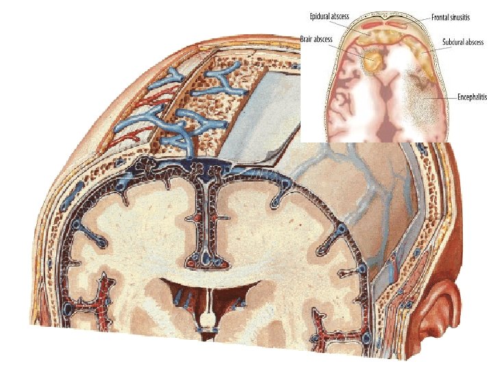
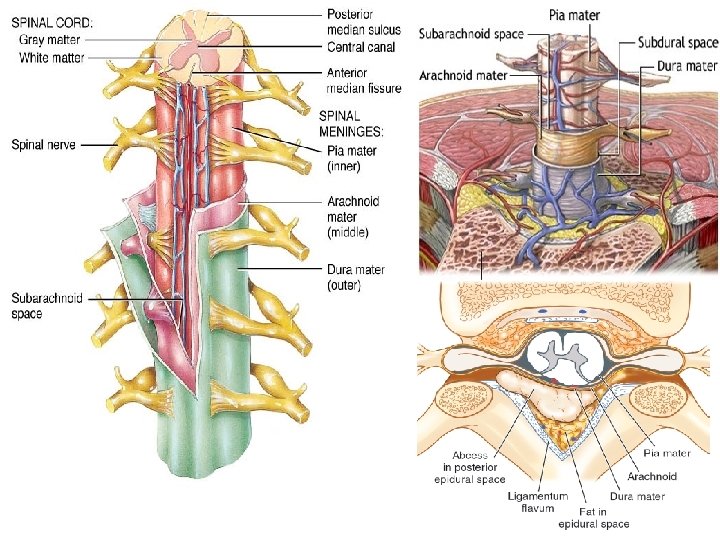
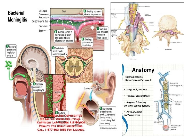
The Meninges The meninges are the 3 connective tissue coverings of the brain and spinal cord. These meninges are: v Pia mater = tender mother v. Arachnoid layer (spider's web) • It is the innermost, highly vascular layer • It is the intermediate layer of the meninges. It does of the meninges. • It consists primarily of 2 layers: • An outer layer of continuous flat cells. • A deeper layer of fine collagenous and elastic fibers. not enter into the sulci of the brain and it is avascular, although the blood vessels course through it in their way to the pia mater. • It consists of: • Numerous arachnoid trabeculae of delicate connective tissue forming a network and covered by flat cells. • The space between the pia and arachnoid is called the subarachnoid space, which is filled with the CSF and traversed by the arachnoid trabeculae. • The arachnoid villi & arachnoid granulations which are pedunculated projections from the arachnoid extending inside the venous sinuses of the dura. They are essential for the normal circulation and reabsorption of CSF as they transport it from the subarachnoid space back to the venous system.
Dura mater (tough mother) • It is the outermost layer, made of dense fibrous connective tissue. The dura is composed of 2 layers: • The outer periosteal layer: it is the periosteum of the inner surface of the cranial vault and the vertebral canal. • The inner meningeal layer: formed of flat cells and small blood vessels. • At certain regions, the 2 layers of the dura are separated to form the dural venous sinuses that drain the CSF. • The subdural space: it is a potential space between the dura and arachnoid, as it appears only after head injury resulting in subdural hemorrhage when blood forces the 2 layers apart.
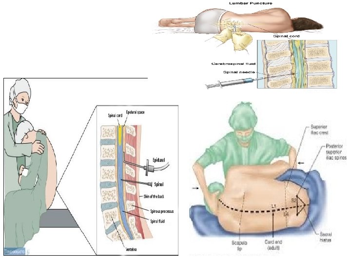
Meninges of the Spinal Cord • Dura matter: ends at the level of the second sacral vertebra. Dura mater of the spinal cord. The spinal dural sheath – only one layer Ø Arachnoid: ends at the level of the second sacral vertebra. Ø Pia matter: ends at the level of SC termination (lower border of the first lumbar vertebra in adult). The spinal cord tapers off into the conus medullaris, from the apex of which a prolongation of the pia mater, the filum terminale, descends to attach to the back of the first coccyx Meningeal Spaces of Spinal Cord 1. Epidural Space: it is located between the dura matter and vertebral periosteum. The epidural space: it is the space between the dura and the bony walls of the vertebral canal. It is filled with fat 2. and venous plexus Subdural Space: it is located between the dura matter and arachnoid matter. Contains thin film of serous fluid. 1. Subarachnoid Space: it is located between the arachnoid and pia matter. Is a wide space contains: • CSF, Spinal blood vessels, Roots of spinal nerves.
• The spinal cord (SC) is an extracranial part of the central nervous system enclosed inside the vertebral column • Protected by bone, meninges, and CSF • Length 45 cm Ø 3 rd month of intrauterine life : the SC fills the whole spinal canal • At birth: it ends at the level of L 3 vertebra. • In adult : Extends from the foramen magnum to the vertebra L 1 - 2 The Spinal Cord
Spinal Cord • The spinal cord is roughly cylindrical with Cervical and lumbar enlargements Keep in position by : v. Continuity with medulla oblongata v Caudal fixation with Filum terminale long filament of Pia matter attaches to the coccyx inferiorly v Ligamentum denticulatum attach the spinal cord to the dura matter ØBelow, the spinal cord tapers off into the conus medullaris, from the apex of which a prolongation of the pia mater, the filum terminale, descends to attach to the back of the first coccyx ØParts of filum terminale : 1. Proximal 15 mm: within dural sheath called filum terminale internum 2. Distal 5 mm: outside the dural sheath is called filum terminale externum
It is formed of 31 segments Spinal segment is part of spinal cord giving rise to a pair of spinal nerve 31 pairs of spinal nerves • Along the entire length of the spinal cord are attached 31 pairs of spinal nerves by the anterior ( motor roots) and the posterior (sensory roots ) • Each posterior nerve root possesses a posterior root ganglion. • Cauda equine : bunch of nerves around the filum terminale
Spinal Cord Internal Structure Gray Matter ? ? • Shape (bilateral structure) Connected by gray & white Commissure • Two deep grooves run the length of the cord – Posterior median sulcus is thickest in sacral region – Anterior median fissure – Central canal – Divided into 3 horns Aggregated into nuclei. • Anterior (ventral) Horn : motor nuclei • Posterior (dorsal) Horn -----sensory nuclei • Lateral Horn ----- autonomic nuclei
Cervical Thoracic Lumber Sacral Size Large Small Outline Oval Rounded Quadrilateral Central canal More anterior Anterior Central Posterior Dorsal horn Thin & divergent Thin Thick Ventral horn Thick White / grey Large amount Little amount Very little Posterior intermedi ate sulcus Present in upper 6 Absent Missing tract Septomarginal Cuneate & Comma from lower 6 Cuneate & Comma + Dorsal spinocerebellar The same
1 -The ventral horn (anterior horn)= ventral horn (Motor nuclei) • it is broad in ? ► ►Cervical, Lumber Large multipolar lower motor neurons. • Grouped into 3 groups of (5) nuclei Medial , Lateral , Central
2 -The dorsal horn: • Small multipolar sensory neurons. • Grouped into 3 nuclei: 1 - Substantia gelatinosa of Rolandi: ► lateral spinothalamic T. 2 - Main sensory): proprius ►nucleus (Nucleus • ventral spinothalamic T. 3. Nucleus dorsalis of Clark: ►T 1 ----L 3 spinocerebellar Ts. 3 -The lateral horn Small multipolar neurons. from T 1 ► ► L 2, 3 = Sympathetic from S 2, 3, 4 ►parasympathetic
The White Matter of the Spinal Cord Occupies the peripheral region The white matter surround the gray matter myelinated N (tract) ? ? Subdivided into 3 columns: Composed of It is divided into three column (fasciculus) of SC on each side: a. Anterior column (VC): lying between the anterior median fissure & the attachment of anterior spinal root. b. Lateral column (LC): lying between attachment of anterior and posterior spinal roots. c. Posterior column (DC): lying between the posterior median fissure & the attachment of dorsal spinal root.
Structure of White Mater The tracts are divided into: 1. Short Tracts =Associative tracts: • Containing short ascending & descending fibers which coordinate the function of the different regions of spinal cord. - They begins and end in the spinal cord. They are found close to the gray matter. 2. Long Tracts: • Ascending tracts. • Descending tracts. The white mater contains nerve fibers =Tract definite origin, termination& function. 1. Ascending tracts of the SC: Carrying sensory impulses from the SC to the brain: e. g. lateral spinothalamic tract. 2. Descending tracts of the SC: Carrying motor impulses from the brain to the SC: e. g. pyramidal tract “corticospinal tract, corticobulber
Short tracts: Short 1 -The fasciculi tracts proprii • Fibers ascend& descend for few segments. • Coordinate the function of different segments of the cord. • 1 - Fasciculi propri = spino-spinal tract 2 - Septomarginal • Along postero-median • septum. Below T 6. . . • • Stretch reflex in lower ½. 3 - Lissauer’s T. • Part of Spinothalamic T. 4 - Comma shape • Fibers between gracile & cuneate 4
Long tracts: I- Ascending (Sensory) Ts.
long ascending Tracts • Finally to sub- cortical level (4) 1 - Anterior &posterior spinocerebellar Ts. 2 -Spinoreticular 3 - Spino- olivary T. 4 - Spinotectal T. • Finally to Cerebral cortex: sensory area(4) 1 - Gracile T. 2 - Cuneate T. 3 - Lateral & 4 - ventral spinothalamic Ts.
Lateral spinothalamic tract • First order neuron. • Impulses from temperature receptors –dorsal root ganglia--- Lissauer, s tract on the same side to end on the cells of substentia gelatenosa of rolandi. • Second order neuron. • It arises from the cells of substentia gelatinosa of rolandi, crosses the midline in front of the central canal and ascends in the opposite side of the spinal cord as Lateral Spinothalamic tract to reach the PVN of the thalamus. • Third order neuron. • Neuron arises from PVN of the thalamus and ascends in the posterior limb of the internal capsule to reach the sensory area of the cerebral cortex.
Ventral spinothalamic T. • First order neuron. • Impulses from touch receptors ----dorsal root ganglia--sensory cells in the posterior horn (Main sensory nucleus) • Second order neuron. • Arise from the sensory cells in the posterior horn(main sensory N), crosses the midline in front of the central canal and ascend in the opposite side of the spinal cord as the ventral spinothalamic tract to reach the posteroventral nucleus of the thalamus (PVN). • Third order neuron. • Arises from the PVN of the thalamus ascend in the posterior limb of the internal capsule to reach the sensory area of the cerebral cortex
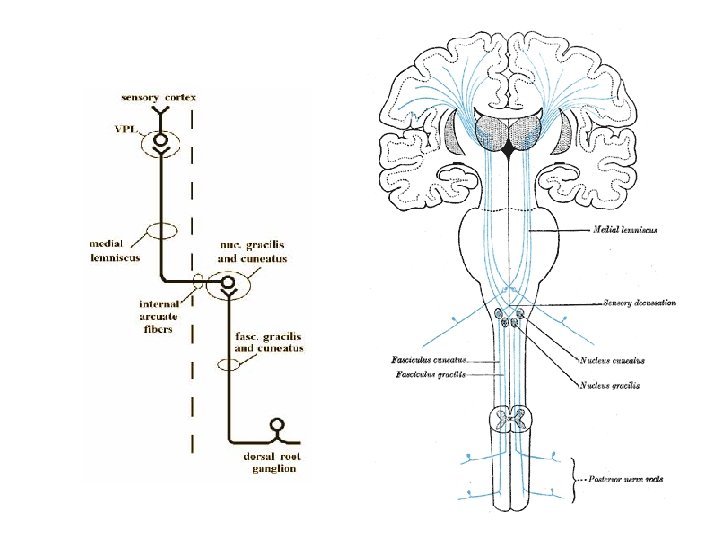
Gracilis & Cuneatus tract • • Posterior White Column-Medial Lemniscal Pathway Modality: Discriminative Touch Sensation (include Vibration) and Conscious Proprioception (Position Sensation) Receptor: Most receptors except free nerve endings Ist Neuron: Dorsal Root Ganglion (Spinal Ganglion) Posterior Root - Posterior White Column • 2 nd Neuron: Dorsal Column Nuclei (Nucleus Gracilis & Cuneatus) Medulla oblongata Internal Arcuate Fiber - Lemniscal Decussation - • Medial Lemniscus 3 rd Neuron: Thalamus Internal Capsule ----- Corona Radiata Termination: Primary Somesthetic Area
Spinocerebellar Tract Posterior Spinocerebellar Tract posterior spinocerebellar fibers receive muscle joint information from the muscle spindles, tendon organs and joint receptors of the trunk and lower limbs. • Posterior (dorsal) root ganglion • second-order neurons nucleus dorsalis (Clarke’s column). • posterior spinocerebellar tract -----inferior cerebellar peduncle and terminates in the cerebellar cortex Anterior Spinocerebellar Tract The anterior spinocerebellar tract conveys muscle joint information from upper part & upper limb. • posterior root ganglion • second-order neurons in the nucleus dorsalis anterior spinocerebellar tract in the lateral white column of the same side enter the cerebellum through the superior cerebellar peduncle and terminate in the cerebellar cortex.
Spino-olivary Tract The spino-olivary tract conveys information to the cerebellum from cutaneous and proprioceptive organs. This pathway provides afferent information for spinovisual reflexes and brings about movements of the eyes and head toward the source of the stimulation. • • • The axons enter the spinal cord from the posterior root ganglion unknown second-order neurons in the posterior gray column The axons from the second-order neurons cross the midline and ascend as the spinoolivary tract in the white matter at the junction of the anterior and lateral columns. third-order neurons in the inferior olivary nuclei in the medulla oblongata The axons of the third-order neurons cross the midline and enter the cerebellum through the inferior cerebellar peduncle. Ø The axons enter the spinal cord from the posterior root ganglion Ø unknown second-order neurons. Ø The axons of the second-order neurons cross the median plane and ascend as the spinotectal tract in the anterolateral white column lying close to the lateral spinothalamic tract. Ø terminate in the superior colliculus of the midbrain
Spinoreticular Tract Spinoreticular tract provides an afferent pathway for the reticular formation, which plays an important role in influencing levels of consciousness. • The axons enter the spinal cord from the posterior root ganglion • unknown second-order neurons in the gray matter • The axons from these second order neurons ascend the spinal cord as the spinoreticular tract in the lateral white column mixed with the lateral spinothalamic tract. • Most of the fibers are uncrossed and terminate by synapsing with neurons of the reticular formation in the medulla oblongata, pons, and midbrain. The spinoreticular tract provides an afferent pathway for the reticular formation, which plays an important role in influencing levels of consciousness.