
The myeloproliferative neoplasms Basics and beyond Martin H
The myeloproliferative neoplasms Basics and beyond…. . Martin H. Ellis MD Hematology Institute and Blood Bank Meir Medical Center Sackler School of Medicine, Tel Aviv, Israel
Outline-Basics • Normal blood cell formation (hematopoiesis) • Spectrum and definition of myeloid neoplasms • Molecular pathogenesis • Polycythemia vera • Essential thrombocytosis • Primary myelofibrosis Incidence Clinical features Laboratory abnormalities Treatment Prognosis
Outline-Beyond the Basics • The MPN continuum • Calreticulin – a deep dive
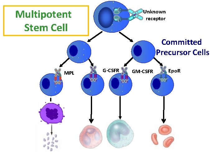
Multipotent Stem Cell Unknown receptor Committed Precursor Cells MPL G-CSFR GM-CSFR Epo. R
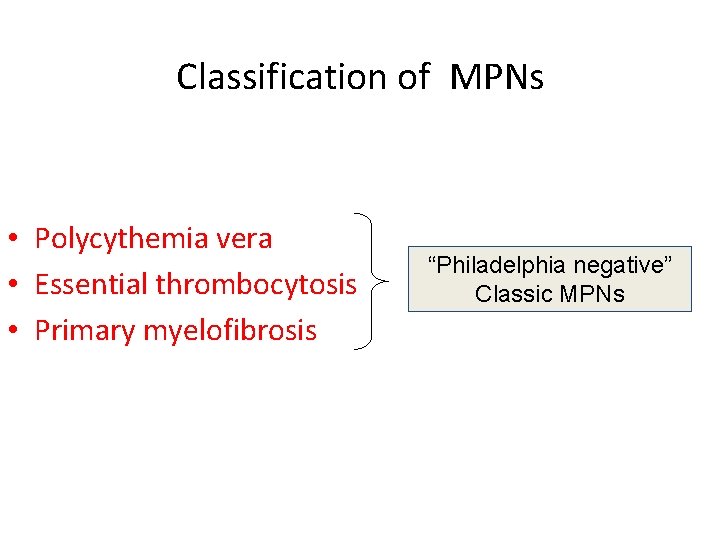
Classification of MPNs • Polycythemia vera • Essential thrombocytosis • Primary myelofibrosis “Philadelphia negative” Classic MPNs
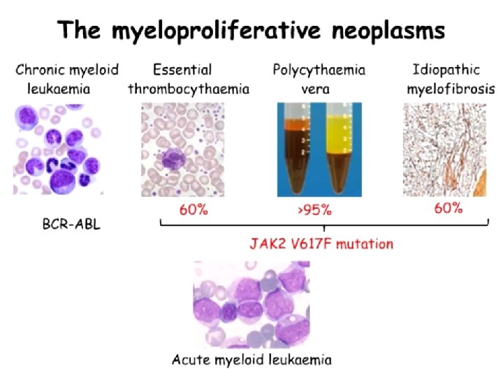
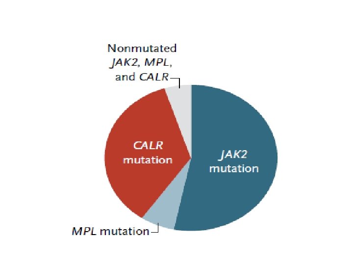
“Driver” mutations are central in the development of the MPNs • JAK 2 V 617 F • MPL • CALR (calreticulin)
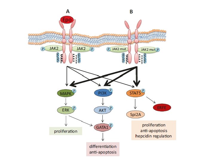
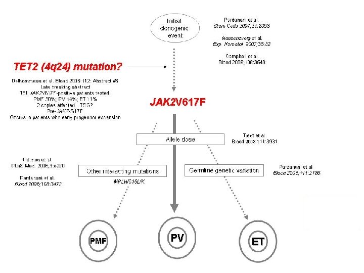
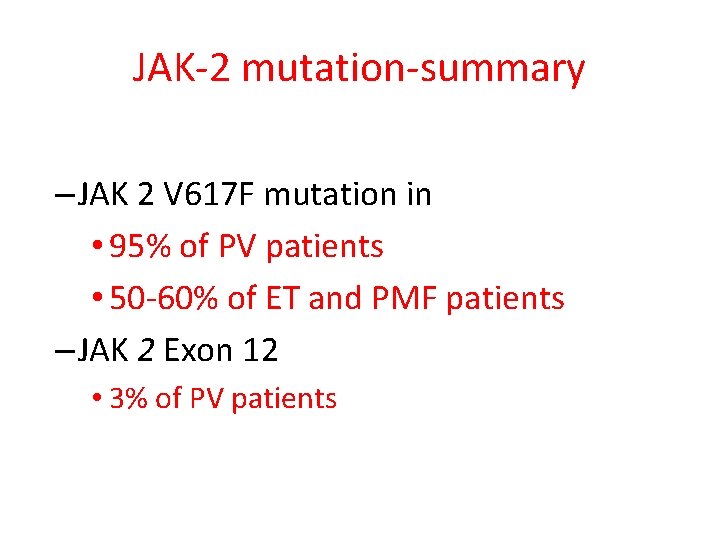
JAK-2 mutation-summary – JAK 2 V 617 F mutation in • 95% of PV patients • 50 -60% of ET and PMF patients – JAK 2 Exon 12 • 3% of PV patients
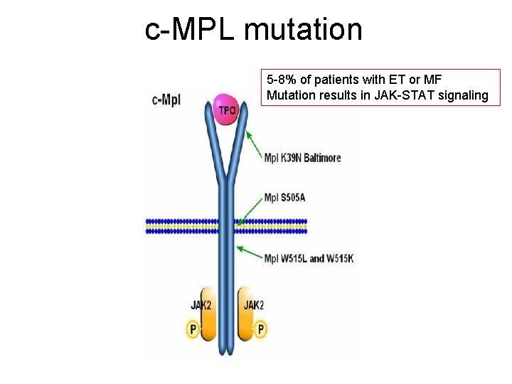
c-MPL mutation 5 -8% of patients with ET or MF Mutation results in JAK-STAT signaling
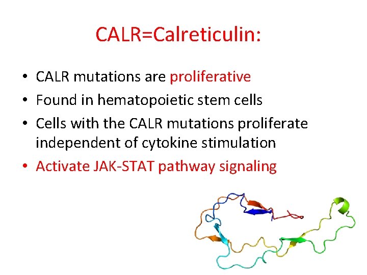
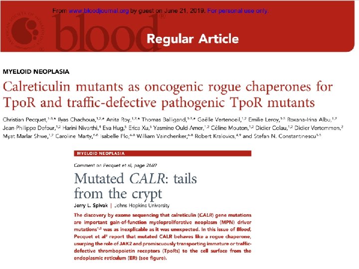
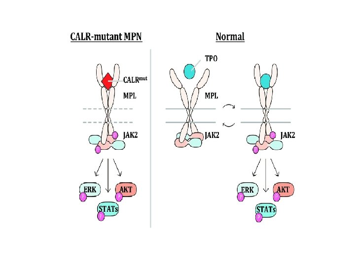
CALR=Calreticulin: • CALR mutations are proliferative • Found in hematopoietic stem cells • Cells with the CALR mutations proliferate independent of cytokine stimulation • Activate JAK-STAT pathway signaling
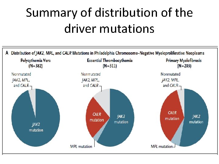
Summary of distribution of the driver mutations
10% of pts with ET or PMF are “triple negative” – Negative for mutations in JAK 2, MPL, CALR • Mutated clone too small to detect? • Other mutations? • Reactive Thrombocytosis?

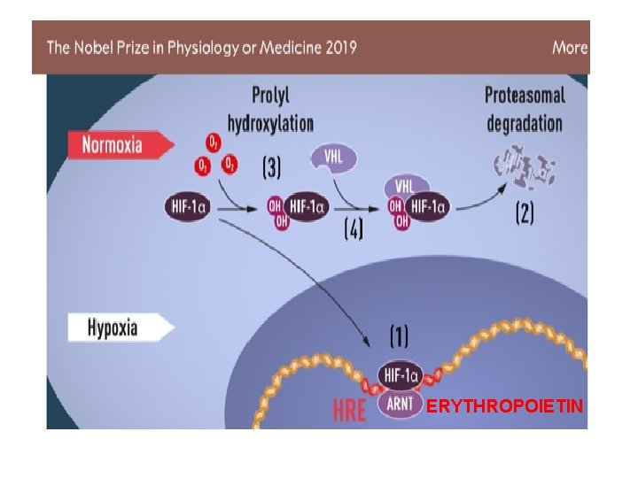
ERYTHROPOIETIN
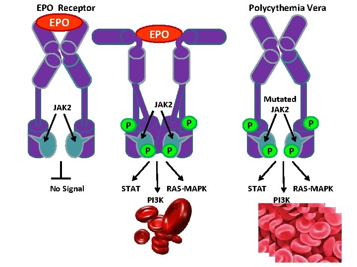
Polycythemia Vera EPO Receptor EPO JAK 2 P P P No Signal Mutated JAK 2 STAT P RAS-MAPK PI 3 K P P STAT RAS-MAPK PI 3 K
Polycythemia Vera
Polycythemia vera Epidemiology and incidence • • • All populations, lower in Japan Average age: 60 yrs, 25% are < 50 yrs, 10% < 40 yrs Men > women Incidence = approximately 2: 100 000 individuals Familial tendency Cause unknown in vast majority
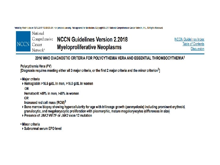
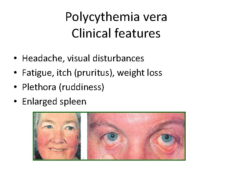
Polycythemia vera Clinical features • • Headache, visual disturbances Fatigue, itch (pruritus), weight loss Plethora (ruddiness) Enlarged spleen
Polycythemia vera Laboratory abnormalities • • • Elevated hemoglobin and hematocrit Elevated red cell mass (radionuclide scan) Leukocytosis and thrombocytosis Blood chemistry abnormalities (uric acid, LDH) Low serum erythropoietin JAK-2 mutation
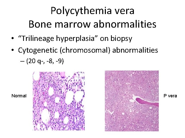
Polycythemia vera Bone marrow abnormalities • “Trilineage hyperplasia” on biopsy • Cytogenetic (chromosomal) abnormalities – (20 q-, -8, -9) Normal P vera
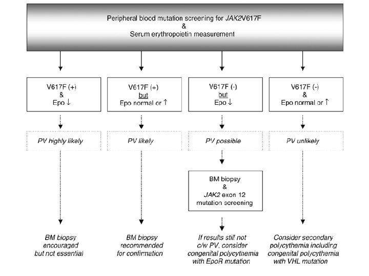
P vera: diagnosis
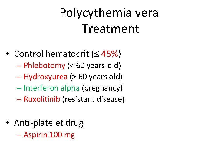
Polycythemia vera Treatment • Control hematocrit (≤ 45%) – Phlebotomy (< 60 years-old) – Hydroxyurea (> 60 years old) – Interferon alpha (pregnancy) – Ruxolitinib (resistant disease) • Anti-platelet drug – Aspirin 100 mg
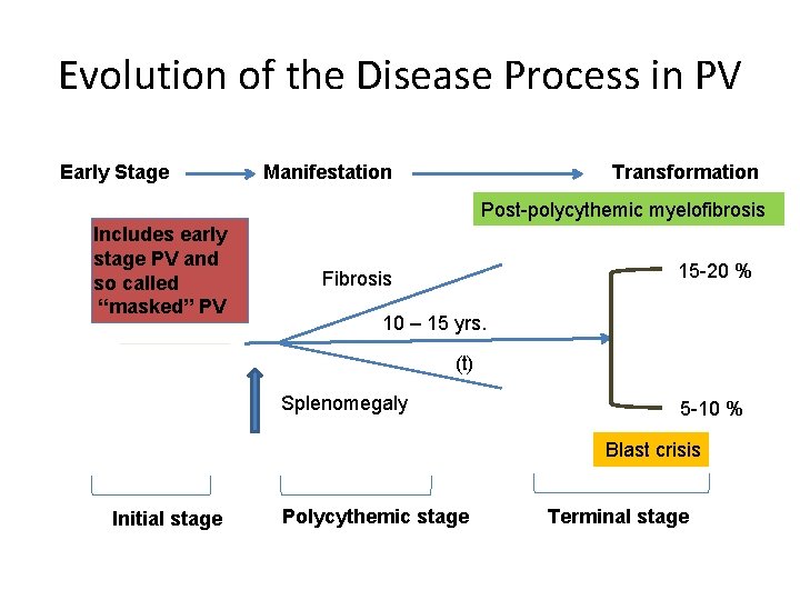
Evolution of the Disease Process in PV Early Stage Manifestation Transformation Post-polycythemic myelofibrosis Includes early stage PV and so called “masked” PV 15 -20 % Fibrosis 10 – 15 yrs. (t) Splenomegaly 5 -10 % Blast crisis Initial stage Polycythemic stage Terminal stage
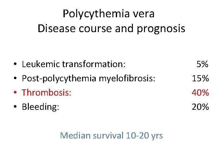
Polycythemia vera Disease course and prognosis • • Leukemic transformation: Post-polycythemia myelofibrosis: Thrombosis: Bleeding: Median survival 10 -20 yrs 5% 15% 40% 20%
Essential Thrombocytosis
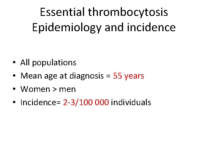
Essential thrombocytosis Epidemiology and incidence • • All populations Mean age at diagnosis = 55 years Women > men Incidence= 2 -3/100 000 individuals
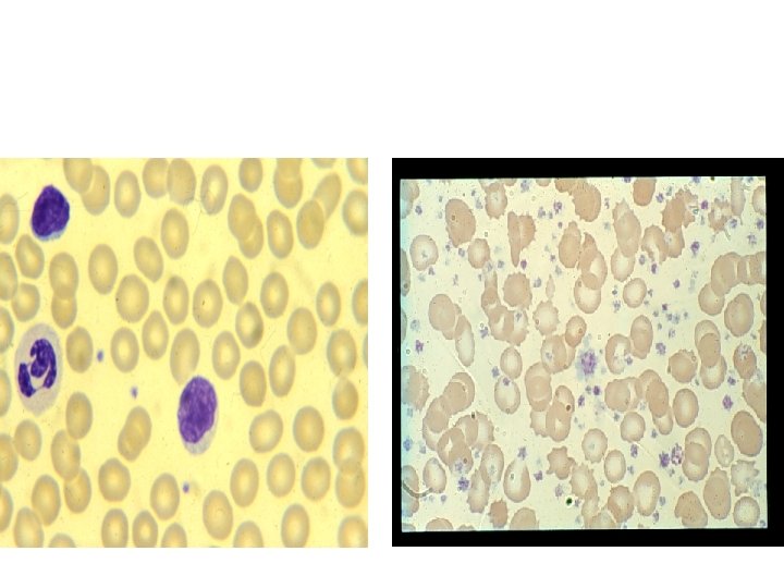
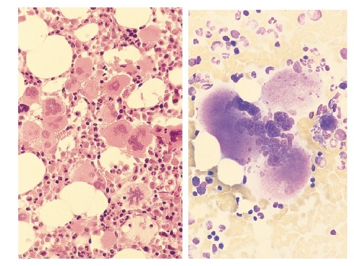
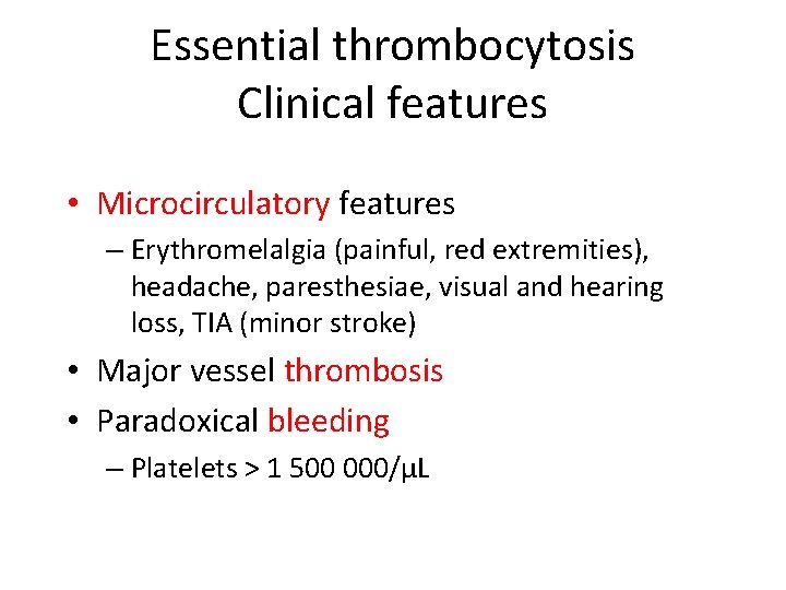
Essential thrombocytosis Clinical features • Microcirculatory features – Erythromelalgia (painful, red extremities), headache, paresthesiae, visual and hearing loss, TIA (minor stroke) • Major vessel thrombosis • Paradoxical bleeding – Platelets > 1 500 000/µL
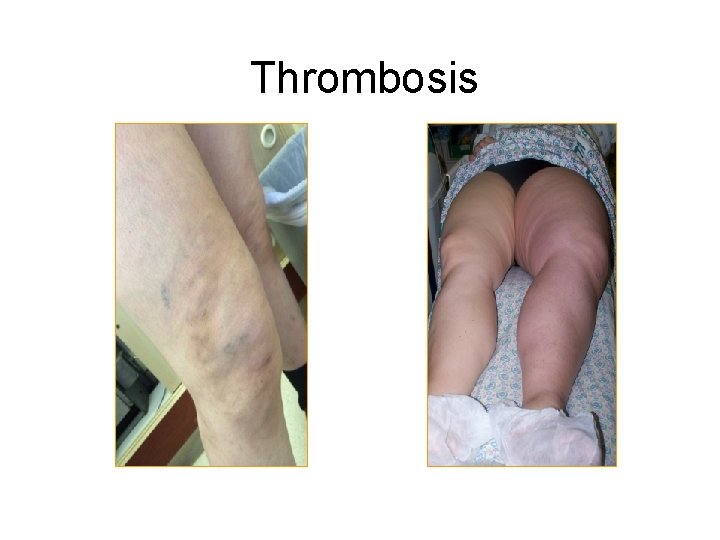
Thrombosis
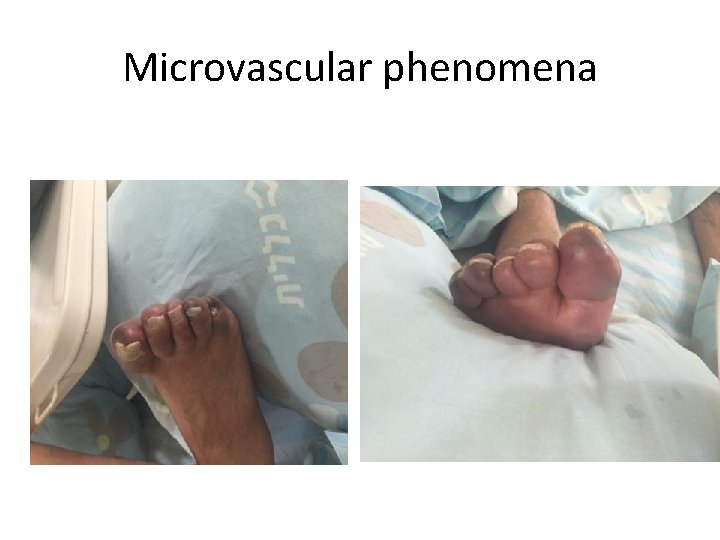
Microvascular phenomena
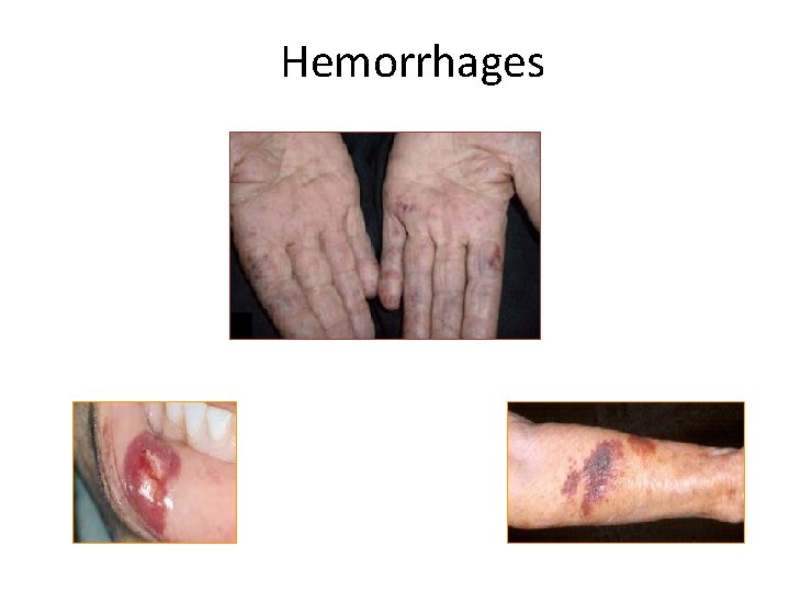
Hemorrhages
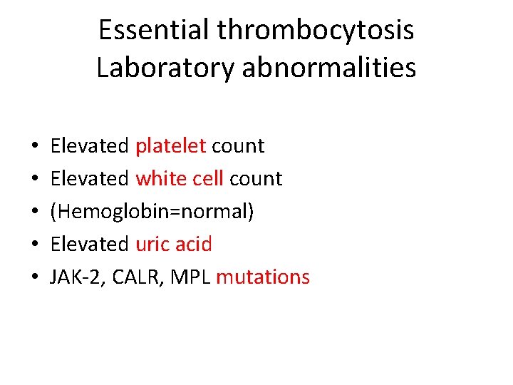
Essential thrombocytosis Laboratory abnormalities • • • Elevated platelet count Elevated white cell count (Hemoglobin=normal) Elevated uric acid JAK-2, CALR, MPL mutations
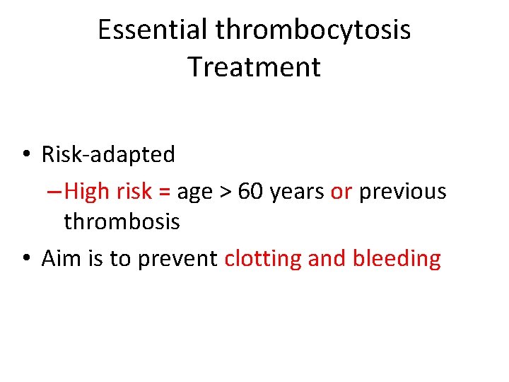
Essential thrombocytosis Treatment • Risk-adapted – High risk = age > 60 years or previous thrombosis • Aim is to prevent clotting and bleeding
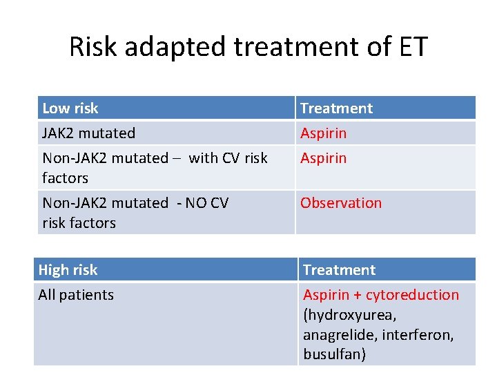
Risk adapted treatment of ET Low risk JAK 2 mutated Non-JAK 2 mutated – with CV risk factors Treatment Aspirin Non-JAK 2 mutated - NO CV risk factors Observation High risk All patients Treatment Aspirin + cytoreduction (hydroxyurea, anagrelide, interferon, busulfan)
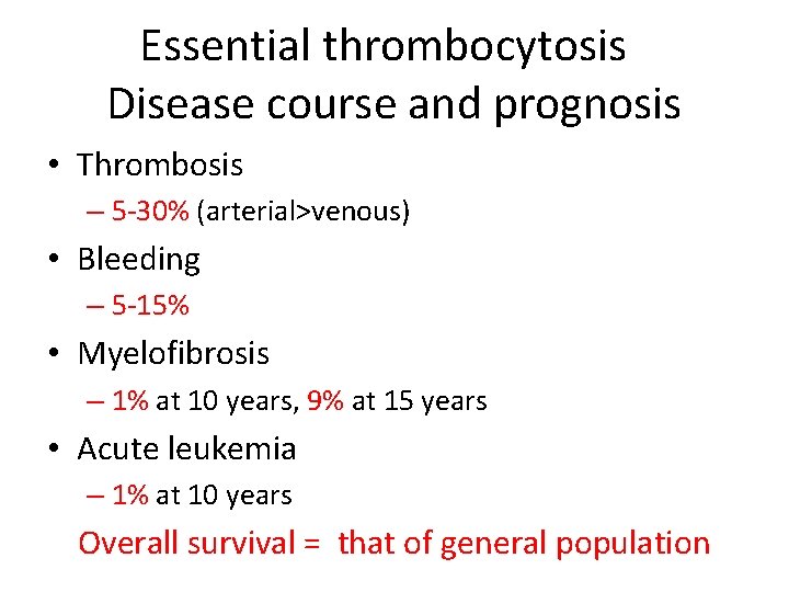
Essential thrombocytosis Disease course and prognosis • Thrombosis – 5 -30% (arterial>venous) • Bleeding – 5 -15% • Myelofibrosis – 1% at 10 years, 9% at 15 years • Acute leukemia – 1% at 10 years Overall survival = that of general population
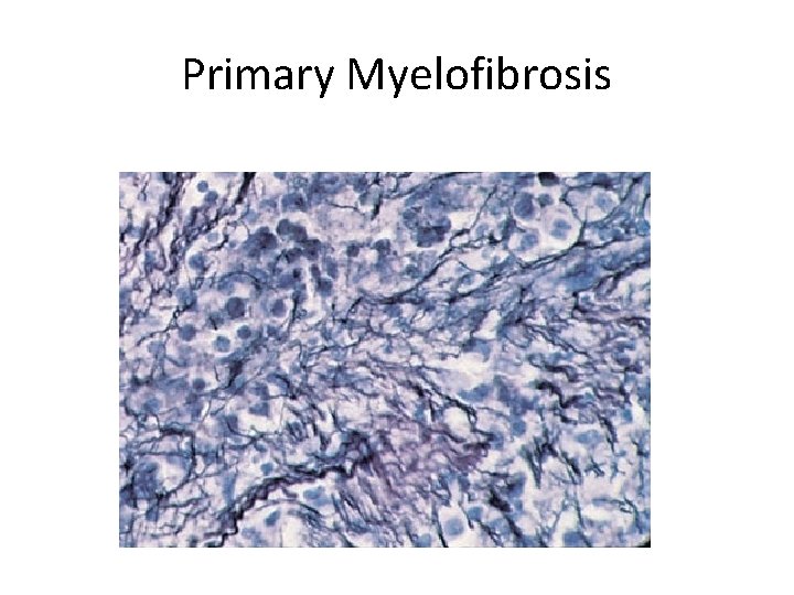
Primary Myelofibrosis
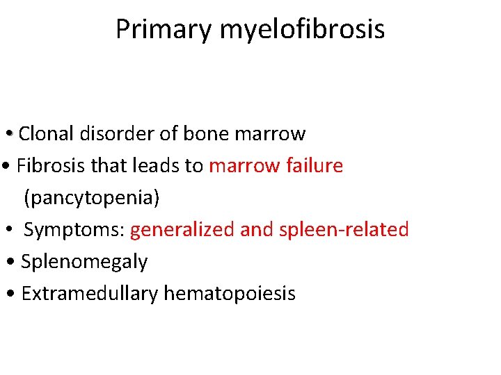
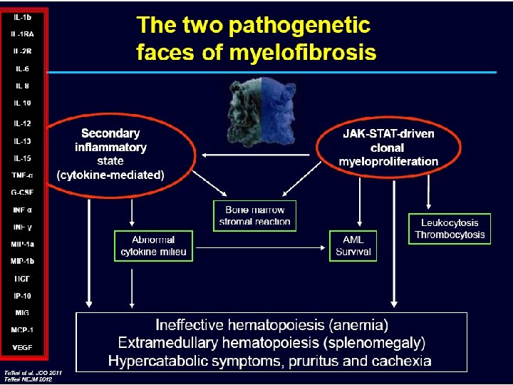
Primary myelofibrosis • Clonal disorder of bone marrow • Fibrosis that leads to marrow failure (pancytopenia) • Symptoms: generalized and spleen-related • Splenomegaly • Extramedullary hematopoiesis
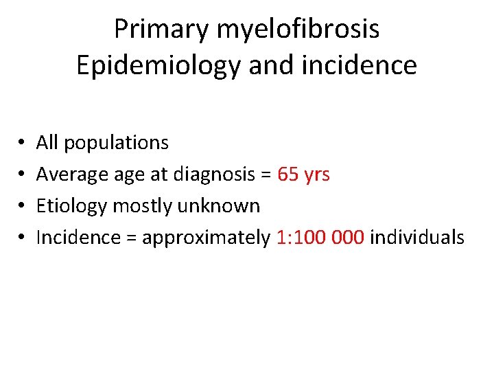
Primary myelofibrosis Epidemiology and incidence • • All populations Average at diagnosis = 65 yrs Etiology mostly unknown Incidence = approximately 1: 100 000 individuals
Primary myelofibrosis Clinical features
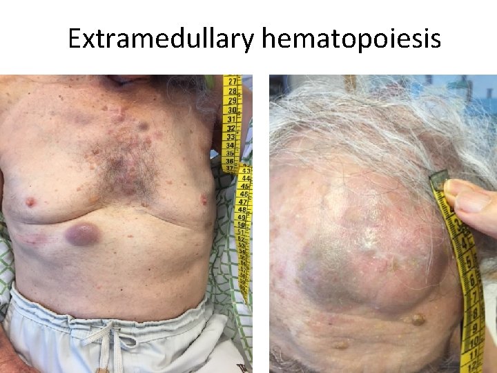
Extramedullary hematopoiesis
Primary myelofibrosis Laboratory abnormalities • • Blood count abnormalities – variable Elevated LDH, uric acid Cytogenetic (chromosomal) abnormalities Mutations
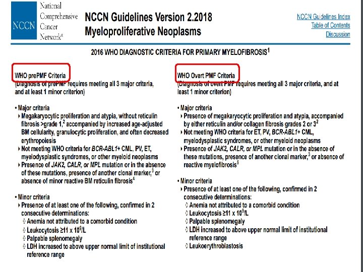
Primary myelofibrosis diagnosis • • • Typical blood picture Splenomegaly Failed bone marrow aspirate “dry-tap” Fibrosis on trephine biopsy Absence of other cause Molecular abnormality in 75% of patients
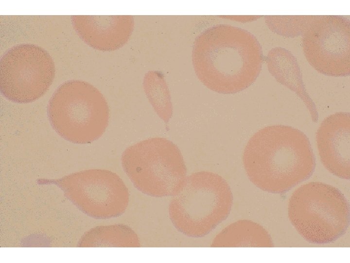
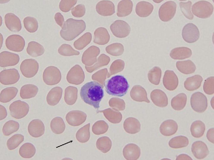
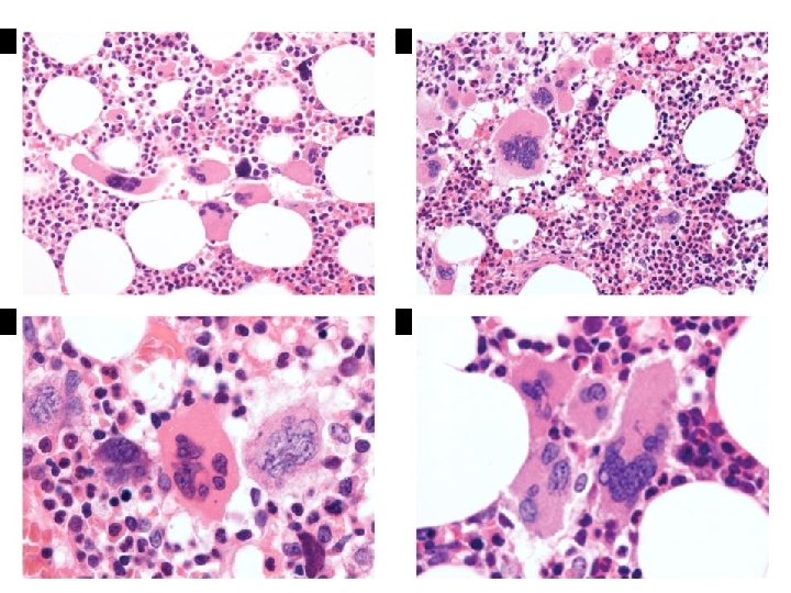
WHO 2014 Diagnostic Criteria Major criteria • Megakaryocyte proliferation and atypia accompanied by fibrosis • Not meeting WHO criteria for CML, PV, MDS, or other myeloid neoplasm • Presence of JAK 2, CALR or MPL mutation Minor criteria • Increased serum LDH • Anemia • Palpable splenomegaly • Leukoerythroblastosis
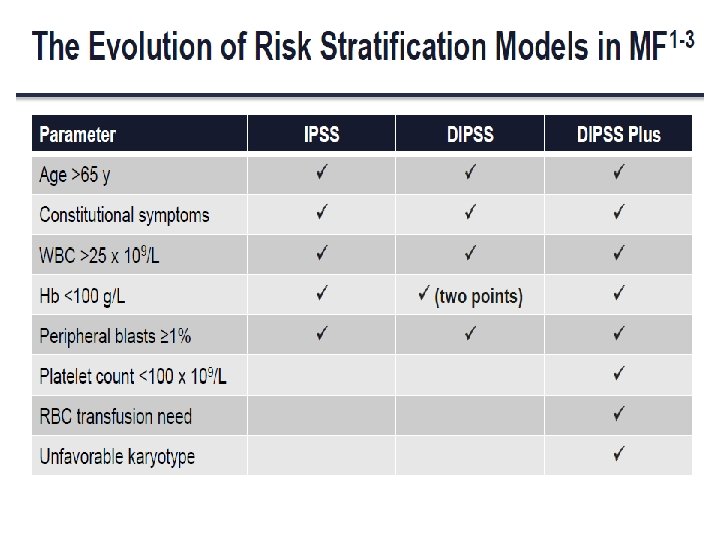
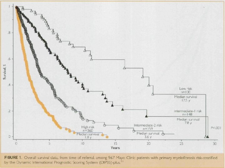
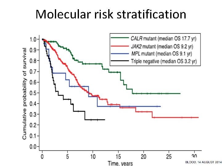
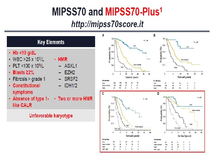
Molecular risk stratification
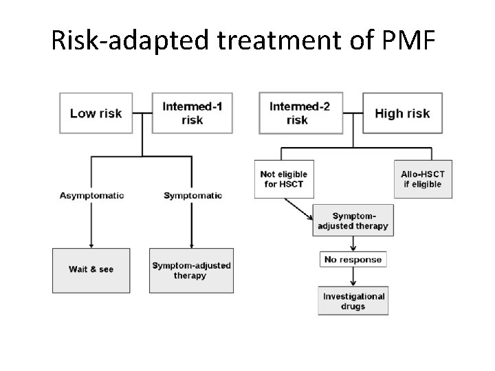
Risk-adapted treatment of PMF
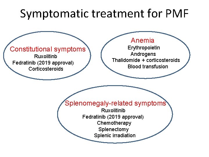
Symptomatic treatment for PMF Anemia Constitutional symptoms Ruxolitinb Fedratinib (2019 approval) Corticosteroids Erythropoietin Androgens Thalidomide + corticosteroids Blood transfusion Splenomegaly-related symptoms Ruxolitinib Fedratinib (2019 approval) Chemotherapy Splenectomy Splenic irradiation
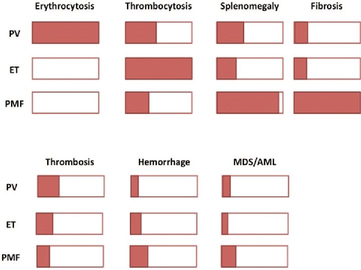
The MPN continuum
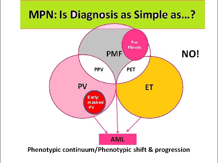
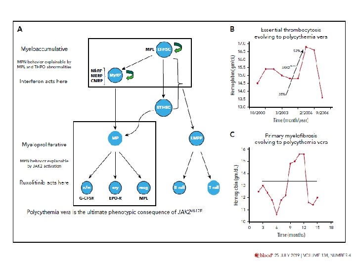
Early/ masked PV
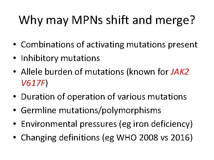
Why may MPNs shift and merge? • Combinations of activating mutations present • Inhibitory mutations • Allele burden of mutations (known for JAK 2 V 617 F) • Duration of operation of various mutations • Germline mutations/polymorphisms • Environmental pressures (eg iron deficiency) • Changing definitions (eg WHO 2008 vs 2016)
New information about mutant Calreticulin
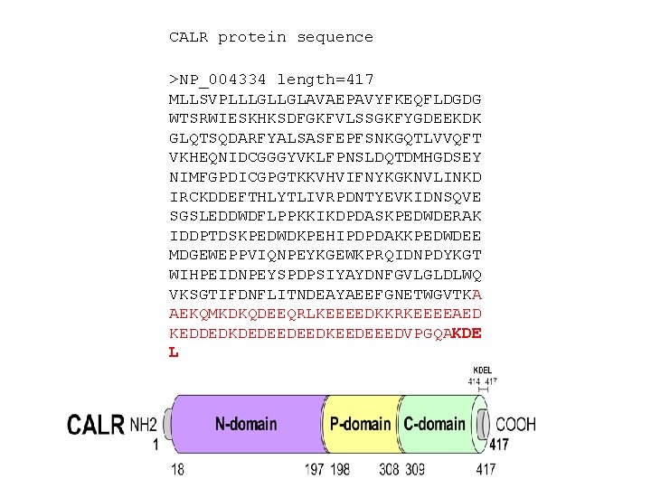
CALR protein sequence >NP_004334 length=417 MLLSVPLLLGLLGLAVAEPAVYFKEQFLDGDG WTSRWIESKHKSDFGKFVLSSGKFYGDEEKDK GLQTSQDARFYALSASFEPFSNKGQTLVVQFT VKHEQNIDCGGGYVKLFPNSLDQTDMHGDSEY NIMFGPDICGPGTKKVHVIFNYKGKNVLINKD IRCKDDEFTHLYTLIVRPDNTYEVKIDNSQVE SGSLEDDWDFLPPKKIKDPDASKPEDWDERAK IDDPTDSKPEDWDKPEHIPDPDAKKPEDWDEE MDGEWEPPVIQNPEYKGEWKPRQIDNPDYKGT WIHPEIDNPEYSPDPSIYAYDNFGVLGLDLWQ VKSGTIFDNFLITNDEAYAEEFGNETWGVTKA AEKQMKDKQDEEQRLKEEEEDKKRKEEEEAED KEDDEDKDEDEEDEEDKEEDEEEDVPGQAKDE L
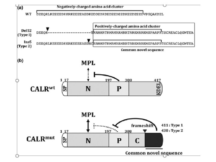
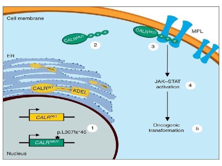
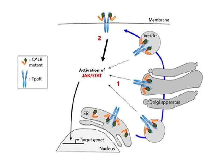
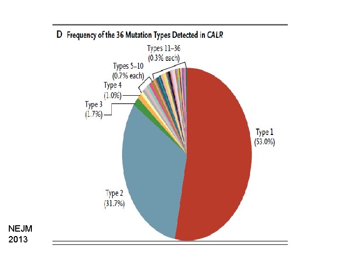
NEJM 2013
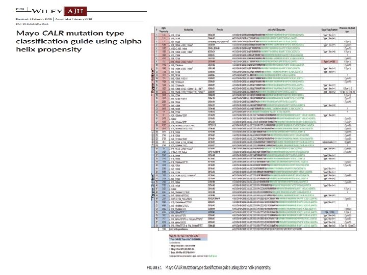
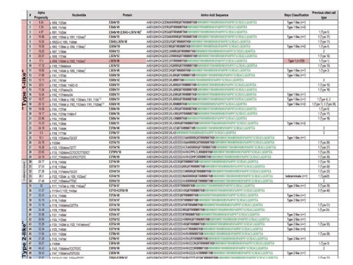
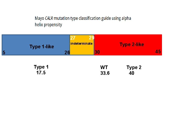
27 indeterminate Type 1 -like 5 26 Type 1 17. 5 29 Type 2 -like 45 30 WT 33. 6 Type 2 40
Meir Medical Center Case: Unusual CALR mutation in a man with ET Courtesy of Dr Tali Tohami
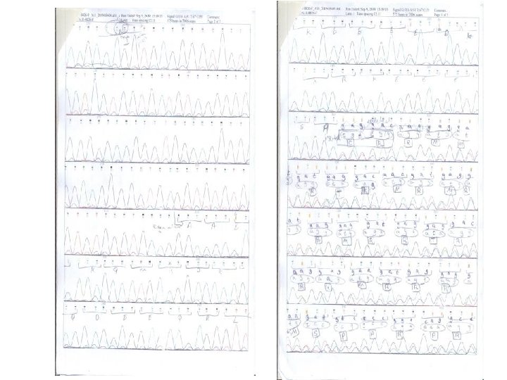
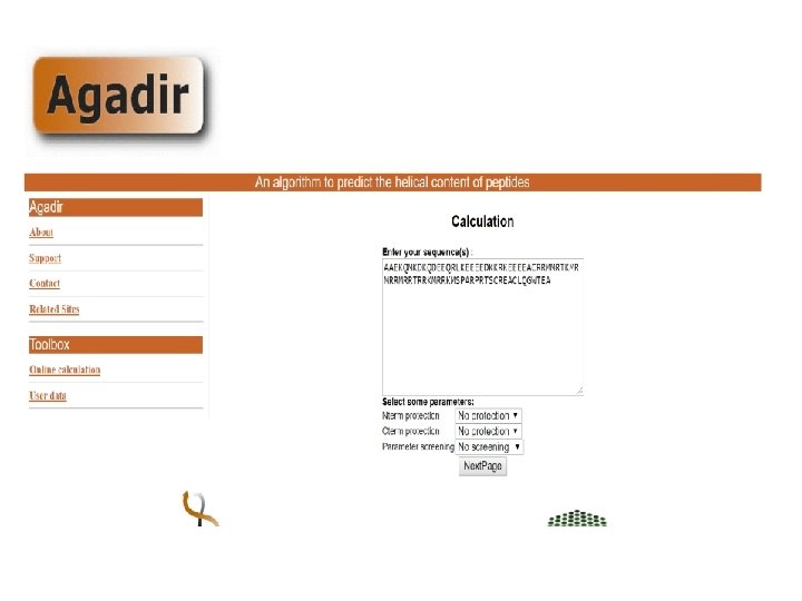
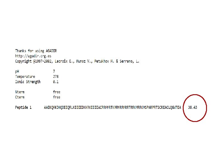
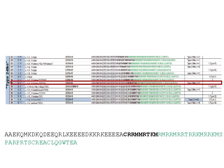
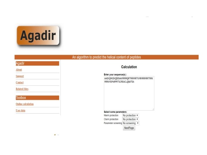
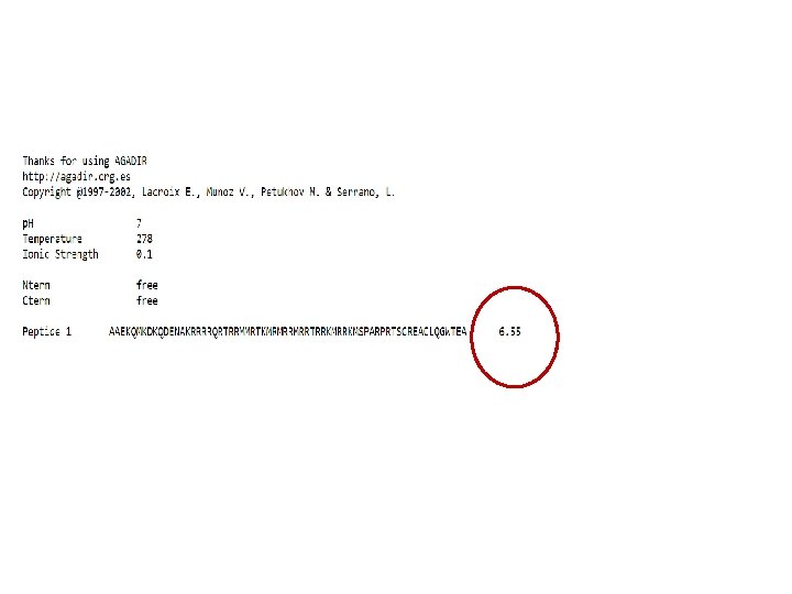
AAEKQMKDKQDEEQRLKEEEEDKKRKEEEEACRRMMRTKMRMRRMRRTRRKMS PARPRTSCREACLQGWTEA
Diagnosis ET with a type-2 –like CALR mutation
- Slides: 87