
The Muscular System Skeletal Muscle Tissue and Muscle
- Slides: 16
The Muscular System Skeletal Muscle Tissue and Muscle Organization Chapter 9 Alireza Ashraf, M. D. Professor of Physical Medicine & Rehabilitation Shiraz Medical school
The Muscular System n Skeketal Muscle, Cardiac Muscle, Smooth Muscle n Share 4 basic properties: 1) Excitability – respond to stimulation by either the nervous system or to circulating hormones 2) Contractility – ability to shorten actively and exert a pull or tension harnessed by CT 3) Extensibility – ability to contract over a range of resting lengths 4) Elasticity – ability of a muscle to rebound toward its original length after a contraction
Skeletal Muscle Functions n Contractile organs directly or indirectly attached to bones n Functions include: 1) Produce skeletal movement: muscle contractions pull on tendons and move the bones - effects range from simple motions to highly coordinated movements of swimming, skiing, or typing 2) Maintain posture and body positions: contraction of secific muscles – involves the contraction of muscles that stabilize joints, without constant muscular contraction, we could not sit upright without collapsing or stand without toppling over
Skeletal Muscle Functions 3) Support soft tissues: the abdominal wall and the floor of the pelvic cavity consist of layers of skeletal muscle - support the weight of visceral organs and protect internal tissues 4) Regulate entering and exiting of material: openings or orifices, of the digestive and urinary tracts are encircled by skeletal muscles - provide voluntary control over swallowing, defecation, and urination 5) Maintain body temperature: muscle contractions require energy some is converted to heat - heat lost by contracting muscles maintains our body temp
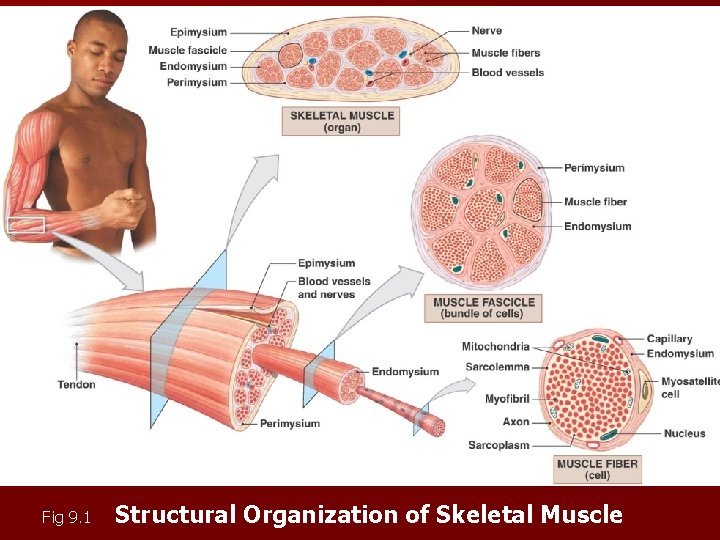
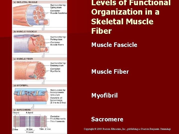
Anatomy of Skeletal Muscles n Epimysium (mys = muscle) - outer layer of dense irregular CT connected to the deep fascia: - surrounds the entire skeletal muscle and separates the muscle from surrounding tissues n Perimysium - CT fibers that surrounds each fascicle: - divides the muscle into a series of internal compartments each containing a bundle of muscle fibers called fascicles - collagen and elastic fibers, BVs and nerve supply n Endomysium – surrounds each skeletal muscle fiber - binds each muscle fiber to its neighbor and supports the capillaries that supply each individual fiber - consists of a network of reticular fibers with scattered myosatellite cells that function in repair of damaged muscle
Copyright © 2009 Pearson Education, Inc. , publishing as Pearson Benjamin Cummings Fig 9. 1 Structural Organization of Skeletal Muscle
Tendons and Aponeuroses n CT fibers of the all 3 are interwoven, those of the perimysium blend into the epimysium n At each end of the muscle, collagen fibers often converge - form a fibrous tendon that attaches muscle to bone, skin, or another muscle - tendons resemble thick cords or cables those that form flattened sheets are called aponeuroses n Tendon fibers are interwoven into the periosteum and matrix of associated bone - provides an extremely strong bond to withstand muscle contractions
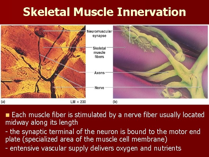
Nerves and Blood Vessels n 3 CT layers contain the nerves and BVs that supply the muscle fibers n Called voluntary muscles because their contractions can be controlled - provided by the nerves of the nervous system n Nerves, bundles of axons, penetrate the epimysium - branch through the perimysium and enter the endomysium to innervate individual fibers n Neuromuscular synapse or myoneural junction – site of chemical communication between a synaptic terminal of the neuron and a skeletal muscle fiber
Skeletal Muscle Innervation Fig 9. 2 Each muscle fiber is stimulated by a nerve fiber usually located midway along its length - the synaptic terminal of the neuron is bound to the motor end plate (specialized area of the muscle cell membrane) - entensive vascular supply delivers oxygen and nutrients n
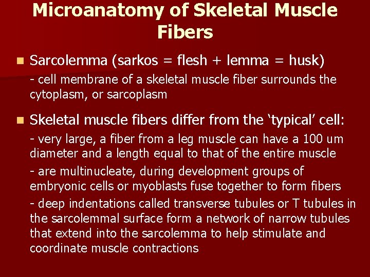
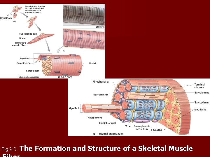
Microanatomy of Skeletal Muscle Fibers n Sarcolemma (sarkos = flesh + lemma = husk) - cell membrane of a skeletal muscle fiber surrounds the cytoplasm, or sarcoplasm n Skeletal muscle fibers differ from the ‘typical’ cell: - very large, a fiber from a leg muscle can have a 100 um diameter and a length equal to that of the entire muscle - are multinucleate, during development groups of embryonic cells or myoblasts fuse together to form fibers - deep indentations called transverse tubules or T tubules in the sarcolemmal surface form a network of narrow tubules that extend into the sarcolemma to help stimulate and coordinate muscle contractions
Fig 9. 3 The Formation and Structure of a Skeletal Muscle
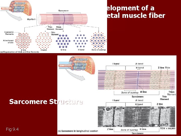
Development of a skeletal muscle fiber Sarcomere Structure Fig 9. 4
Levels of Functional Organization in a Skeletal Muscle Fiber Muscle Fascicle Muscle Fiber Myofibril Sacromere Copyright © 2009 Pearson Education, Inc. , publishing as Pearson Benjamin Cummings
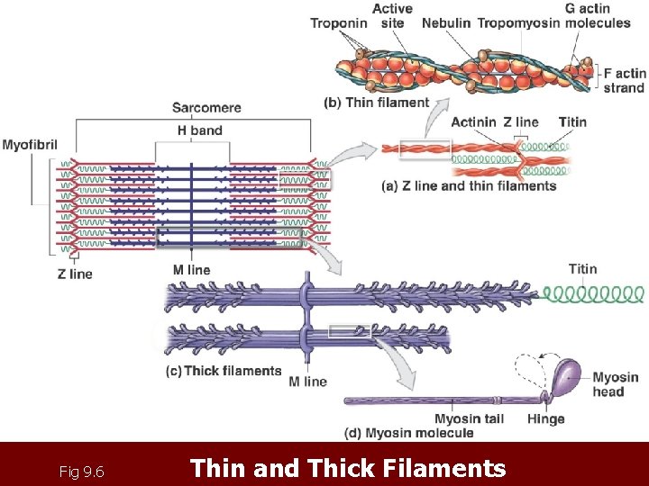
Fig 9. 6 Thin and Thick Filaments
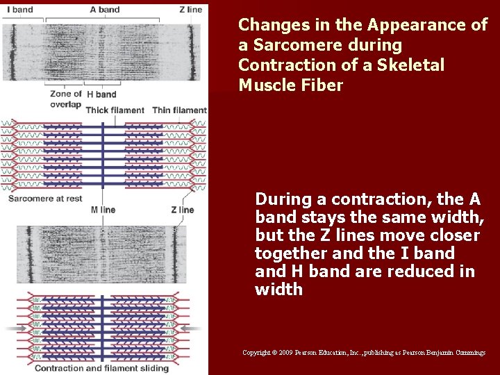
Changes in the Appearance of a Sarcomere during Contraction of a Skeletal Muscle Fiber During a contraction, the A band stays the same width, but the Z lines move closer together and the I band H band are reduced in width Copyright © 2009 Pearson Education, Inc. , publishing as Pearson Benjamin Cummings
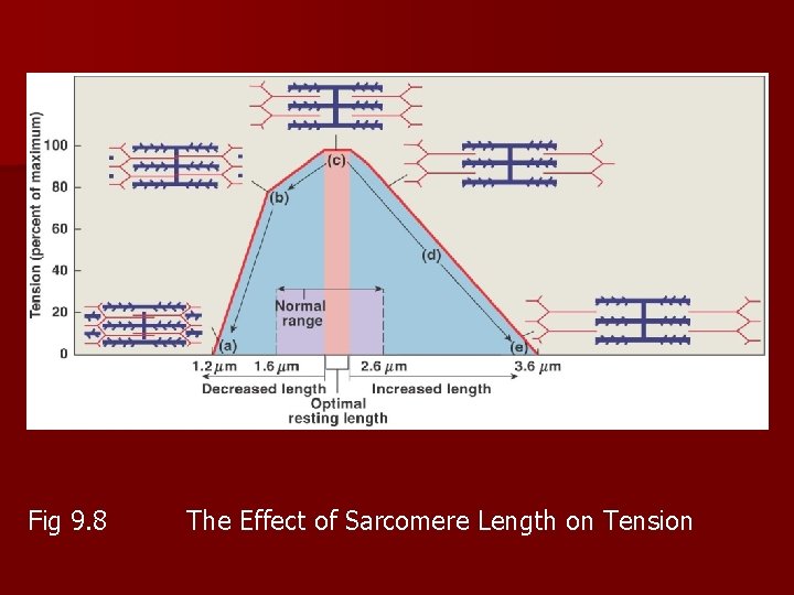
Fig 9. 8 The Effect of Sarcomere Length on Tension