The Muscular System More than 600 muscles in


 • CNS")


- Slides: 33
The Muscular System • More than 600 muscles in the human body • attached to bones by tendons • Exert force by converting chemical energy (food) into mechanical energy (muscle contraction)
Types of Muscles 1. • • 2. • • • 3. • • Skeletal Voluntary striated Most in human body e. g biceps Cardiac Involuntary striated e. g. heart only Smooth Involuntary/not striated Surrounds internal organs Contracts slowly but can sustain a contraction for a long period of time e. g. intestine lining
Properties of Muscle Fibres 1. Irritability: muscle responds to stimulus 2. Contractibility: shorten in length 3. Elasticity: Ability to stretch and return to its normal position 4. Extensibility: ability to extend in length 5. Conductivity: can transmit nerve impulses
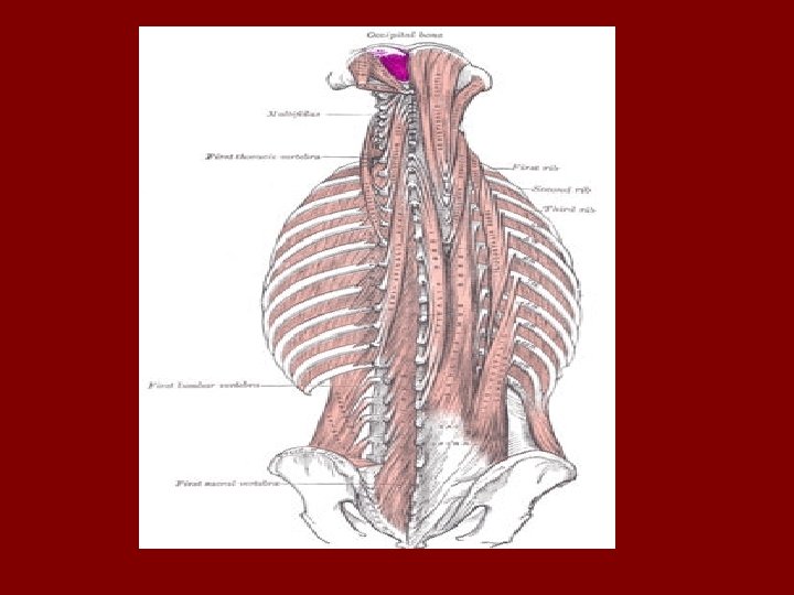
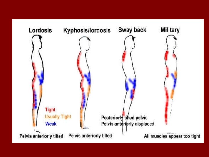
Skeletal Muscles Functions 1. Movement • Locomotion or movement of parts 2. Heat Production • Responsible for a major share of total body heat and maintaining homeostasis of temperature 3. Posture • Partial contractions of man skeletal muscles
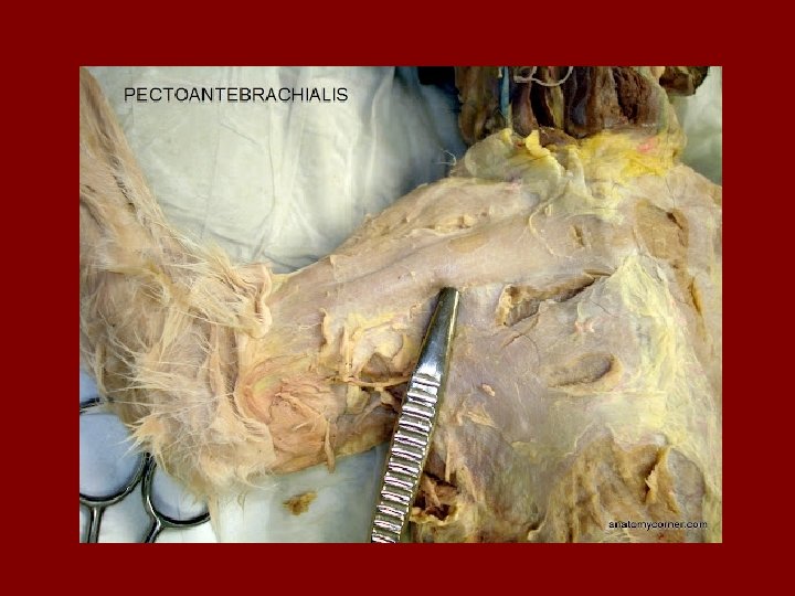
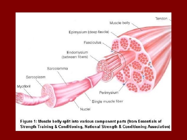
Muscle Structure Macroscopic Level: • Muscles are surrounded by fascia • Fascia is a thin, tight sheath of connective tissue • Protects muscles, and prevent friction between muscles
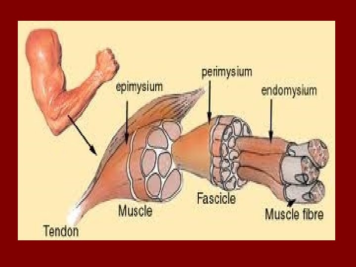
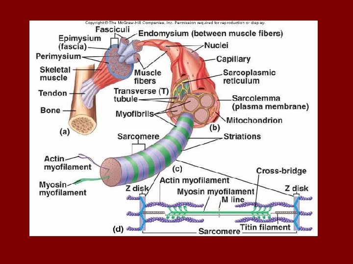
Microscopic Level: • Epimysium: large, strong sheath that envelopes entire muscle and becomes tendon (deep fascia) • Perimysium: connective tissue that binds groups of fasciles • Fasicles: bundles of individual muscles fibres • Endomysium: sheath of connective tissue that surrounds individual muscle fibres
• Sarcolemma: beneath endomysium/acts as a plasma membrane • Sarcoplasm: muscle cell cytoplasm- inside sarcolemma (contains proteins, stores gylcogen and myoglobin)
Myofibril: thread like structure inside muscle fibre/house myosin and actin • Sarcomere: site of muscular contraction and house for myosin and actin – Myosin • Cellular protein • has a head and tail • Head is attachment site for actin – Actin • Cellular protein • Binding site for myosin • 2 proteins – Troponin – binding site for calcium – Tropomyosin – covers the binding site on actin
The Neuromuscular System • The NMS is linkage – Muscular System – Nervous System (nervous impulses originating in the CNS and sent through PNS) – Use and practice improve the coordination between the two systems
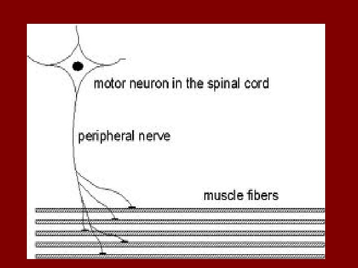
• Motor Unit – Includes a neuron/nerve called the ‘motor neuron’, the axon and the muscle fibers it stimulates – Where the motor neuron and muscle fiber meet is called the neuromuscular junction
How the Motor Unit Works 1. Electrical impulse travels along nerve pathway 2. Once at neuromuscular junction, a chemical neurotransmitter is then released (acetylcholine) 3. ACh is detected by receptors on the surface of the muscle fiber resulting in muscle contraction SO…. . Electrical energy-chemical energy-mechanical work
‘The All or None Principle’ • When a motor unit is stimulated to contract, it does to its full potential • Motor units can be small or large – small: produce fine motor movements (threading a needle) – large: produce big motor movements (kicking a ball)
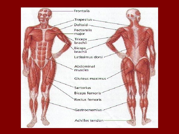
Naming Muscles 1. Action of the Muscle: flexion, extension • e. g. flexor carpi ulnaris 2. Direction of Fibres: rectus, transversus • e. g. rectus abdominus 3. Location of the Muscle: anterior, posterior • e. g. tibialis anterior 4. Number of Divisions: 2 or 3 heads • e. g. biceps brachii 5. Shape of Muscles: deltoid, trapezius • e. g. trapezius looks like a trapezoid 6. Point of Attachment: sternum, clavicle, mastoid • e. g. sternocleidomastoid
How Muscles Attach to Bone • Indirectly: epimysium extends as a tendon and then connects with periosteum • Directly: epimysium adheres to and fuses with periosteum
• Origin: point where muscle attaches to more stationary bones of the axial skeleton • Insertion: point where muscle attaches to bone that is moved most
• Agonistic pairs: skeletal muscles arranged in opposing pairs (e. g. flexors and extensors) • Agonist: prime mover • Synergists: assist the agonist, but not primarily responsible • Antagonist: muscle that counteracts the agonist; lengthen when agonist contracts • Fixators: Stabilize the joint • See Table 3. 3 in text
Types of Contractions 1. Concentric- muscle fibres shorten – Bicep shortens when lifting an object 2. Eccentric- muscle fibres lengthen – Bicep places weight on the grounds 3. Isometric- muscle fibres stay the same – Trying to lift something too heavy
Contraction during Exercise 1. Isotonic Exercise: – Shortening and lengthening contraction 2. Isometric Exercise: – Muscles maintain length – Good for developing specific muscle groups (rehab) 3. Isokinetic Exercise: – Isometric and Isotonic exercise combined – E. g. advanced high performance machines http: //www. youtube. com/watch? v=By-Of-s 1 z. N 8
How do our skeletal muscles contract?
The Sliding Filament Theory Diagram of a Sarcomere
The Process Begins…. . • Message sent by brain (raise your hand) • CNS • PNS • Neuromuscular junction (motor unit) • ACh released (acetylcholine) • Action potential generated opening up sarcolemma (membrane) • Calcium ions is released into sarcoplasm due to ATP……
• What is ATP: – An energy carrying molecule that results from food metabolism – Energy source behind the release of calcium – Release of calcium is what triggers contraction – Also detaches myosin from actin – As you work, more and more ATP is used, and you must replace through food and metabolism
At Rest • Calcium ion within sarcoplasmic reticulum • ATP bound to myosin (thick filaments) • Actin intact Contraction • Calcium binds to troponin • Tropomysoin swivels • Binding sites are exposed • Crossbridges from myosin bind to new sites on actin • ATP breakdown = energy as heat and moves crossbridges causing sliding action • Actin is drawn to center of sarcomere • Crossbridge cycle repeats • Z line is drawn together • Sarcomere shortens
Back to Resting State… • A Ch release halted and remaining is inactive • Calcium ions back to sarcoplasmic reticulum by active transport • Shape of actin is restored – no binding site available as they are covered by tropomyosin • Crossbridges breakway
Visuals http: //www. youtube. com/watch? v=Ed. Hz. KYDxr. Kc http: //www. youtube. com/watch? v=Vlchs 4 om. FDM&feature=related