The Muscular System Function of Muscles Produce movement


 tubules- The sarcolemma folds deep into")


- Slides: 37
The Muscular System
Function of Muscles · Produce movement · Maintain posture · Stabilize joints · Generate heat · Dilate and constrict eyes/passages
5 Properties of muscle tissue • • 1. Excitable –capable of responding to nerve stimulation 2. Elasticity- Can return to original length 3. Contractable - shorten 4. Adaptable- hypertrophy / atrophy
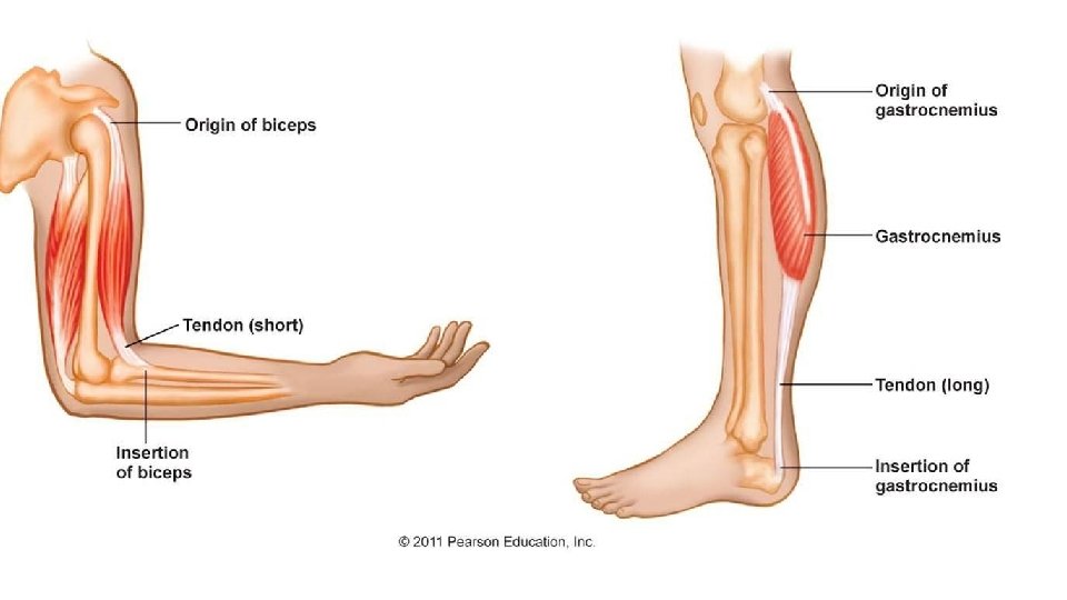
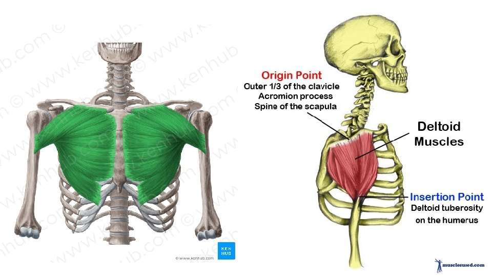
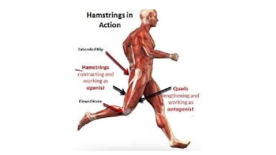
Muscle Rules • • Always cross joint Can only contract to get shorter Antagonistic muscle sets Origin and insertion (picture)
Blood and Nerve supply • One major nerve and artery goes into belly of muscle
Myosin
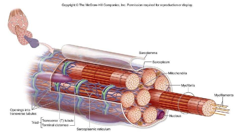
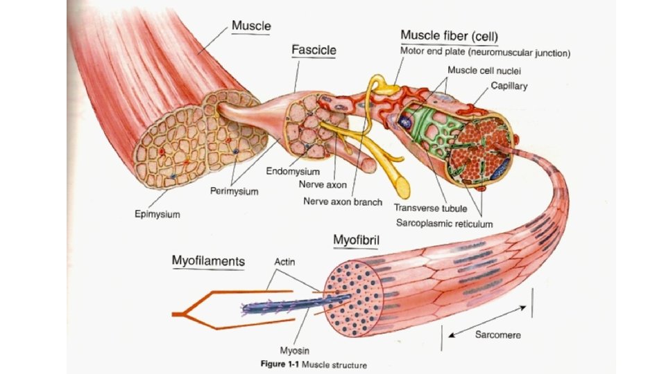
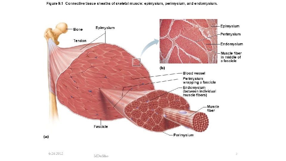
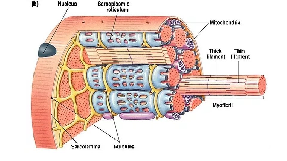
Connective Tissue Wrappings of Skeletal Muscle · Endomysium – around single muscle fiber · Perimysium – around a fascicle (bundle) of fibers Figure 6. 1
Connective Tissue Wrappings of Skeletal Muscle · Epimysium – covers the entire skeletal muscle Figure 6. 1
Unique features of skeletal muscle • Multiple nuclei- fused cells during development • High numbers of mitochondria- ATP for muscle contraction • Sarcoplasm- the cytoplasm (lots of glycogen and myoglobin)
Unique Properties of muscle cells • Sarcolemma- Plasma Membrane – Unique because it can transfer electrical charge (action potential) from neurons
Unique Properties of muscle cells • Transverse (T) tubules- The sarcolemma folds deep into the muscle cell. – Important because they allow the nerve signal to reach all of the sarcomeres.
Unique properties of muscle cells • Sarcoplasmic reticulum – Stores calcium in the muscle cell – Modified Smooth ER – “Sweater” around the myofibrils
3 types of muscle
The Muscular System · Muscles are responsible for all types of body movement – they contract or shorten and are the machine of the body · Three basic muscle types are found in the body · Skeletal muscle · Cardiac muscle · Smooth muscle
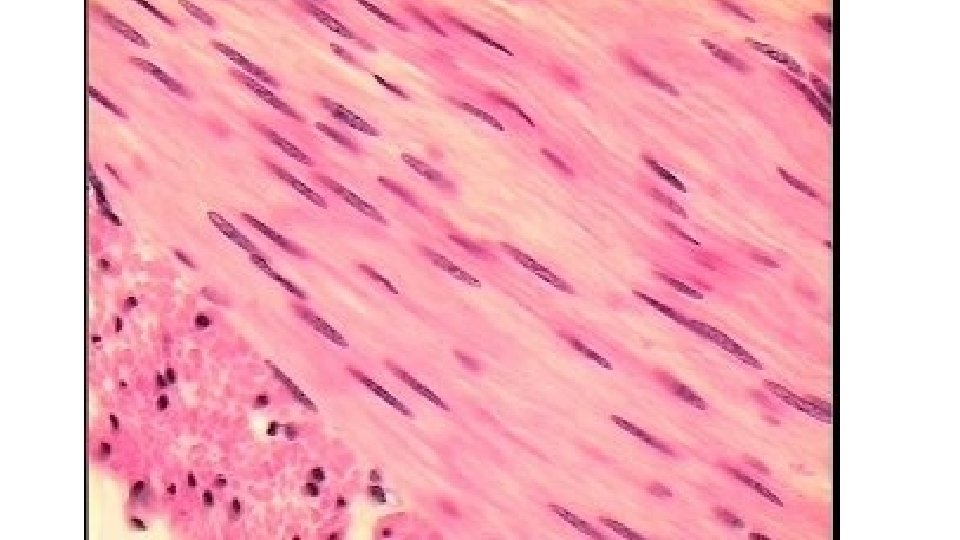
Smooth Muscle Characteristics · Has no striations · Spindle-shaped cells · Single nucleus · Involuntary – no conscious control · Found mainly in the walls of hollow organs · Slow, sustained and tireless Figure 6. 2 a
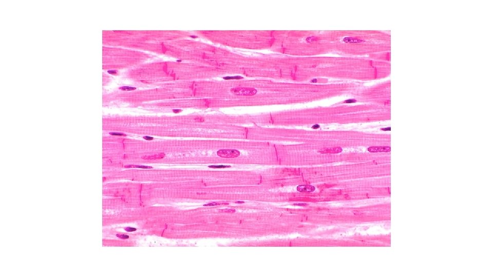
Cardiac Muscle Characteristics · Has striations · Usually has a single nucleus · Joined to another muscle cell at an intercalated disc · Involuntary · Found only in the heart · Steady pace! Figure 6. 2 b
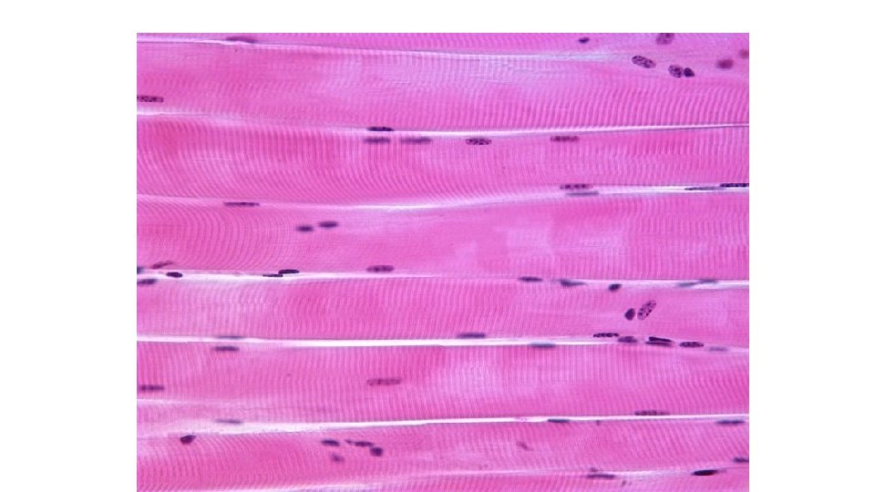
Skeletal Muscle Characteristics · Most are attached by tendons to bones · Cells are multinucleated · Striated – have visible banding · Voluntary – subject to conscious control · Cells are surrounded and bundled by connective tissue = great force, but tires easily
Turn to your neighbor and describe the levels of organization of a skeletal muscle fiber Objective: Analyze and interpret the events of the sliding filament theory of muscle contraction
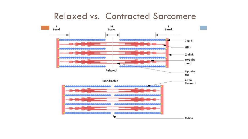
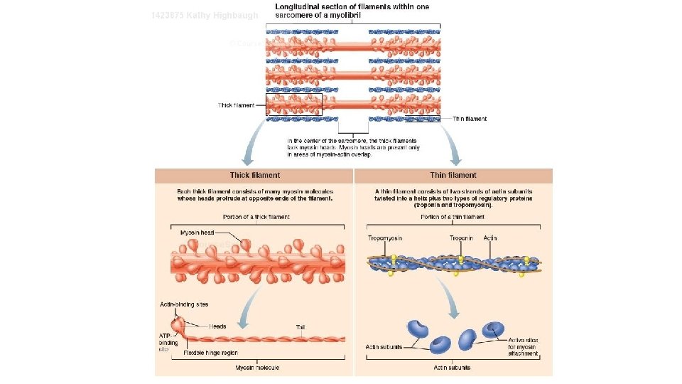
The Sarcomere “myofilaments” • Actin- “thin filament” – Has binding sites for myosin heads • These sites are blocked by tropomysosin • Calcium binding to Troponin unblocks the binding sites and allows the myosin heads to bind. • Myosin – “Thick filament” - Has two round “heads” - ATPase on the heads to break down ATP for energy
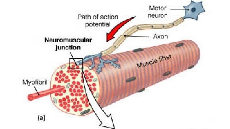
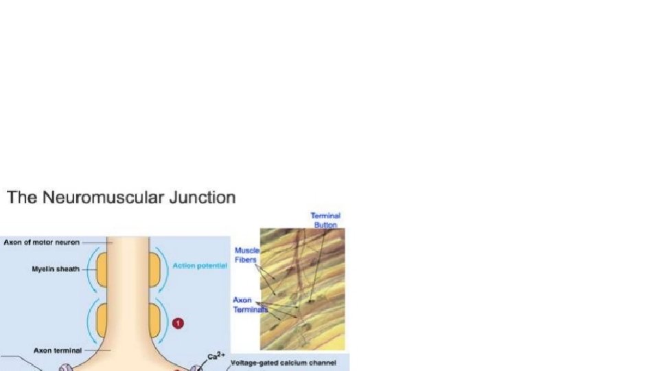
Sliding Filament Theory • Phase 1: Neuron stimulates muscle fiber – Occurs at the Neuromuscular junction • Nerves are called motor (somatic) neurons – Enter in the belly of muscle and branch out – Each muscle cell has one NMJ – Space (50 nm) between the nerve fiber and muscle cell » Synaptic Cleft – ACh – Acetylcholine – a neurotransmitter located at the ends of the motor neurons. • Stored in synaptic vesicles
Events leading to an Action Potential • 1. Action potential sent from brain to muscle cell. • 2. Voltage gated Ca++ channels open in nerve at the NMJ- Calcium flows in • 3. Ca+ influx causes Ach to be resynaptic cleft and binds to sarcolemma receptors. • 5. Binding opens Na+ and K+ channels, more Na+ flows in than K+ out = depolarization • leased by exocytosis • 4. Ach moves across 6. AP travels down T tubules to SR which triggers release of Calcium
Sliding Filament • https: //www. youtube. com/watch? v=Ktv-Ca. Ot 6 UQ
Myasthenia gravis Not enough Ach receptors, probably autoimmune disease.