
The Mouth The mouth cavity is divided into
- Slides: 26
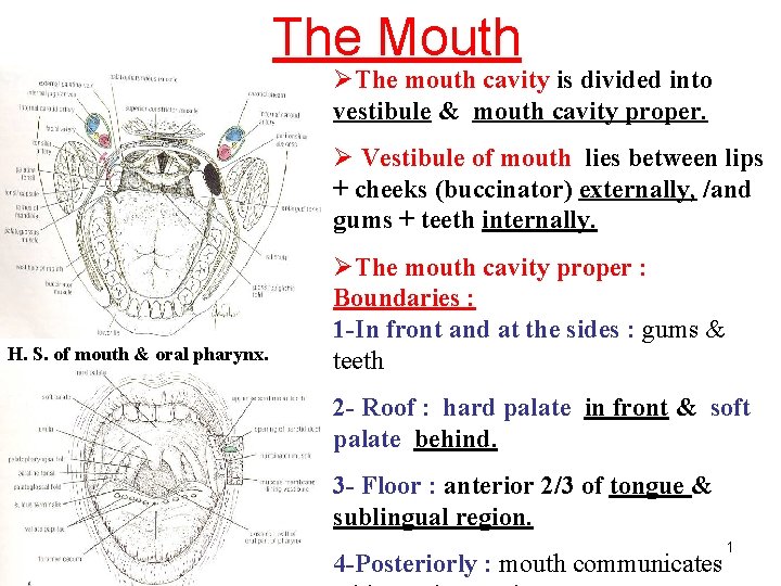
The Mouth ØThe mouth cavity is divided into vestibule & mouth cavity proper. Ø Vestibule of mouth lies between lips + cheeks (buccinator) externally, /and gums + teeth internally. H. S. of mouth & oral pharynx. ØThe mouth cavity proper : Boundaries : 1 -In front and at the sides : gums & teeth 2 - Roof : hard palate in front & soft palate behind. 3 - Floor : anterior 2/3 of tongue & sublingual region. 4 -Posteriorly : mouth communicates 1
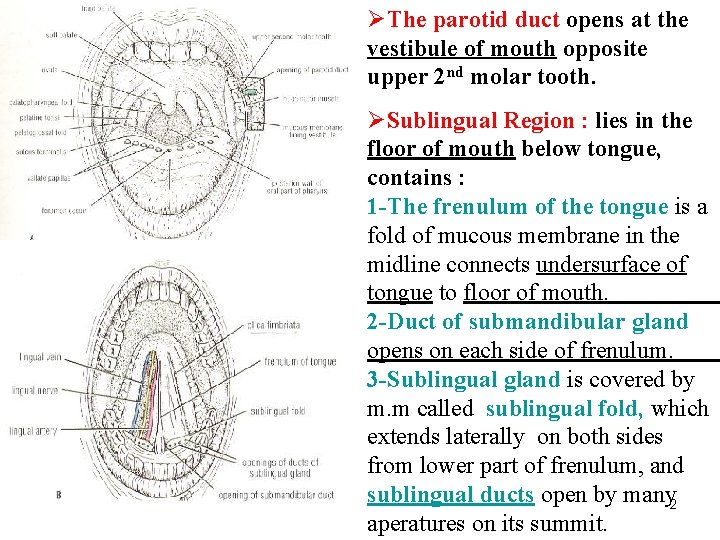
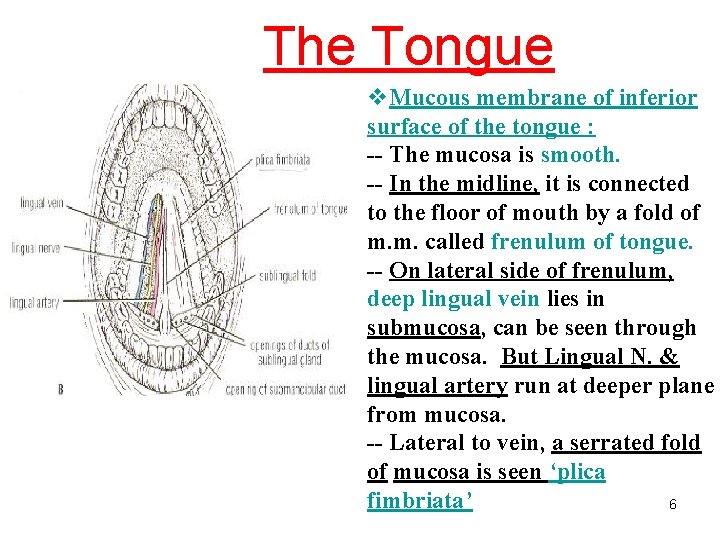
ØThe parotid duct opens at the vestibule of mouth opposite upper 2 nd molar tooth. ØSublingual Region : lies in the floor of mouth below tongue, contains : 1 -The frenulum of the tongue is a fold of mucous membrane in the midline connects undersurface of tongue to floor of mouth. 2 -Duct of submandibular gland opens on each side of frenulum. 3 -Sublingual gland is covered by m. m called sublingual fold, which extends laterally on both sides from lower part of frenulum, and sublingual ducts open by many 2 aperatures on its summit.
Sensory nerve supply of mucous membrane of mouth : • Roof (palate) : greater palatine & Nasopalatine nerves from Maxillary N. • Floor (anterior 2/3 of tongue) : lingual N. of mandibular N. (general sensation), + chorda tympani N. of facial N. (taste sensation). • Cheek : buccal nerve, a branch of mandibular N. 3
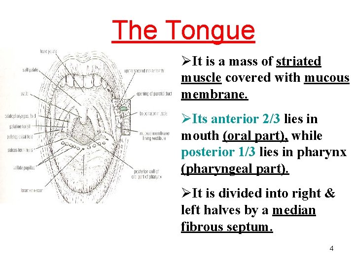
The Tongue ØIt is a mass of striated muscle covered with mucous membrane. ØIts anterior 2/3 lies in mouth (oral part), while posterior 1/3 lies in pharynx (pharyngeal part). ØIt is divided into right & left halves by a median fibrous septum. 4
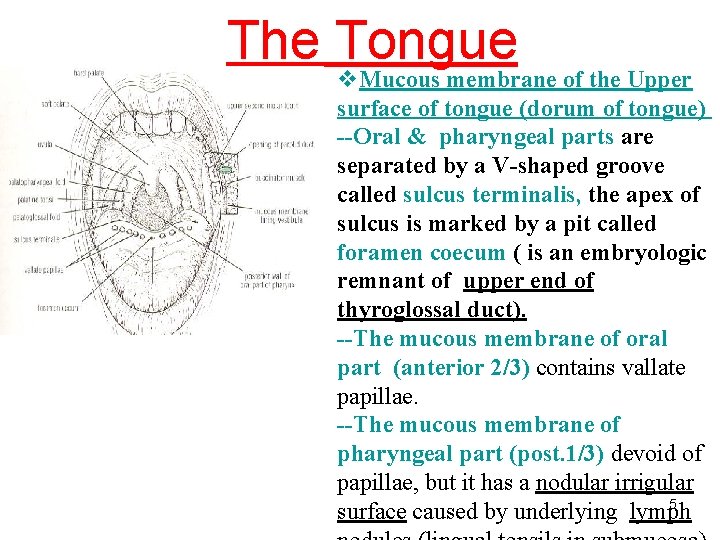
The Tongue v. Mucous membrane of the Upper surface of tongue (dorum of tongue) --Oral & pharyngeal parts are separated by a V-shaped groove called sulcus terminalis, the apex of sulcus is marked by a pit called foramen coecum ( is an embryologic remnant of upper end of thyroglossal duct). --The mucous membrane of oral part (anterior 2/3) contains vallate papillae. --The mucous membrane of pharyngeal part (post. 1/3) devoid of papillae, but it has a nodular irrigular 5 surface caused by underlying lymph
The Tongue v. Mucous membrane of inferior surface of the tongue : -- The mucosa is smooth. -- In the midline, it is connected to the floor of mouth by a fold of m. m. called frenulum of tongue. -- On lateral side of frenulum, deep lingual vein lies in submucosa, can be seen through the mucosa. But Lingual N. & lingual artery run at deeper plane from mucosa. -- Lateral to vein, a serrated fold of mucosa is seen ‘plica fimbriata’ 6
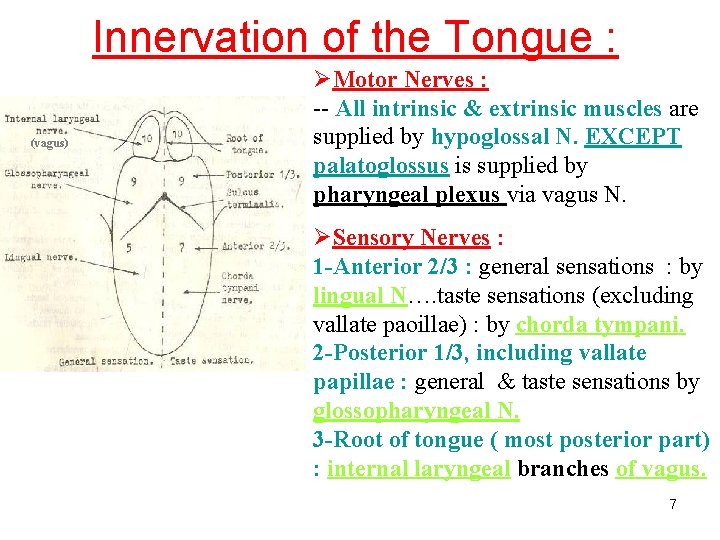
Innervation of the Tongue : (vagus) ØMotor Nerves : -- All intrinsic & extrinsic muscles are supplied by hypoglossal N. EXCEPT palatoglossus is supplied by pharyngeal plexus via vagus N. ØSensory Nerves : 1 -Anterior 2/3 : general sensations : by lingual N…. taste sensations (excluding vallate paoillae) : by chorda tympani. 2 -Posterior 1/3, including vallate papillae : general & taste sensations by glossopharyngeal N. 3 -Root of tongue ( most posterior part) : internal laryngeal branches of vagus. 7
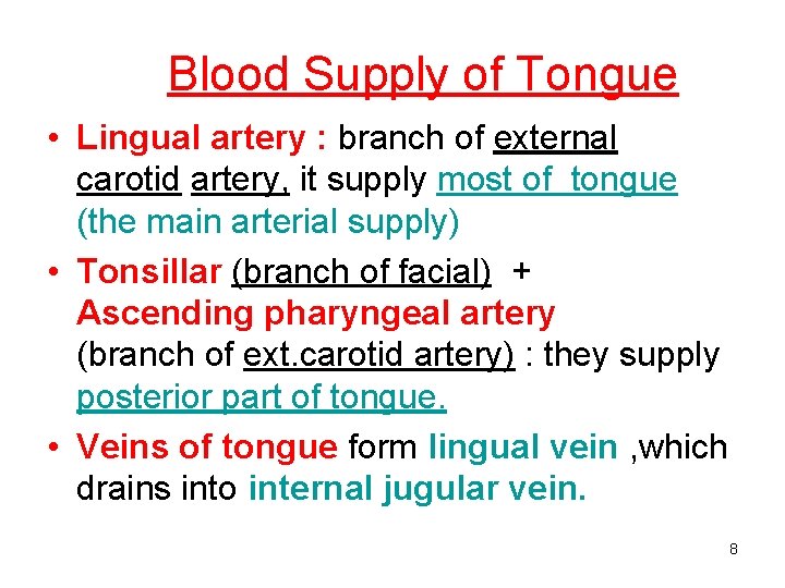
Blood Supply of Tongue • Lingual artery : branch of external carotid artery, it supply most of tongue (the main arterial supply) • Tonsillar (branch of facial) + Ascending pharyngeal artery (branch of ext. carotid artery) : they supply posterior part of tongue. • Veins of tongue form lingual vein , which drains into internal jugular vein. 8
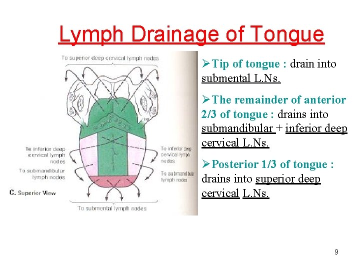
Lymph Drainage of Tongue ØTip of tongue : drain into submental L. Ns. ØThe remainder of anterior 2/3 of tongue : drains into submandibular + inferior deep cervical L. Ns. ØPosterior 1/3 of tongue : drains into superior deep cervical L. Ns. 9
Muscles of Tongue • Intrinsic muscles : -They are not attached to bone. -They consist of longitudinal, transverse & vertical fibers (inside the tongue). -Origin : median septum & submucosa. -Insertion : mucous membrane. -Nerve supply : hypoglossal N. -function : Alter the shape of tongue. 10
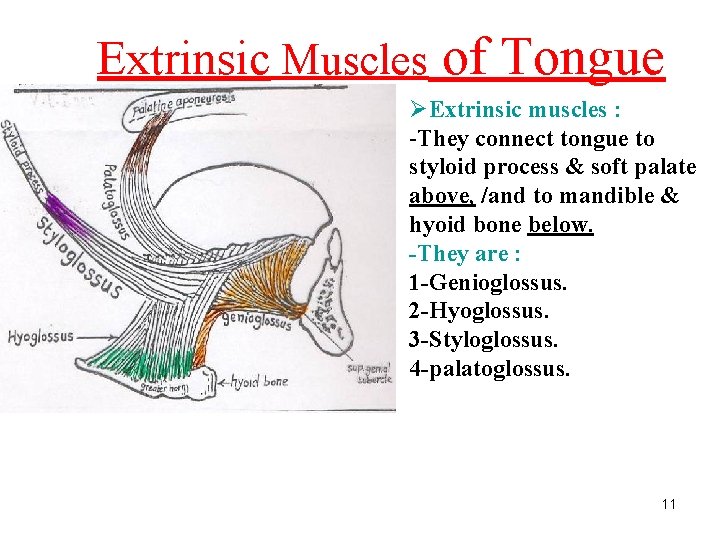
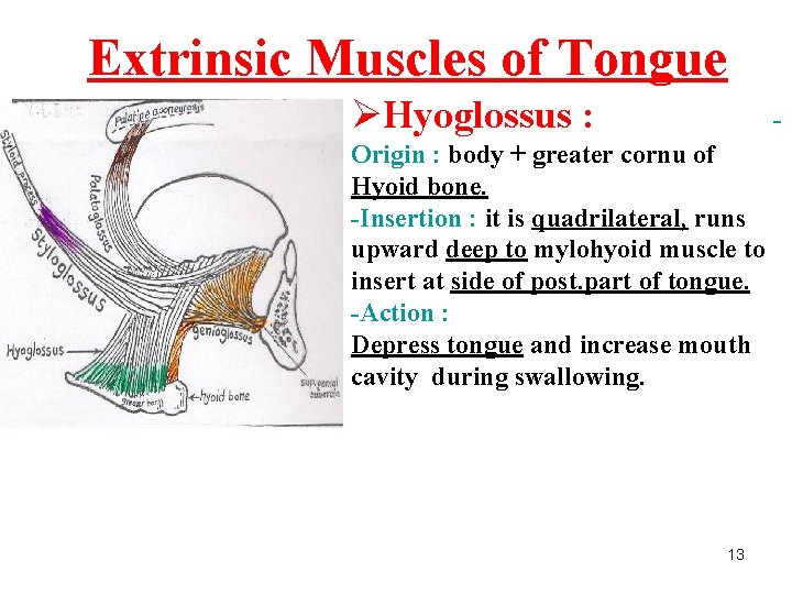
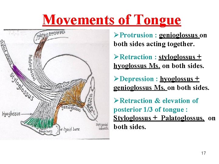
Extrinsic Muscles of Tongue ØExtrinsic muscles : -They connect tongue to styloid process & soft palate above, /and to mandible & hyoid bone below. -They are : 1 -Genioglossus. 2 -Hyoglossus. 3 -Styloglossus. 4 -palatoglossus. 11
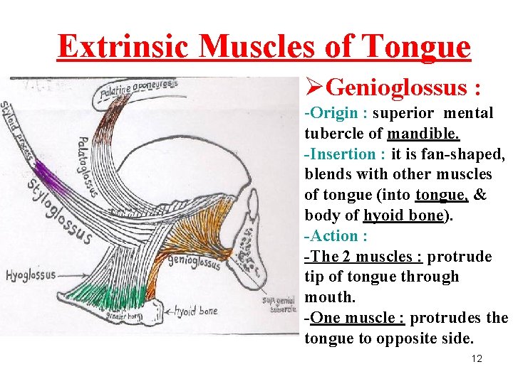
Extrinsic Muscles of Tongue ØGenioglossus : -Origin : superior mental tubercle of mandible. -Insertion : it is fan-shaped, blends with other muscles of tongue (into tongue, & body of hyoid bone). -Action : -The 2 muscles : protrude tip of tongue through mouth. -One muscle : protrudes the tongue to opposite side. 12
Extrinsic Muscles of Tongue ØHyoglossus : - Origin : body + greater cornu of Hyoid bone. -Insertion : it is quadrilateral, runs upward deep to mylohyoid muscle to insert at side of post. part of tongue. -Action : Depress tongue and increase mouth cavity during swallowing. 13
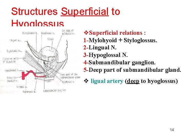
Structures Superficial to Hyoglossus v. Superficial relations : 1 -Mylohyoid + Styloglossus. 2 -Lingual N. 3 -Hypoglossal N. 4 -Submandibular ganglion. 5 -Deep part of submandibular gland. v ligual artery (deep to hyoglossus) 14
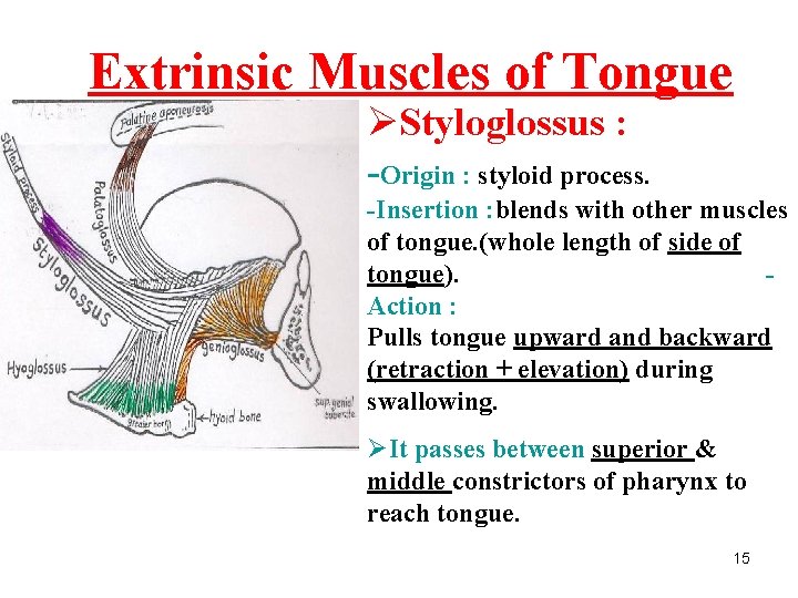
Extrinsic Muscles of Tongue ØStyloglossus : -Origin : styloid process. -Insertion : blends with other muscles of tongue. (whole length of side of tongue). Action : Pulls tongue upward and backward (retraction + elevation) during swallowing. ØIt passes between superior & middle constrictors of pharynx to reach tongue. 15
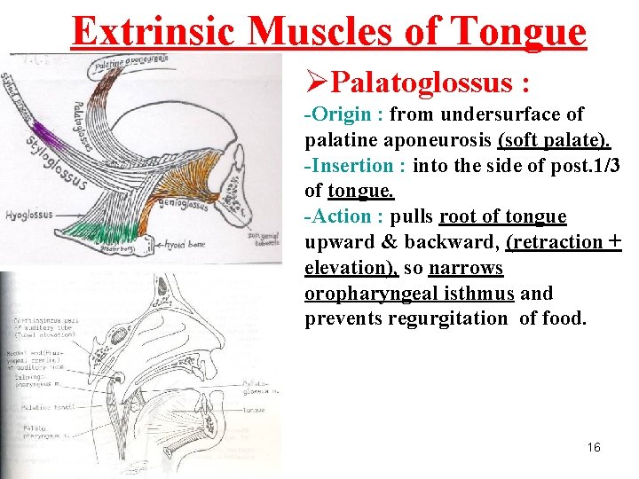
Extrinsic Muscles of Tongue ØPalatoglossus : -Origin : from undersurface of palatine aponeurosis (soft palate). -Insertion : into the side of post. 1/3 of tongue. -Action : pulls root of tongue upward & backward, (retraction + elevation), so narrows oropharyngeal isthmus and prevents regurgitation of food. 16
Movements of Tongue ØProtrusion : genioglossus on both sides acting together. ØRetraction : styloglossus + hyoglossus Ms. on both sides. ØDepression : hyoglossus + genioglossus Ms. on both sides. ØRetraction & elevation of posterior 1/3 of tongue : Styloglossus + Palatoglossus, on both sides. 17
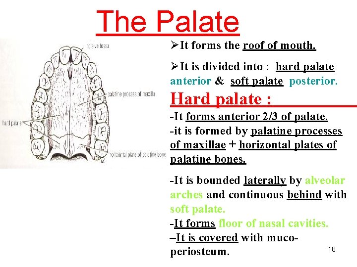
The Palate ØIt forms the roof of mouth. ØIt is divided into : hard palate anterior & soft palate posterior. Hard palate : -It forms anterior 2/3 of palate. -it is formed by palatine processes of maxillae + horizontal plates of palatine bones. -It is bounded laterally by alveolar arches and continuous behind with soft palate. -It forms floor of nasal cavities. –It is covered with muco 18 periosteum.
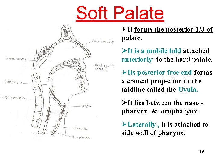
Soft Palate ØIt forms the posterior 1/3 of palate. ØIt is a mobile fold attached anteriorly to the hard palate. Soft palate ØIts posterior free end forms a conical projection in the midline called the Uvula. ØIt lies between the naso pharynx & oropharynx. ØLaterally , it is attached to side wall of pharynx. 19
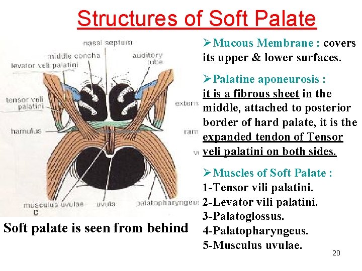
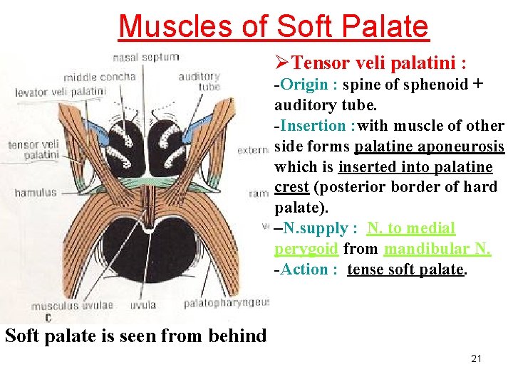
Structures of Soft Palate ØMucous Membrane : covers its upper & lower surfaces. Palatine aponeurosis Soft palate is seen from behind ØPalatine aponeurosis : it is a fibrous sheet in the middle, attached to posterior border of hard palate, it is the expanded tendon of Tensor veli palatini on both sides. ØMuscles of Soft Palate : 1 -Tensor vili palatini. 2 -Levator vili palatini. 3 -Palatoglossus. 4 -Palatopharyngeus. 5 -Musculus uvulae. 20
Muscles of Soft Palate ØTensor veli palatini : -Origin : spine of sphenoid + auditory tube. -Insertion : with muscle of other side forms palatine aponeurosis which is inserted into palatine crest (posterior border of hard palate). –N. supply : N. to medial perygoid from mandibular N. -Action : tense soft palate. Soft palate is seen from behind 21
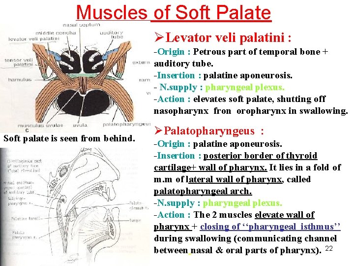
Muscles of Soft Palate ØLevator veli palatini : -Origin : Petrous part of temporal bone + auditory tube. -Insertion : palatine aponeurosis. - N. supply : pharyngeal plexus. -Action : elevates soft palate, shutting off nasopharynx fron oropharynx in swallowing. Soft palate is seen from behind. ØPalatopharyngeus : -Origin : palatine aponeurosis. -Insertion : posterior border of thyroid cartilage+ wall of pharynx. It lies in a fold of m. m of lateral wall of pharynx, called palatopharyngeal arch. -N. supply : pharyngeal plexus. -Action : The 2 muscles elevate wall of pharynx + closing of ‘‘pharyngeal isthmus’’ during swallowing (communicating channel between nasal & oral parts of pharynx). 22
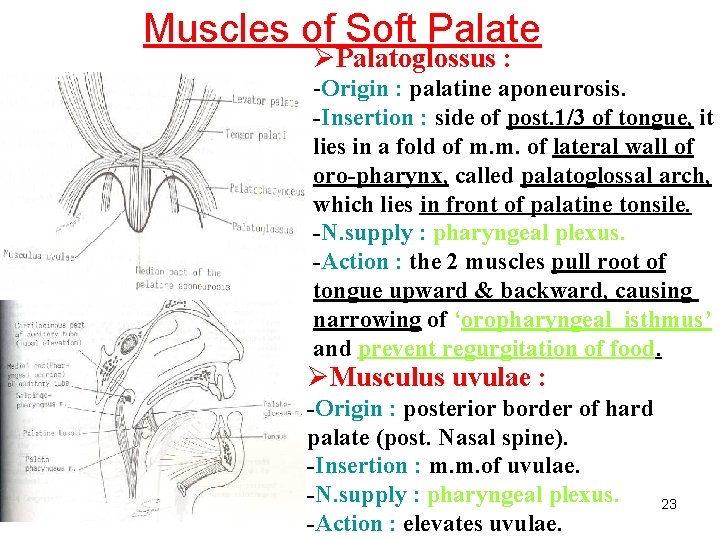
Muscles of Soft Palate ØPalatoglossus : -Origin : palatine aponeurosis. -Insertion : side of post. 1/3 of tongue, it lies in a fold of m. m. of lateral wall of oro-pharynx, called palatoglossal arch, which lies in front of palatine tonsile. -N. supply : pharyngeal plexus. -Action : the 2 muscles pull root of tongue upward & backward, causing narrowing of ‘oropharyngeal isthmus’ and prevent regurgitation of food. ØMusculus uvulae : -Origin : posterior border of hard palate (post. Nasal spine). -Insertion : m. m. of uvulae. -N. supply : pharyngeal plexus. 23 -Action : elevates uvulae.
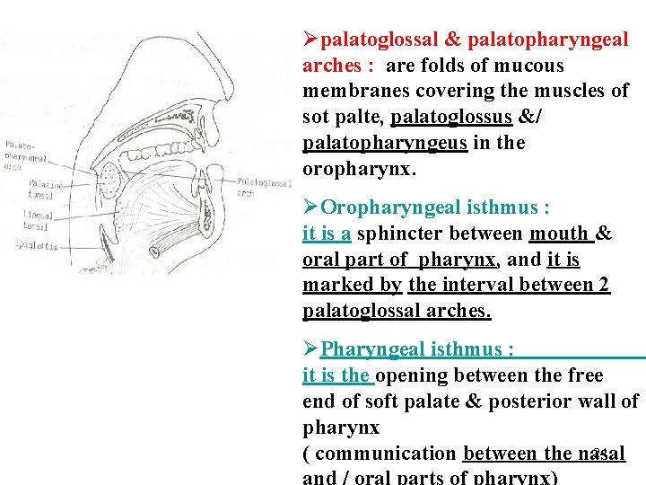
Øpalatoglossal & palatopharyngeal arches : are folds of mucous membranes covering the muscles of sot palte, palatoglossus &/ palatopharyngeus in the oropharynx. ØOropharyngeal isthmus : it is a sphincter between mouth & oral part of pharynx, and it is marked by the interval between 2 palatoglossal arches. ØPharyngeal isthmus : it is the opening between the free end of soft palate & posterior wall of pharynx 24 ( communication between the nasal
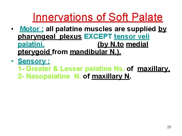
Innervations of Soft Palate • Motor : all palatine muscles are supplied by pharyngeal plexus EXCEPT tensor veli palatini. (by N. to medial pterygoid from mandibular N. ). • Sensory : 1 - Greater & Lesser palatine Ns. of maxillary, 2 - Nasopalatine N. of maxillary N. 25
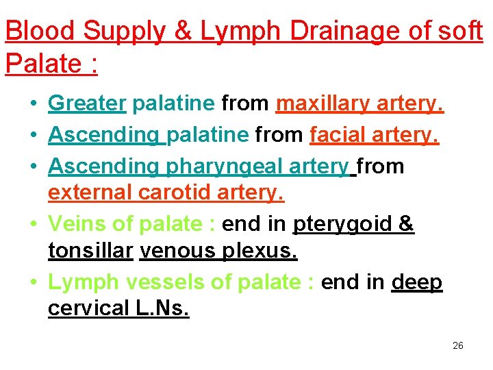
Blood Supply & Lymph Drainage of soft Palate : • Greater palatine from maxillary artery. • Ascending palatine from facial artery. • Ascending pharyngeal artery from external carotid artery. • Veins of palate : end in pterygoid & tonsillar venous plexus. • Lymph vessels of palate : end in deep cervical L. Ns. 26