
The Molecular Genetics of Immunoglobulins Dr Colin R
The Molecular Genetics of Immunoglobulins ©Dr. Colin R. A. Hewitt
The molecular genetics of immunoglobulins How can the bifunctional nature of antibodies be explained genetically? Dreyer & Bennett (1965) For a single isotype of antibody there may be: • A single C region gene encoded in the GERMLINE and separate from the V region genes • Multiple choices of V region genes available • A mechanism to rearrange V and C genes in the genome so that they can fuse to form a complete Immunoglobulin gene. This was genetic heresy as it violated then accepted notion that DNA was identical in every cell of an individual
Genetic models of the 1960’s were also unable to explain: • How B cells shut down the Ig genes on just one of their chromosomes. All other genes known at the time were expressed co-dominantly. B cells expressed a light chain from one parent only and a heavy chain from one parent only (evidence from allotypes). • A genetic mechanism to account for increased antibody affinity in an immune response • How a single specificity of antibody sequentially switched isotype. • How the same specificity of antibody was secreted and simultaneously expressed on the cell surface of a B cell.
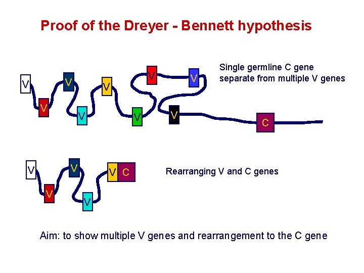
Proof of the Dreyer - Bennett hypothesis V V V C V V Single germline C gene separate from multiple V genes C Rearranging V and C genes V Aim: to show multiple V genes and rearrangement to the C gene
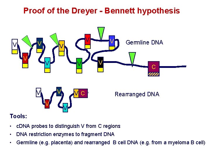
Proof of the Dreyer - Bennett hypothesis V V V C V Germline DNA V C Rearranged DNA V Tools: • c. DNA probes to distinguish V from C regions • DNA restriction enzymes to fragment DNA • Germline (e. g. placenta) and rearranged B cell DNA (e. g. from a myeloma B cell)
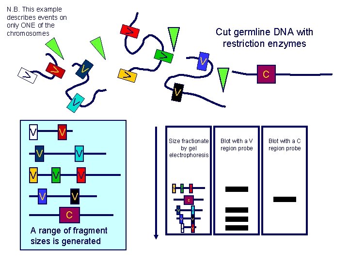
V V V V V C Size fractionate by gel electrophoresis V V V V Cut germline DNA with restriction enzymes V N. B. This example describes events on only ONE of the chromosomes V V C A range of fragment sizes is generated V C V V V Blot with a V region probe Blot with a C region probe

Evidence for gene recombination V V V V Cut myeloma B cell V DNA with restriction enzymes V V C VC Size fractionate by gel electrophoresis Blot with a V region probe Blot with a C region probe V C V V V C Size fractionate by gel electrophoresis V V Blot with a V region probe Blot with a C region probe V C V V V V and C probes detect the same fragment Some V regions missing C fragment is larger cf germline V V V - compare the pattern of bands with germline DNA

Ig gene sequencing complicated the model Structures of germline VL genes were similar for Vk, and Vl, However there was an anomaly between germline and rearranged DNA: VL CL ~ 95 aa ~ 100 aa L L V L CL ~ 208 aa Where do the extra 13 amino acids come from? L V L JL ~ 95 aa CL ~ 100 aa Extra amino acids provided by one of a small set of J or JOINING regions

Further diversity in the Ig heavy chain L VH JH DH CH Heavy chain: between 0 and 8 additional amino acids between JH and CH The D or DIVERSITY region Each heavy chain requires three recombination events: VH to JH, VHJH to DH and VHJHDH to CH L VL JL CL Each light chain requires two recombination events: VL to JL and VLJL to CL
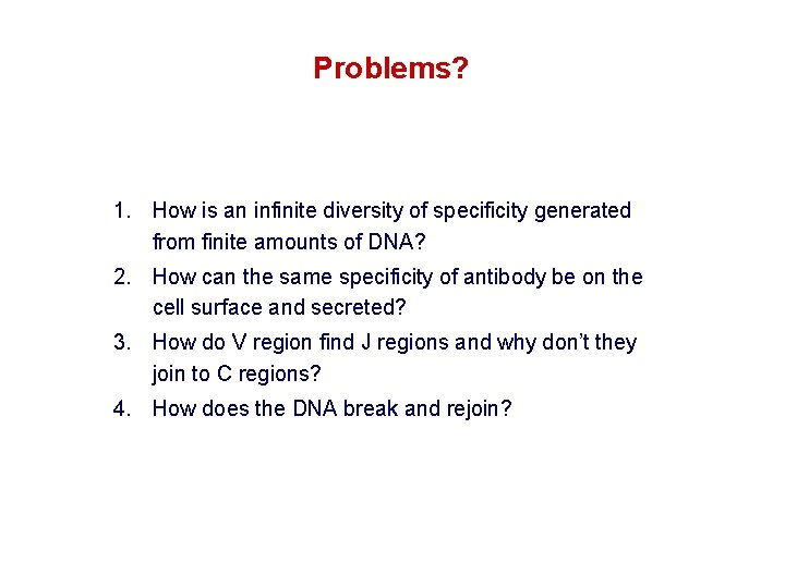
Problems? 1. How is an infinite diversity of specificity generated from finite amounts of DNA? 2. How can the same specificity of antibody be on the cell surface and secreted? 3. How do V region find J regions and why don’t they join to C regions? 4. How does the DNA break and rejoin?
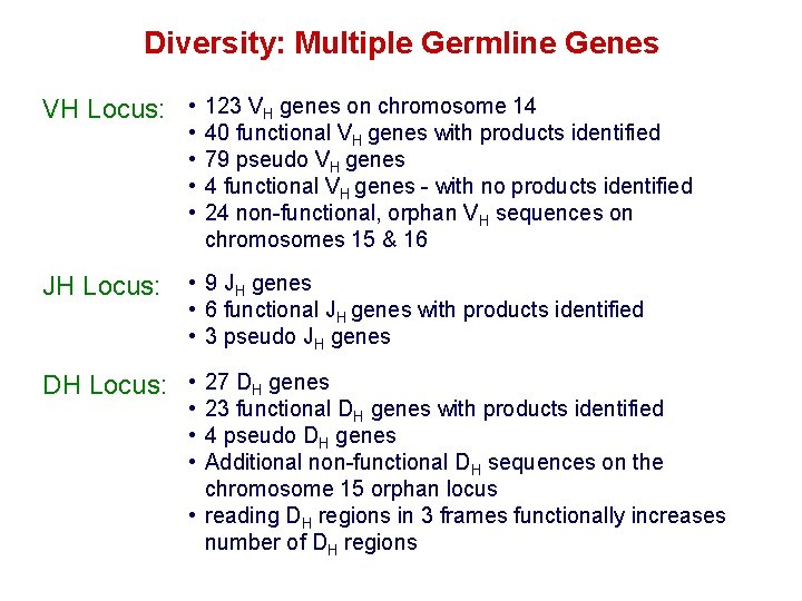
Diversity: Multiple Germline Genes VH Locus: • 123 VH genes on chromosome 14 • • 40 functional VH genes with products identified 79 pseudo VH genes 4 functional VH genes - with no products identified 24 non-functional, orphan VH sequences on chromosomes 15 & 16 JH Locus: • 9 JH genes • 6 functional JH genes with products identified • 3 pseudo JH genes DH Locus: • 27 DH genes • 23 functional DH genes with products identified • 4 pseudo DH genes • Additional non-functional DH sequences on the chromosome 15 orphan locus • reading DH regions in 3 frames functionally increases number of DH regions

Reading D segment in 3 frames Analysis of D regions from different antibodies One D region can be used in any of three frames Different protein sequences lead to antibody diversity GGGACAGGGGGC Gly. Thr. Gly Frame 1 GGGACAGGGGGC Gly. Gln. Gly Frame 2 GGGACAGGGGGC Asp. Arg. Gly Frame 3
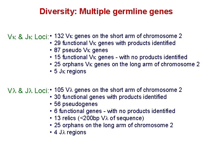
Diversity: Multiple germline genes Vk & Jk Loci: • 132 Vk genes on the short arm of chromosome 2 • • • 29 functional Vk genes with products identified 87 pseudo Vk genes 15 functional Vk genes - with no products identified 25 orphans Vk genes on the long arm of chromosome 2 5 Jk regions Vl & Jl Loci: • 105 Vl genes on the short arm of chromosome 2 • • • 30 functional genes with products identified 56 pseudogenes 6 functional genes - with no products identified 13 relics (<200 bp Vl of sequence) 25 orphans on the long arm of chromosome 2 4 Jl regions

Estimates of combinatorial diversity Using functional V D and J genes: 40 VH x 27 DH x 6 JH = 6, 480 combinations D can be read in 3 frames: 6, 480 x 3 = 19, 440 combinations 29 Vk x 5 Jk = 145 combinations 30 Vl x 4 Jl = 120 combinations = 265 different light chains If H and L chains pair randomly as H 2 L 2 i. e. 19, 440 x 265 = 5, 151, 600 possibilities Due only to COMBINATORIAL diversity In practice, some H + L combinations are unstable. Certain V and J genes are also used more frequently than others. Other mechanisms add diversity at the junctions between genes JUNCTIONAL diversity
Problems? 1. How is an infinite diversity of specificity generated from finite amounts of DNA? Mathematically, Combinatorial Diversity can account for some diversity – how do the elements rearrange? 2. How can the same specificity of antibody be on the cell surface and secreted? 3. How do V region find J regions and why don’t they join to C regions? 4. How does the DNA break and rejoin?
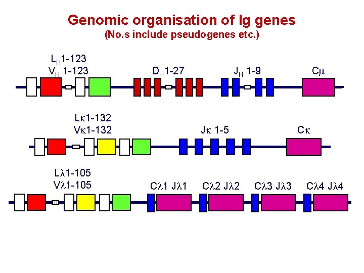
Genomic organisation of Ig genes (No. s include pseudogenes etc. ) LH 1 -123 VH 1 -123 DH 1 -27 Lk 1 -132 Vk 1 -132 Ll 1 -105 Vl 1 -105 JH 1 -9 Jk 1 -5 Cl 1 Jl 1 Cl 2 Jl 2 Cm Ck Cl 3 Jl 3 Cl 4 Jl 4
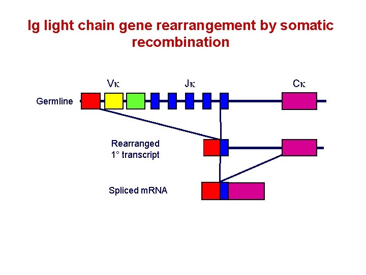
Ig light chain gene rearrangement by somatic recombination Vk Germline Rearranged 1° transcript Spliced m. RNA Jk Ck
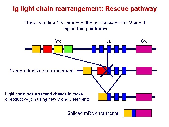
Ig light chain rearrangement: Rescue pathway There is only a 1: 3 chance of the join between the V and J region being in frame Vk Jk Non-productive rearrangement Light chain has a second chance to make a productive join using new V and J elements Spliced m. RNA transcript Ck
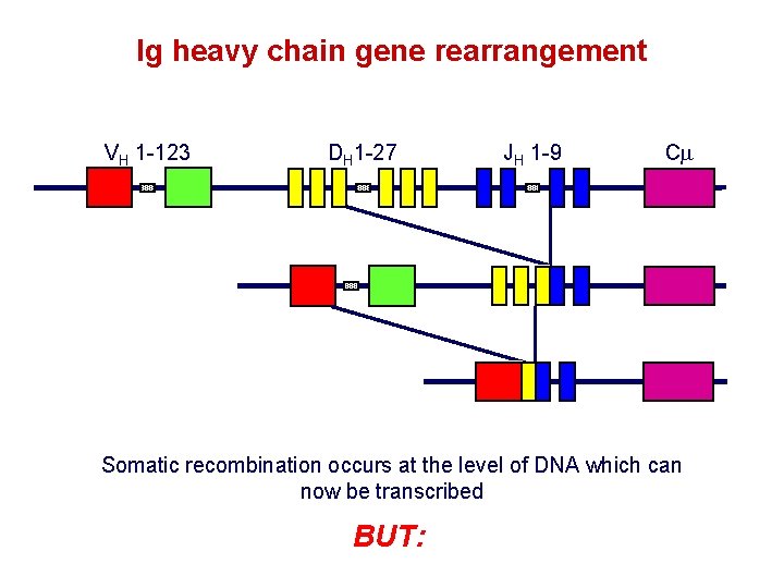
Ig heavy chain gene rearrangement VH 1 -123 DH 1 -27 JH 1 -9 Cm Somatic recombination occurs at the level of DNA which can now be transcribed BUT:
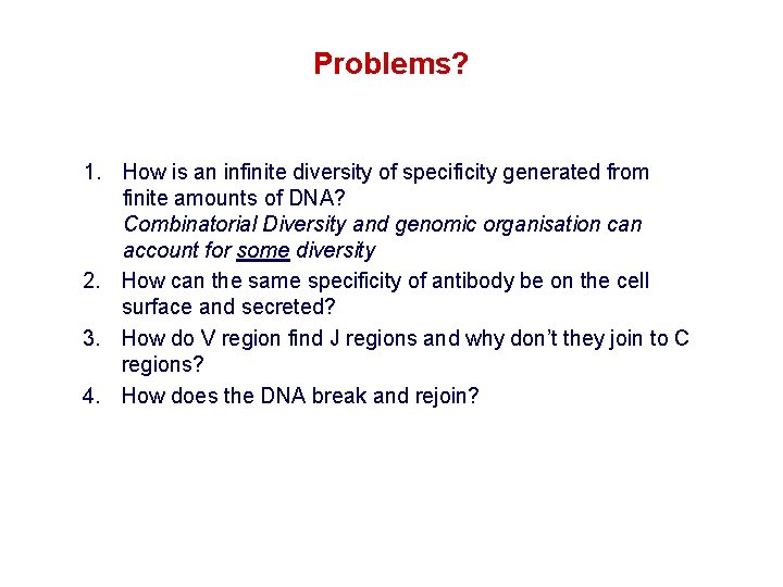
Problems? 1. How is an infinite diversity of specificity generated from finite amounts of DNA? Combinatorial Diversity and genomic organisation can account for some diversity 2. How can the same specificity of antibody be on the cell surface and secreted? 3. How do V region find J regions and why don’t they join to C regions? 4. How does the DNA break and rejoin?
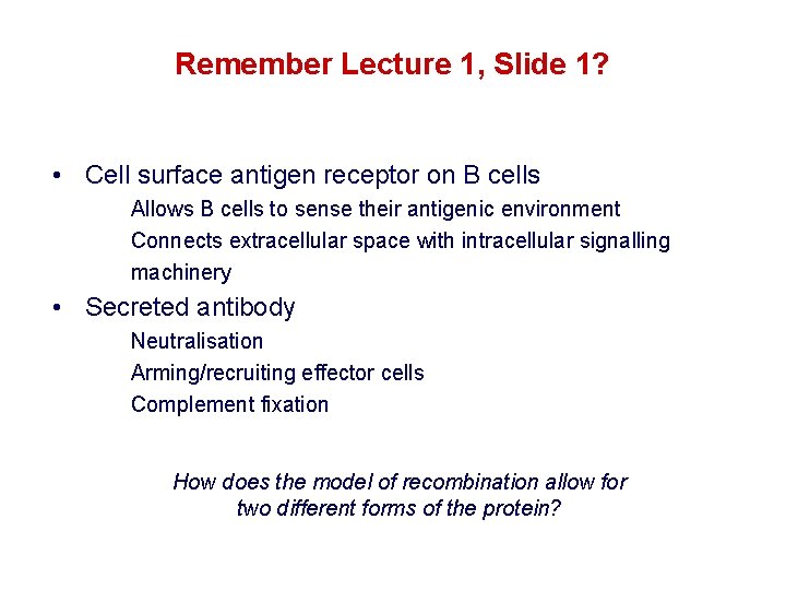
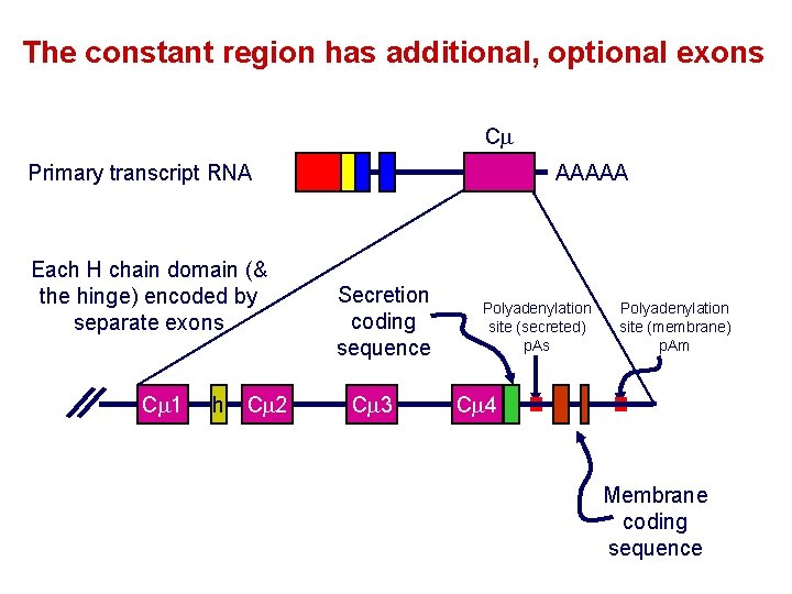
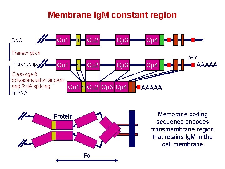
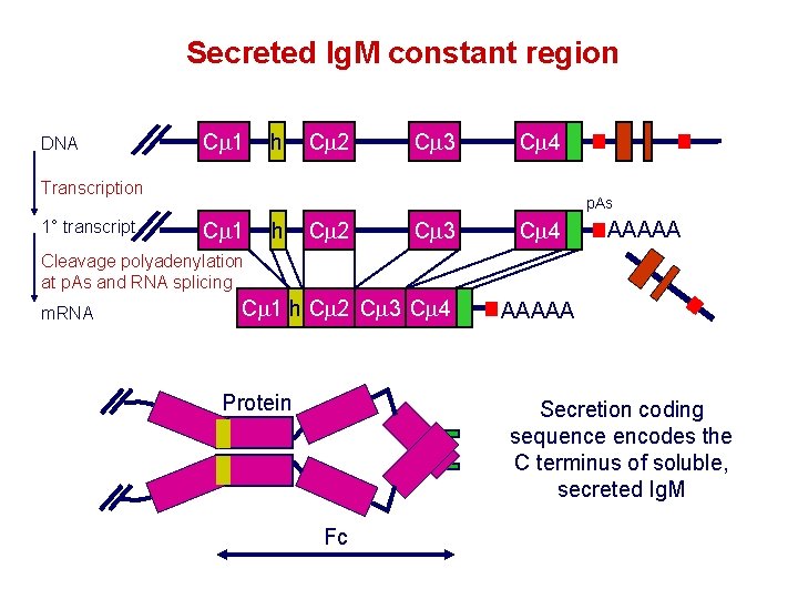
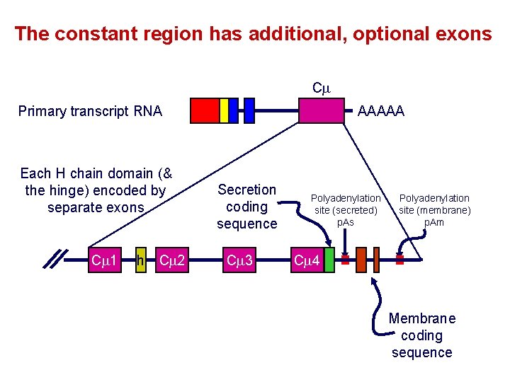
Remember Lecture 1, Slide 1? • Cell surface antigen receptor on B cells Allows B cells to sense their antigenic environment Connects extracellular space with intracellular signalling machinery • Secreted antibody Neutralisation Arming/recruiting effector cells Complement fixation How does the model of recombination allow for two different forms of the protein?
The constant region has additional, optional exons Cm Primary transcript RNA Each H chain domain (& the hinge) encoded by separate exons Cm 1 h Cm 2 AAAAA Secretion coding sequence Cm 3 Polyadenylation site (secreted) p. As Polyadenylation site (membrane) p. Am Cm 4 Membrane coding sequence
Membrane Ig. M constant region DNA Cm 1 h Cm 2 Cm 3 Cm 4 Transcription 1° transcript p. Am Cm 1 Cleavage & polyadenylation at p. Am and RNA splicing m. RNA h Cm 2 Cm 3 Cm 1 h Cm 2 Cm 3 Cm 4 AAAAA Membrane coding sequence encodes transmembrane region that retains Ig. M in the cell membrane Protein Fc
Secreted Ig. M constant region DNA Cm 1 h Cm 2 Cm 3 Cm 4 Transcription 1° transcript p. As Cm 1 h Cm 2 Cm 3 Cm 4 AAAAA Cleavage polyadenylation at p. As and RNA splicing m. RNA Cm 1 h Cm 2 Cm 3 Cm 4 Protein AAAAA Secretion coding sequence encodes the C terminus of soluble, secreted Ig. M Fc
The constant region has additional, optional exons Cm Primary transcript RNA Each H chain domain (& the hinge) encoded by separate exons Cm 1 h Cm 2 AAAAA Secretion coding sequence Cm 3 Polyadenylation site (secreted) p. As Polyadenylation site (membrane) p. Am Cm 4 Membrane coding sequence
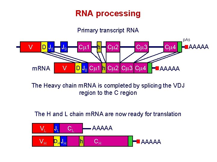
RNA processing Primary transcript RNA p. As V D J 8 Cm 1 J 9 Cm 2 Cm 3 D J 8 Cm 1 h Cm 2 Cm 3 Cm 4 V m. RNA h Cm 4 AAAAA The Heavy chain m. RNA is completed by splicing the VDJ region to the C region The H and L chain m. RNA are now ready for translation VL JL VH DH JH AAAAA CL h CH AAAAA
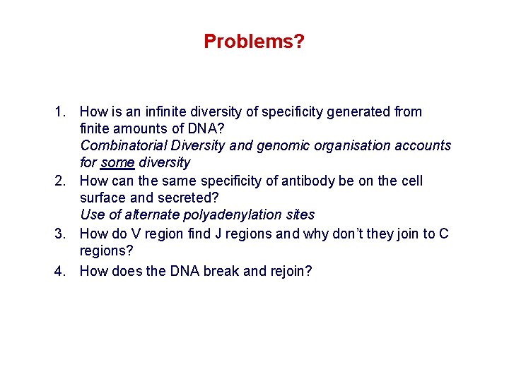
Problems? 1. How is an infinite diversity of specificity generated from finite amounts of DNA? Combinatorial Diversity and genomic organisation accounts for some diversity 2. How can the same specificity of antibody be on the cell surface and secreted? Use of alternate polyadenylation sites 3. How do V region find J regions and why don’t they join to C regions? 4. How does the DNA break and rejoin?
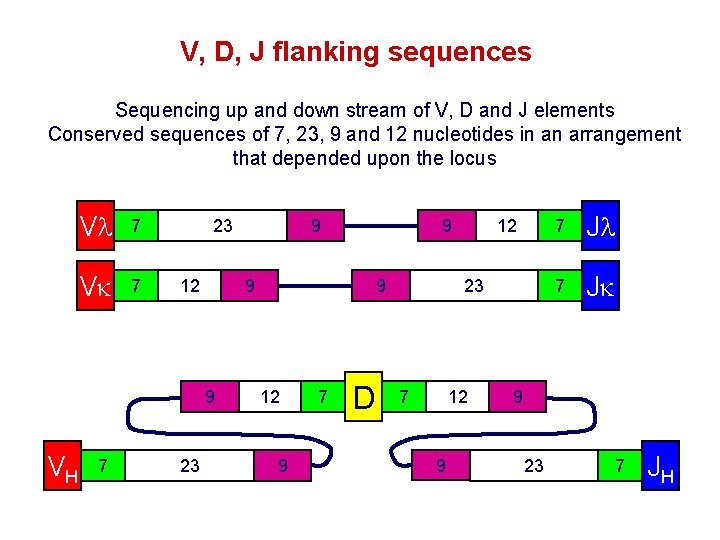
V, D, J flanking sequences Sequencing up and down stream of V, D and J elements Conserved sequences of 7, 23, 9 and 12 nucleotides in an arrangement that depended upon the locus Vl 7 Vk 7 23 12 7 23 9 12 9 7 12 9 9 9 VH 9 D 23 7 12 9 7 Jl 7 Jk 9 23 7 JH
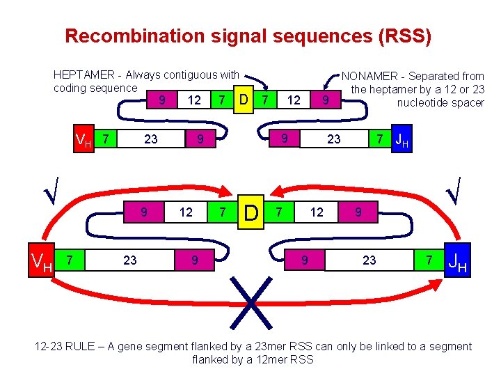
Recombination signal sequences (RSS) HEPTAMER - Always contiguous with coding sequence 9 VH 7 23 √ VH 9 7 12 23 7 D 7 9 9 12 7 D NONAMER - Separated from the heptamer by a 12 or 23 nucleotide spacer 7 JH 23 7 12 9 √ 9 23 7 JH 12 -23 RULE – A gene segment flanked by a 23 mer RSS can only be linked to a segment flanked by a 12 mer RSS
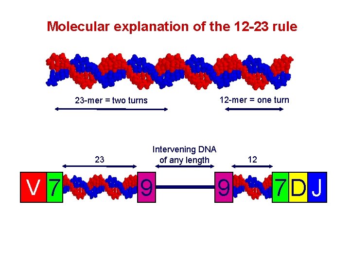
Molecular explanation of the 12 -23 rule 12 -mer = one turn 23 -mer = two turns 23 V 7 Intervening DNA of any length 9 12 9 7 D J
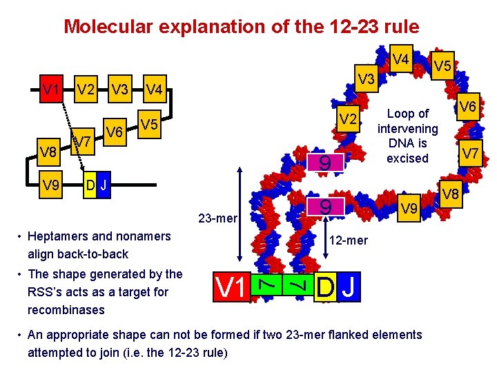
Molecular explanation of the 12 -23 rule V 4 V 1 V 8 V 9 V 3 V 2 V 7 V 6 V 3 V 4 V 2 V 5 9 9 • Heptamers and nonamers align back-to-back V 9 V 1 7 12 -mer 7 • The shape generated by the RSS’s acts as a target for recombinases V 6 Loop of intervening DNA is excised DJ 23 -mer V 5 DJ • An appropriate shape can not be formed if two 23 -mer flanked elements attempted to join (i. e. the 12 -23 rule) V 7 V 8
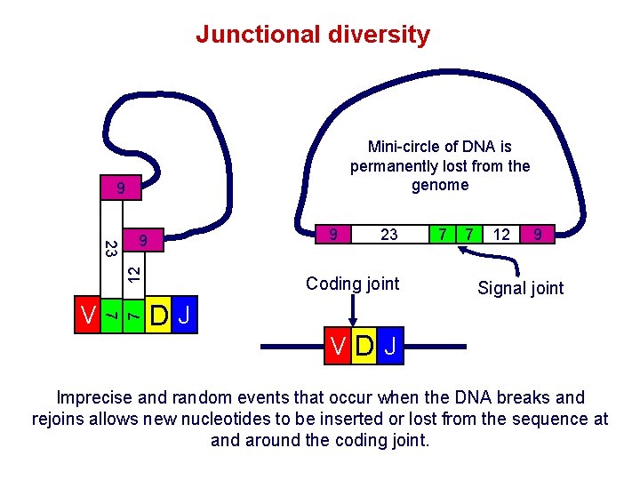
Junctional diversity Mini-circle of DNA is permanently lost from the genome 9 7 V 7 12 23 9 9 23 Coding joint DJ 7 7 12 9 Signal joint VDJ Imprecise and random events that occur when the DNA breaks and rejoins allows new nucleotides to be inserted or lost from the sequence at and around the coding joint.
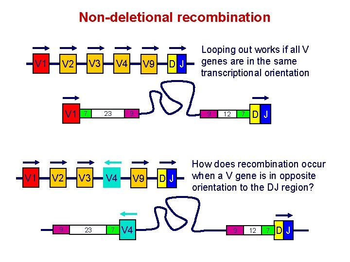
Non-deletional recombination V 1 V 1 V 3 V 2 7 V 2 V 3 9 23 V 4 7 V 9 DJ 9 V 4 Looping out works if all V genes are in the same transcriptional orientation 9 DJ 12 7 D J How does recombination occur when a V gene is in opposite orientation to the DJ region? 9 12 7 DJ
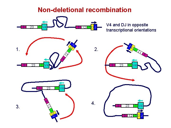
Non-deletional recombination V 4 and DJ in opposite transcriptional orientations J 9 12 7 D J 2. 9 12 7 D 1. 9 7 V 4 23 9 9 23 9 12 3. 23 7 V 4 4. 9 23 7 V 4 J 7 D 9 12 7 D J 9 7 V 4 12 23 7 D J 9
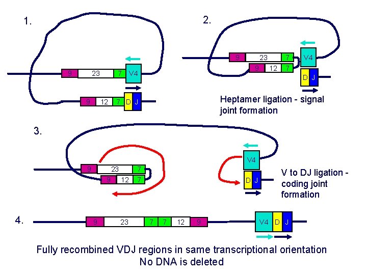
2. 1. 9 9 23 7 12 9 23 9 V 4 12 7 7 V 4 D J Heptamer ligation - signal joint formation 7 D J 3. V 4 9 23 9 4. 9 12 23 7 7 D J 7 7 12 9 V to DJ ligation coding joint formation V 4 D J Fully recombined VDJ regions in same transcriptional orientation No DNA is deleted
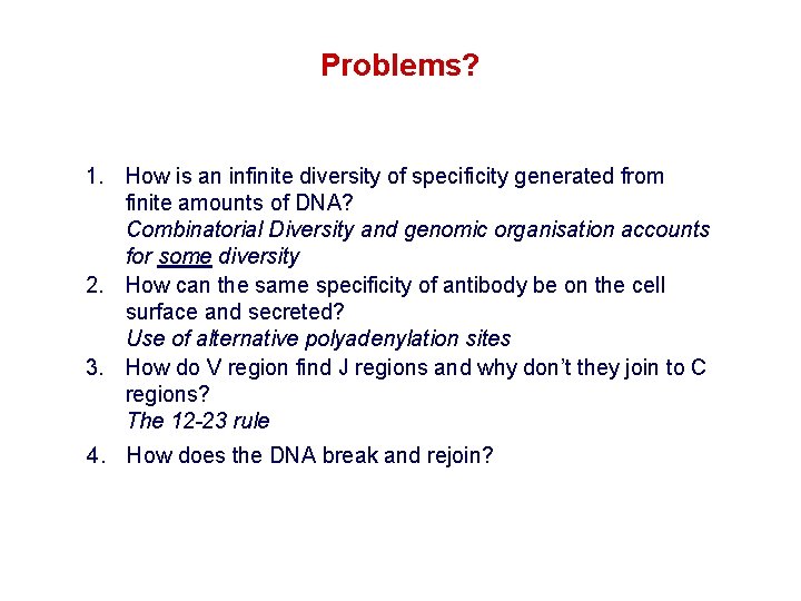
Problems? 1. How is an infinite diversity of specificity generated from finite amounts of DNA? Combinatorial Diversity and genomic organisation accounts for some diversity 2. How can the same specificity of antibody be on the cell surface and secreted? Use of alternative polyadenylation sites 3. How do V region find J regions and why don’t they join to C regions? The 12 -23 rule 4. How does the DNA break and rejoin?
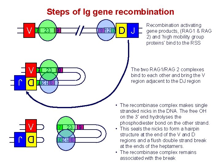
Steps of Ig gene recombination V 7 23 D J 9 12 7 7 23 9 12 7 9 7 23 D J The two RAG 1/RAG 2 complexes bind to each other and bring the V region adjacent to the DJ region 9 9 12 7 V 9 Recombination activating gene products, (RAG 1 & RAG 2) and ‘high mobility group proteins’ bind to the RSS 9 • The recombinase complex makes single stranded nicks in the DNA. The free OH on the 3’ end hydrolyses the phosphodiester bond on the other strand. • This seals the nicks to form a hairpin structure at the end of the V and D regions and a flush double strand break at the ends of the heptamers. • The recombinase complex remains associated with the break D J
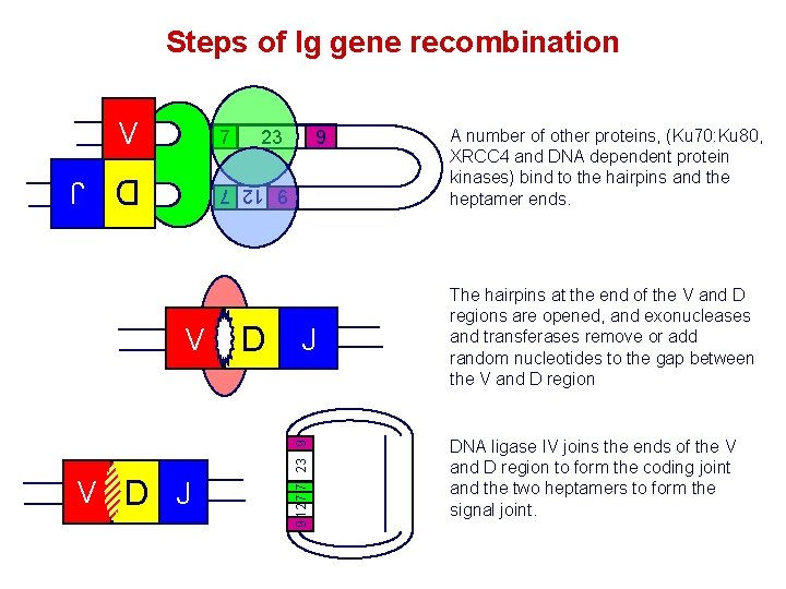
Steps of Ig gene recombination V 7 23 9 D J 9 12 7 V D J 9 12 7 7 23 9 V A number of other proteins, (Ku 70: Ku 80, XRCC 4 and DNA dependent protein kinases) bind to the hairpins and the heptamer ends. The hairpins at the end of the V and D regions are opened, and exonucleases and transferases remove or add random nucleotides to the gap between the V and D region DNA ligase IV joins the ends of the V and D region to form the coding joint and the two heptamers to form the signal joint.

Junctional diversity: P nucleotide additions 7 23 V AT GTGACAC J DTA CACTGTG 9 9 12 7 7 7 9 23 12 9 The recombinase complex makes single stranded nicks at random sites close to the ends of the V and D region DNA. U U V TC CACAGTG AG GTGTCAC TC AG TC CACAGTG AG GTGTCAC 7 GTGACAC CACTGTG 7 V V AT AT J JDTA 9 23 12 9 The 2 nd strand is cleaved and hairpins form between the complimentary bases at ends of the V and D region. D J
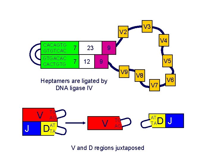
V 3 V 2 V 4 CACAGTG GTGTCAC 7 GTGACAC CACTGTG 7 9 23 12 V 5 9 V AT J DTA V U TC AG V 7 TC AG AT TA U U U Heptamers are ligated by DNA ligase IV V 8 V and D regions juxtaposed V 6 D J
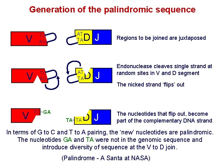
V U TC AG TC~GA AG AT TA D J AT TA U V TC AG U V U Generation of the palindromic sequence AT TA~TA D J Regions to be joined are juxtaposed Endonuclease cleaves single strand at random sites in V and D segment The nicked strand ‘flips’ out D J The nucleotides that flip out, become part of the complementary DNA strand In terms of G to C and T to A pairing, the ‘new’ nucleotides are palindromic. The nucleotides GA and TA were not in the genomic sequence and introduce diversity of sequence at the V to D join. (Palindrome - A Santa at NASA)
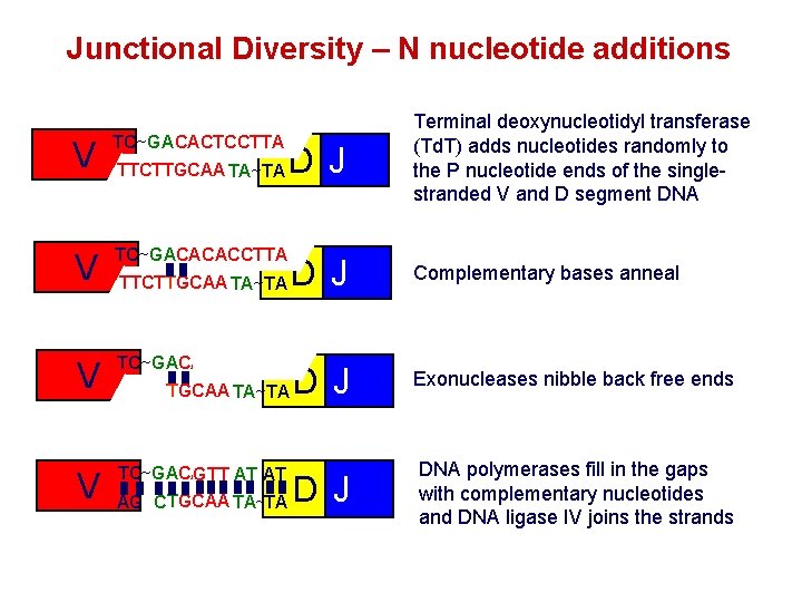
Junctional Diversity – N nucleotide additions V TC~GA CACTCCTTA AT AG TTCTTGCAA TA~TA V TC~GACACACCTTA V TC CACACCTTA TC~GA GTT AT AG TTCTTGCAA TA~TA TA AG C D J TTCTTGCAA TA~TA Terminal deoxynucleotidyl transferase (Td. T) adds nucleotides randomly to the P nucleotide ends of the singlestranded V and D segment DNA Complementary bases anneal D J Exonucleases nibble back free ends D J DNA polymerases fill in the gaps with complementary nucleotides and DNA ligase IV joins the strands
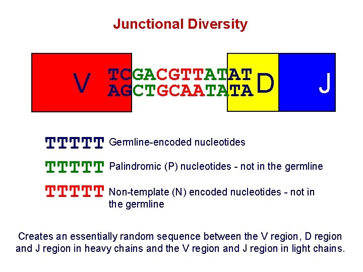
Junctional Diversity V TCGACGTTATAT AGCTGCAATATA D J TTTTT Germline-encoded nucleotides TTTTT Palindromic (P) nucleotides - not in the germline TTTTT Non-template (N) encoded nucleotides - not in the germline Creates an essentially random sequence between the V region, D region and J region in heavy chains and the V region and J region in light chains.
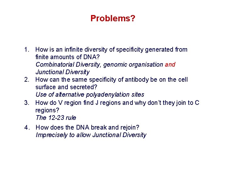
Problems? 1. How is an infinite diversity of specificity generated from finite amounts of DNA? Combinatorial Diversity, genomic organisation and Junctional Diversity 2. How can the same specificity of antibody be on the cell surface and secreted? Use of alternative polyadenylation sites 3. How do V region find J regions and why don’t they join to C regions? The 12 -23 rule 4. How does the DNA break and rejoin? Imprecisely to allow Junctional Diversity

Why do V regions not join to J or C regions? VH DH JH C IF the elements of Ig did not assemble in the correct order, diversity of specificity would be severely compromised 2 x DIVERSITY Full potential of the H chain for diversity needs V -D-J-C joining - in the correct order 1 x DIVERSITY Were V-J joins allowed in the heavy chain, diversity would be reduced due to loss of the imprecise join between the V and D regions

Somatic hypermutation FR 1 CDR 1 FR 2 CDR 2 FR 3 CDR 3 FR 4 100 Variability 80 60 40 20 20 40 60 80 100 120 Amino acid No. Wu - Kabat analysis compares point mutations in Ig of different specificity. What about mutation throughout an immune response to a single epitope? How does this affect the specificity and affinity of the antibody?
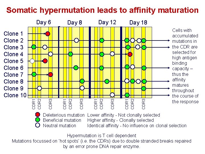
Somatic hypermutation leads to affinity maturation Day 6 Day 8 Day 12 Day 18 CDR 3 CDR 1 CDR 2 Clone 1 Clone 2 Clone 3 Clone 4 Clone 5 Clone 6 Clone 7 Clone 8 Clone 9 Clone 10 Cells with accumulated mutations in the CDR are selected for high antigen binding capacity – thus the affinity matures throughout the course of the response Deleterious mutation Lower affinity - Not clonally selected Beneficial mutation Higher affinity - Clonally selected Identical affinity - No influence on clonal selection Neutral mutation Hypermutation is T cell dependent Mutations focussed on ‘hot spots’ (i. e. the CDRs) due to double stranded breaks repaired by an error prone DNA repair enzyme.
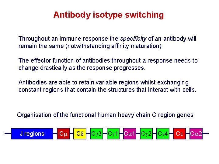
Antibody isotype switching Throughout an immune response the specificity of an antibody will remain the same (notwithstanding affinity maturation) The effector function of antibodies throughout a response needs to change drastically as the response progresses. Antibodies are able to retain variable regions whilst exchanging constant regions that contain the structures that interact with cells. Organisation of the functional human heavy chain C region genes J regions Cm Cd Cg 3 Cg 1 Ca 1 Cg 2 Cg 4 Ce Ca 2
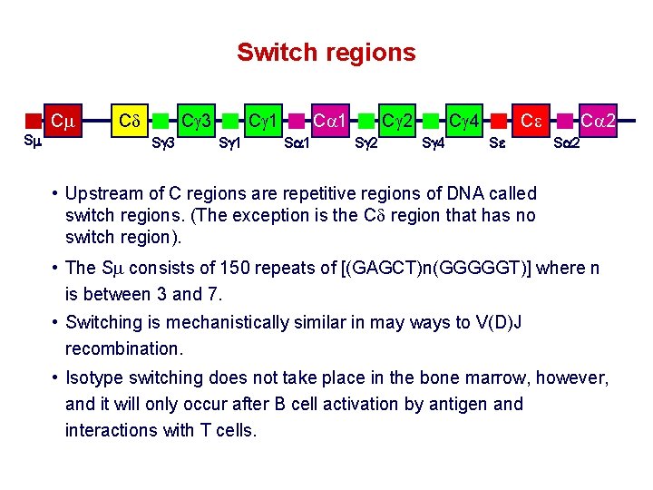
Switch regions Cm Sm Cd Cg 3 Sg 3 Cg 1 Sg 1 Ca 1 Sa 1 Cg 2 Sg 2 Cg 4 Sg 4 Ce Se Ca 2 Sa 2 • Upstream of C regions are repetitive regions of DNA called switch regions. (The exception is the Cd region that has no switch region). • The Sm consists of 150 repeats of [(GAGCT)n(GGGGGT)] where n is between 3 and 7. • Switching is mechanistically similar in may ways to V(D)J recombination. • Isotype switching does not take place in the bone marrow, however, and it will only occur after B cell activation by antigen and interactions with T cells.
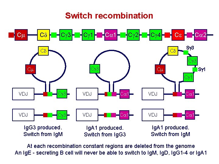
Switch recombination Cm Cd Cg 3 Cg 1 Ca 1 Cg 2 Cg 4 Cd Ce Cd Ca 2 Sg 3 Cg 3 Cm Sg 1 Cm Cg 1 VDJ Cg 3 VDJ Ca 1 Ig. G 3 produced. Switch from Ig. M Ig. A 1 produced. Switch from Ig. G 3 Ig. A 1 produced. Switch from Ig. M At each recombination constant regions are deleted from the genome An Ig. E - secreting B cell will never be able to switch to Ig. M, Ig. D, Ig. G 1 -4 or Ig. A 1
- Slides: 50