The molecular basis of lung diseases The alveolar

The molecular basis of lung diseases

The alveolar system is made up of 2 types of epithelium cell 1. Pneumocytes I 2. Pneumocytes II Type I are relatively large, extremely flattened cells and make up most of the alveolar wall. Type II pneumocytes secrete Surfactant, a mixture of lipids and proteins, which helps to reduce surface tension of the alveoli and prevent the alveolus from collapsing

• Lung surfactant is synthesized by type II cells and is then stored and secreted in intracellular organelles called lamellar bodies. • Surfactant is composed of 1 - 80% phospholipids 2 - 10% neutral lipids (cholesterol) 3 - 10% proteins • The predominant phospholipids are A. Phosphatidylcholine (PC) which makes up 70 -80% B. Phosphatidylglycerol (PG) which makes 5 -10%. • Majority of phosphatidylcholine (PC) is in the form of dipalmitoylphosphatidylcholine (DPPC), which is thought to be responsible for the lowering of surface tension and to provide alveolar stability because of its low surface compressibility. • The choline residue of DPPC is polar and hydrophilic and associates with the liquid of the alveoli, • while the palmitic acid (a fatty acid) residue is nonpolar and hydrophobic and points towards the air.

• The surfactant-associated proteins include four particular proteins and are referred to as SP-A, SP-B, SP-C, and SP-D • 1. SP-A is hydrophilic protein, and the major component of the surfactant complex. • Functions of SP-A: – SP-A and SP-D can bind to a variety of pathogens, and causing pathogen neutralization and clearing by leukocytes. – SP-A contribute to regulating the secretion and recycling of phospholipids by type II cells

• 2 - SP-B is a small hydrophobic protein. • SP-B is the only surfactant protein essential for life. • Infants with hereditary SP-B deficiency suffer from Respiratory Distress Syndrome (RDS). • SP-B has a critical role in the biophysical properties of surfactant, including surface tension reduction • SP-B also play a role in adsorption and spreading of phospholipids to the air-fluid interface.

• 3 - SP-C is a small hydrophobic protein. • Similar to SP-B, SP-C enhancing the adsorption and spreading of phospholipids to the air-fluid interface and takes part in the maintenance of the film. • Recently, SP-C has been shown to bind bacterial lipopolysacherides (LPS), and it is thus also considered to have a role in pulmonary host defence. • 4 - SP-D is a hydrophilic protein and has structural and functioning similarities with SP-A. The main role of SP-D has been thought to be in pulmonary host defence
 • due to insufficient amount of surfactant •")
• Respiratory distress syndrome (RDS) • due to insufficient amount of surfactant • during the third trimester of pregnancy, the fetal lung begins to secrete the surfactant that accumulates into amniotic fluid. • Pre-term infants (birth of a baby of less than 37 weeks gestational age) are associated with insufficient surfactant production. • When an infant is born prematurely, not enough surfactant has been formed in the alveoli causing the lungs to collapse and making it very difficult for the baby to get enough air. • The overall incidence of RDS is approximately 1% of all infants. The incidence decreases from nearly 100% at 24– 26 gestational weeks to 15– 40% at 29– 32 weeks and virtually zero at about term. • Respiratory distress syndrome due to an insufficient amount of surfactant can also occur in adults whose surfactantproducing pneumocytes have been damaged or destroyed, for example, as an adverse side effect of immunosuppressive medication or chemotherapy drug use.

• Hereditary RDS due to SP-B deficiency: • SP-B protein is encoded by a single gene on human chromosome 2. • The common SP-B mutation is a net 2 base pair (bp) insertion into codon 121 of the SP-B m. RNA, causing a frame-shift and premature termination signal that results in complete absence of pro. SP-B and mature SP-B. • Hereditary SP-B deficiency is an autosomal recessive disorder in which affected infants develop severe respiratory disease that clinically and radiographically resembles RDS in premature infants. • However, infants with hereditary SP-B deficiency develop progressive respiratory failure that dose not respond to all treatment and death except if lung transplantation is performed.

• Molecular basis of emphysema • The walls of alveoli consist of the elastic protein elastin. • The alveoli are exposed to low levels of neutrophil elastase released from neutrophils as part of the body’s defence against microorganisms. Elastase is a powerful protease that degrades elastin of alveolar walls • Elastase activity is opposed by the action of α 1 antitrypsine (α 1 -AT) • α 1 -AT consist of a single polypeptide chain of 394 amino acid. • The gene for α 1 -AT is located on chromosome 14. • α 1 -AT binds to the active site of elastase and thus blocks its activity. • Most of the α 1 -AT found in plasma is synthesized and secreted by the liver. The remainder is synthesized by several tissues, including bronchial epithelial cells and alveolar macrophages, which may be important in the prevention of local tissue injury by elastase. • Because lung tissue cannot regenerate, emphysema results from the destruction of the connective tissue of alveolar walls.

• Emphysema resulting from α 1 -AT deficiency: 1 - The most widespread mutation of α 1 -AT deficiency is a single purine base mutation in codon from GAG to AAG, resulting in the substitution of lysine (positively charged) for glutamic acid (negatively charged) at position 342 of the protein. • This amino acid substitution alter the charge attraction between the amino acids at position 342 and 290 present in the normal form of α 1—AT and prevent the formation of a fold in the molecule. • This change in the tertiary structure promotes dimerization of α 1 -AT in the endoplasmic reticulum that obstructs secretion of the protein from the cell and result in low level of α 1 -AT. • An individual must inherit two abnormal α 1 -AT alleles to be at risk for the development of emphysema. • In a heterozygote, with one normal and one defective gene, the levels of α 1 -AT are sufficient to protect the alveoli from damage.

2 - Methionine 358 of α 1 -AT is necessary for α 1 AT binding to the elastase oxidation of this methionine destroys α 1 -AT binding capacity. • Cigarette smoke oxidizes Met-358, thereby increasing the risk for emphysema. • Smoking also causes lung retention of neutrophils by increasing adhesiveness of neutrophils and pulmonary microvascular endothelium and also by increasing neutrophils stiffness so the cant get through pulmonary capillary normally. • The deficiency of α 1 -AT can be reversed by weekly intravenous administration of α 1 -AT.

• Cystic fibrosis • is an inherited autosomal recessive disease that causes thick, sticky mucus to build up in the lungs. • The gene defective in cystic fibrosis codes for CFTR (cystic fibrosis transmembrane conductance regulator), a membrane protein that pumps Cl- out of cells. • If this Cl- pump is defective, Cl- ions remain in cells, which then take up water from the surrounding mucus by osmosis. The mucus thickens and accumulates in various organs, including the lungs, where its presence favours infections such as pneumonia. Left untreated, children with cystic fibrosis seldom survive past the age of 5 years.

• The gene responsible for cystic fibrosis resides on the long arm of chromosome 7. • The most common mutation is a three-base deletion that results in the loss of a phenylalanine residue from the CFTR protein. • Because the mutant allele is three bases shorter than the normal allele, it is possible to distinguish them from each other by the size of the PCR products obtained by amplifying that portion of the DNA and using gel electrophoresis.
 has no cure.")
• How Is Cystic Fibrosis Treated? • Cystic fibrosis (CF) has no cure. However, treatments have greatly improved in recent years. The goals of CF treatment are to prevent and control lung infections and loosen and remove thick, sticky mucus from the lungs • Treatment for lung problems includes: • Antibiotics to prevent and treat lung and sinus infections. They may be taken by mouth, or given in the veins or by breathing treatments. • Inhaled medicines to help open the airways • Enzyme replacement therapy to thin mucus and make it easier to cough up • Flu vaccine and pneumococcal vaccine yearly

• Immobile Cilia Syndrome is an autosomal recessive disease of abnormalities of ciliary structure and function • Air is frequently contaminated with a variety of pollutants, particles and bacteria that become deposited in the airways. • The airways have developed the defence mechanism of mucociliary clearance. • The beating activity of the cilia moves mucus out of areas vulnerable to infection or inflammation. • The trachea to the terminal bronchioles are lined with a ciliated epithelium • Mucus is a viscous, elastic secretion, therefore the effective and cooperative ciliary motility remove these mucus. • Immotile cilia syndrome (ICS) also known as Primary ciliary dyskinesia (PCD) is a rare, autosomal recessive disease of abnormalities of ciliary structure and function. {Later studies showed that disorganized motion, rather than immotile cilia, resulted in the uncoordinated and ineffective ciliary beat, hence the term ciliary dyskinesia syndrome (CDS)} • Structural and functional defects of cilia result in the lack of effective ciliary motility, causing abnormal mucociliary clearance. This leads to respiratory secretions begin to collect, thicken, and promote infection. Without treatment, permanent lung damage develops at an early age. Without treatment some Immobile Cilia Syndrome patients may require lung transplantation.

• Normal and abnormal ciliary ultrastructure • A cilium consists of nine double separate microtubule that surround a central pair of microtubules, this structure is known as 9+2. • Dynein arms are motor protein that converts the chemical energy in ATP into movement. • The nine microtubule doublets of cilia and flagella are linked by proteins. The motion of cilia results from the sliding of the microtubules past each other. This sliding is driven by dynein.
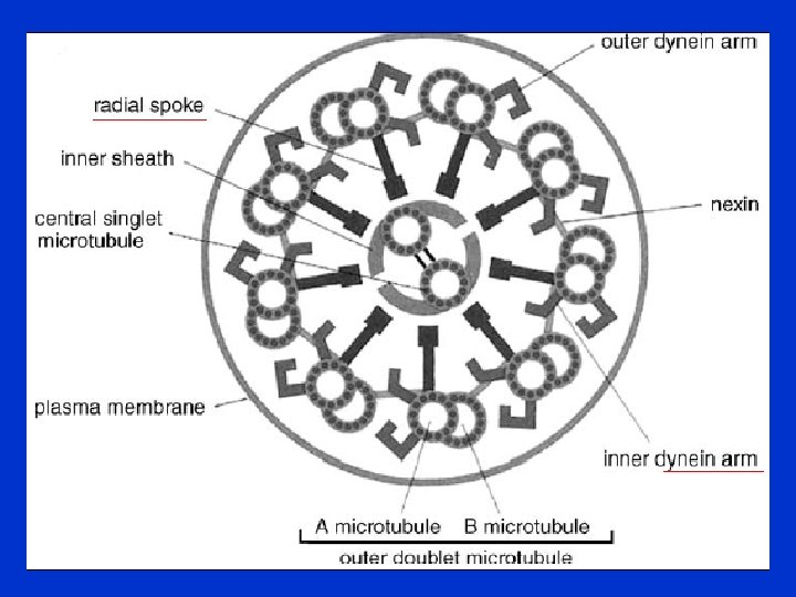

• Defects in the ciliary component include: 1 - Dynein arm defects involve total or a partial absence of either both inner or both outer dynein arms or involve just the inner or outer arms. Sometimes, shortened dynein arms are the only defect. 2 - Radial spoke defects exhibit either a total absence of radial spokes or an absence of radial spoke heads. 3 - Microtubular transposition defects occur in the form of absence of the central pair of tubules with transposition of the outer doublet to the centre. • How is Immobile Cilia Syndrome Treated? • The main goal of treatment in Immobile Cilia Syndrome is to minimize the damage caused by chronic infection and/or inflammation. Airway clearance therapy, including secretion removal and bronchodilation, and aggressive use of antibiotics are the most common forms of treatment.

Surface Energy and Tension in Liquids • The hydrogen bonding between molecules down into a liquid are shared with all neighbouring atoms. • Those on the surface have no neighbouring atoms above, and exhibit stronger attractive forces upon their nearest neighbours on the surface. • This enhancement of the intermolecular attractive forces at the surface is called surface tension.

Surface Tension: Intermolecular Forces

• The surface tension of water supports this water strider. The nonpolar surfaces of its feet also help to repel the water. • Surface tension The result of inward intermolecular forces of attraction among liquid particles that must be overcome to expand the surface area.
- Slides: 21