The Molecular Basis of Inheritance The Genetic Material

• The Molecular Basis of Inheritance

The Genetic Material • Frederick Griffith investigated virulence of Streptococcus pneumoniae § Concluded that virulence could be passed from a dead strain to a nonvirulent living strain § Transformation • Further research by Avery et al. § Discovered that DNA is the transforming substance § DNA from dead cells was being incorporated into the genome of living cells 2

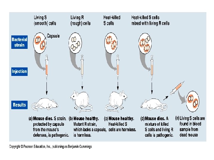
The Genetic Material • Griffith’s Transformation Experiment § Mice were injected with two strains of pneumococcus: an encapsulated (S) strain and a non-encapsulated (R) strain. • The S strain is virulent (the mice died); it has a mucous capsule and forms “shiny” colonies. • The R strain is not virulent (the mice lived); it has no capsule and forms “dull” colonies. 3

Griffith’s Transformation Experiment Copyright © The Mc. Graw-Hill Companies, Inc. Permission required for reproduction or display. capsule Injected live R strain has no capsule and mice do not die. Injected live S strain has capsule and causes mice to die. a. b. Injected heatkilled S strain does not cause mice to die. c. Injected heat-killed S strain plus live R strain causes mice to die. Live S strain is withdrawn from dead mice. d. 4
 • bacteriophages (phages) • DNA,")
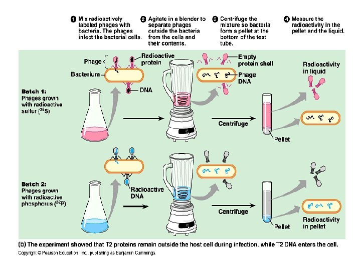
Searching for Genetic Material • Hershey and Chase (1952) • bacteriophages (phages) • DNA, not protein, is the hereditary material • Experiment: sulfur(S) is in protein, phosphorus (P) is in DNA; only P was found in host cell

The Genetic Material • Transformation of organisms today: § Result is the so-called genetically modified organisms (GMOs) • Invaluable tool in modern biotechnology today • Commercial products that are currently much used • Green fluorescent protein (GFP) can be used as a marker – A jellyfish gene codes for GFP – The jellyfish gene is isolated and then transferred to a bacterium, or the embryo of a plant, pig, or mouse. – When this gene is transferred to another organism, the organism glows in the dark 8

The Genetic Material • DNA contains: § Two Nucleotides with purine bases • Adenine (A) • Guanine (G) § Two Nucleotides with pyrimidine bases • Thymine (T) • Cytosine (C) 9

The Genetic Material • Chargaff’s Rules: § The amounts of A, T, G, and C in DNA: • Are constant among members of the same species • Vary from species to species § In each species, there are equal amounts of: • A and T • G and C § All this suggests that DNA uses complementary base pairing to store genetic information § Each human chromosome contains, on average, about 140 million base pairs § The number of possible nucleotide sequences is 4140, 000 10

Nucleotide Composition of DNA Copyright © The Mc. Graw-Hill Companies, Inc. Permission required for reproduction or display. NH 2 adenine (A) N C C CH HC N O HO P O C O 5 4 CH 2 thymine (T) N N nitrogen-containing base HN C H H C 3 OH H C 1 C H 2 H P O O guanine (G) C HN P O O 5 4 a. Purine nucleotides C N O HO CH O 5 4 CH 2 O C H H C 3 OH H C 1 C H 2 H sugar = deoxyribose NH 2 cytosine (C) N CH H 2 N C phosphate C CH 3 C N O HO C C O O O CH 2 C H H C 3 OH HO H C 1 C H 2 H N O O P O b. Pyrimidine nucleotides CH CH C O N C N O 5 4 CH 2 O C H H C 3 OH H C 1 C H 2 H DNA Composition in Various Species (%) Species A T G C Homo sapiens (human) Drosophila melanogaster (fruit fly) Zea mays (corn) Neurospora crassa (fungus) 31. 0 27. 3 25. 6 23. 0 31. 5 27. 6 25. 3 23. 3 19. 1 22. 5 24. 5 27. 1 18. 4 22. 5 24. 6 26. 6 Escherichia coli (bacterium) Bacillus subtilis (bacterium) 24. 6 28. 4 24. 3 29. 0 25. 5 21. 0 25. 6 21. 6 c. Chargaff’s data 11

The Genetic Material • X-Ray diffraction: § Rosalind Franklin studied the structure of DNA using X-rays. § She found that if a concentrated, viscous solution of DNA is made, it can be separated into fibers. § Under the right conditions, the fibers can produce an X-ray diffraction pattern • She produced X-ray diffraction photographs. • This provided evidence that DNA had the following features: – DNA is a helix. – Some portion of the helix is repeated. 12

X-Ray Diffraction of DNA Copyright © The Mc. Graw-Hill Companies, Inc. Permission required for reproduction or display. Rosalind Franklin diffraction pattern diffracted X-rays a. X-ray beam Crystalline DNA b. c. © Photo Researchers, Inc. ; c: © Science Source/Photo Researchers, Inc. 13
 § Double helix model")
The Genetic Material • The Watson and Crick Model (1953) § Double helix model is similar to a twisted ladder • Sugar-phosphate backbones make up the sides • Hydrogen-bonded bases make up the rungs § Complementary base pairing ensures that a purine is always bonded to a pyrimidine (A with T, G with C) § Received a Nobel Prize in 1962 14

Watson and Crick Model of DNA Copyright © The Mc. Graw-Hill Companies, Inc. Permission required for reproduction or display. 3. 4 nm 0. 34 nm 2 nm b. d. C a. G 5′ end sugar-phosphate backbone T 3′ end P A C G S P S A T P P T S 3′ end A S P 5′ end P G C P complementary base pairing c. C G P sugar hydrogen bonds a: © Photodisk Red/Getty RF; d: © A. Barrington Brown/Photo Researchers 15

12. 2 Replication of DNA • DNA replication is the process of copying a DNA molecule. • Semiconservative replication - each strand of the original double helix (parental molecule) serves as a template (mold or model) for a new strand in a daughter molecule. 16

Semiconservative Replication Copyright © The Mc. Graw-Hill Companies, Inc. Permission required for reproduction or display. 5′ 3′ G G C C A A region of parental DNA double helix T C G T A G A A G G T C C G C T C G C A T A T T A G T C C A T G C T A C C G A T C G region of replication: new nucleotides are pairing with those of parental strands G A old strand G A A 5′ DNA polymerase enzyme T G T region of completed replication A A T A G T A C G C new strand G A 3′ old strand daughter DNA double helix new strand daughter DNA double helix 17

Replication of DNA • Replication requires the following steps: § Unwinding, or separation of the two strands of the parental DNA molecule § Complementary base pairing between a new nucleotide and a nucleotide on the template strand § Joining of nucleotides to form the new strand • Each daughter DNA molecule contains one old strand one new strand 18
: beginning of replication •")
DNA Replication: a closer look • Origin of replication (“bubbles”): beginning of replication • Replication fork: ‘Y’-shaped region where new strands of DNA are elongating • Helicase: catalyzes the untwisting of the DNA at the replication fork • DNA polymerase: catalyzes the elongation of new DNA
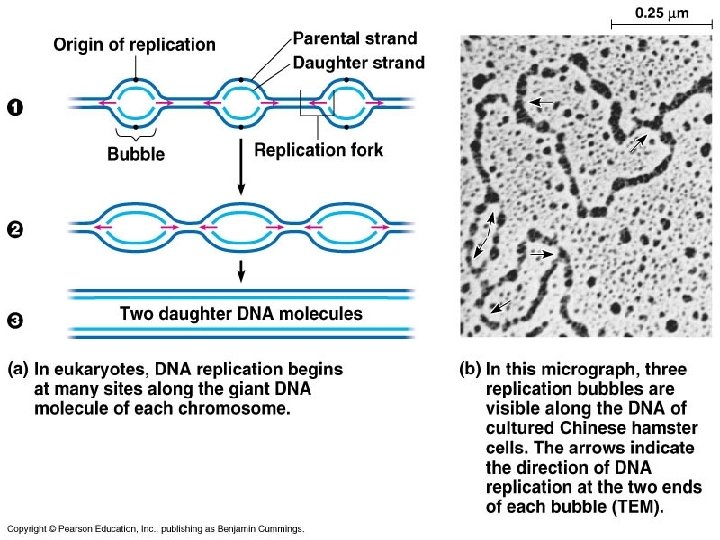

Replication of DNA • Eukaryotic Replication § DNA replication begins at numerous points along each linear chromosome § DNA unwinds and unzips into two strands § Each old strand of DNA serves as a template for a new strand § Complementary base-pairing forms a new strand paired with each old strand • Requires enzyme DNA polymerase 21

Replication of DNA • Eukaryotic Replication § Replication bubbles spread bidirectionally until they meet § The complementary nucleotides are joined to form new strands. Each daughter DNA molecule contains an old strand a new strand. § Replication is semiconservative: • One original strand is conserved in each daughter molecule, i. e. , each daughter double helix has one parental strand one new strand. 22

Aspects of DNA Replication Copyright © The Mc. Graw-Hill Companies, Inc. Permission required for reproduction or display. OH 5′ P Is attached here base is attached here CH 2 4′ C H O OH C 1′ H H C 2′ 3′ C OH 1 H H De ox yrib os e m o lec u le 2 5′ end P DNA polymerase attaches a new nucleotide to the 3 ′ carbon of the previous nucleotide. T A P P G C C G 3′ e nd P 5′ P T A P C G 3 ′end P P 5′ end template strand 3′ template strand P new strand lagging strand DNA polymerase leading new strand 3′ Direction of replication 5 4 3 RNA primer template strand 6 helicase at replication fork Okazaki fragment 3′ 5′ 5′ parental DNA helix 7 DNA ligase 3′ Replication fork introduces complications DNA polymerase 23

Replication of DNA • Accuracy of Replication § DNA polymerase is very accurate, yet makes a mistake about once per 100, 000 base pairs. • Capable of identifying and correcting errors 24
; •")
DNA Replication, II • Antiparallel nature: sugar/phosphate backbone runs in opposite directions (Crick); • one strand runs 5’ to 3’, while the other runs 3’ to 5’; • DNA polymerase only adds nucleotides at the free 3’ end, forming new DNA strands in the 5’ to 3’ direction only

Figure 16. 12 The two strands of DNA are antiparallel

DNA Replication, III • Leading strand: synthesis toward the replication fork (only in a 5’ to 3’ direction from the 3’ to 5’ master strand) • Lagging strand: synthesis away from the replication fork (Okazaki fragments); joined by DNA ligase (must wait for 3’ end to open; again in a 5’ to 3’ direction) • Initiation: Primer (short RNA sequence~w/primase enzyme), begins the replication process

Figure 16. 15 The main proteins of DNA replication and their functions

DNA Repair • Mismatch repair: DNA polymerase • Excision repair: Nuclease • Telomere ends: telomerase

Figure 16. 17 Nucleotide excision repair of DNA damage

Figure 16. 18 The end-replication problem

Figure 16. 19 b Telomeres and telomerase
- Slides: 32