
The mode of action of the calcium channel
- Slides: 6
The mode of action of the calcium channel blocker Mibefradil on human lens epithelial cells – first insights Joint congress of SOE / AAO 2007 Vienna, Austria 9 -12 June 2007 “The mode of action of the calcium channel blocker mibefradil on human lens epithelial cells – first insights“ Arne Weidmann 1, Ria Beck 2, Barbara Nebe 1 1 Dept. of Internal Medicine, 2 Dept. of Ophthalmology e-poster presentation Weidmann A. et al University of Rostock
The mode of action of the calcium channel blocker Mibefradil on human lens epithelial cells – first insights Introduction Inhibition of cell adhesion and migration of residual lens epithelial cells is an important therapeutic aspect in posterior capsule opacification after cataract surgery. Our former results demonstrated that mibefradil, known as a T-type calcium channel blocker with the potential to block potassium channels, reduced adhesion and proliferation of primary human lens epithelial cells and induced their apoptosis. The objective of these new experiments was to find out the mode of action of mibefradil. Therefore we investigated if the specific T-type channel blocker Ethosuximide (ETX) and the nonselective potassium channel blocker 4 -Aminopyridine (4 -AP) have similar inhibitory effects on cell functions compared to mibefradil Materials and Methods Channel antagonists Primary human lens epithelial cells (h. LEC) and The T-type calcium channel antagonists mibefradil dihydro-chloride (Sigma), ethosuximide (Sigma) and the nonselective potassium channel blocker 4 -aminopyridine (Sigma) were used. The substance was solved in distilled water. Cell culture Primary human lens epithelial cells (h. LEC) and the human lens epithelial cell line HLE-B 3 (ATCC No: CRL-11421) were cultured in DMEM with 10 % FCS and 1 % gentamicin at 37°C with 5 % CO 2. Weidmann A. et al University of Rostock
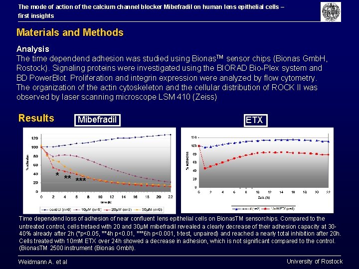
The mode of action of the calcium channel blocker Mibefradil on human lens epithelial cells – first insights Materials and Methods Analysis The time dependend adhesion was studied using Bionas. TM sensor chips (Bionas Gmb. H, Rostock). Signaling proteins were investigated using the BIORAD Bio-Plex system and BD Power. Blot. Proliferation and integrin expression were analyzed by flow cytometry. The organization of the actin cytoskeleton and the cellular distribution of ROCK II was observed by laser scanning microscope LSM 410 (Zeiss) Results Mibefradil ETX * ** *** Time dependend loss of adhesion of near confluent lens epithelial cells on Bionas. TM sensorchips. Compared to the untreated control, cells tretaed with 20 and 30µM mibefradil revealed a clearly decrease of their adhesion capacity at 3040% already after 2 h (*p<0. 05, **4 h p<0. 01, ***6 h p<0. 001, t-test, unpaired) and reached a nearly total inhibition after 20 h. Cells treated with 10 m. M ETX over 24 h showed a decrease in adhesion, which is not significant compared to the control. (Bionas. TM 2500 instrument (Bionas Gmbh). Weidmann A. et al University of Rostock
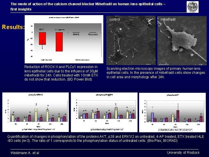
The mode of action of the calcium channel blocker Mibefradil on human lens epithelial cells – first insights control mibefradil Results: Reduction of ROCK II and PLCγ 1 expression in lens epithelial cells due to the influence of 30µM mibefradil for 24 h. Cells treated with 10 m. M ETX do not show that reduction. (BD Power Blot) Scanning electron microscopy images of primary human lens epithelial cells. In the presence of mibefradil cells show changes in cell area and morphology after 24 h. * * * * ** ** * * * * Quantification of changes in phosphorylation of the proteins AKT, p 38 and ERK 1/2 on untreated, 4 -AP treated, ETX treated HLE -B 3 cells (n=3). The ratio of 1 corresponds to the phosphorylation status of untreated cells. (Bio-Plex, BIORAD) Weidmann A. et al University of Rostock
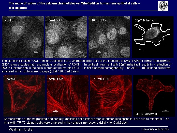
The mode of action of the calcium channel blocker Mibefradil on human lens epithelial cells – first insights control 5 m. M 4 -AP 10 m. M ETX 30µM Mibefradil The signalling protein ROCK II in lens epithelial cells. Untreated cells, cells at the presence of 5 m. M 4 -APand 10 m. M Ethosuximide (ETX) show cytoplasmatic and nuclear localisation of ROCK II. In contrast, treatment with 30µM mibefradil results in a reduction of ROCK II expression in the cells. Moreover the protein ROCK II is not disposed homogenously. The ALEXA 488 stained cells were analyzed in the confocal microscope (LSM 410, Carl Zeiss). control 5 m. M 4 -AP 10 m. M ETX 30µM Mibefradil Demonstration of the fragmented and partially abolished actin cytoskeleton of human lens epithelial cells due to mibefradil. The phalloidin TRITC stained cells were analyzed in the confocal microscope (LSM 410, Carl Zeiss). Weidmann A. et al University of Rostock
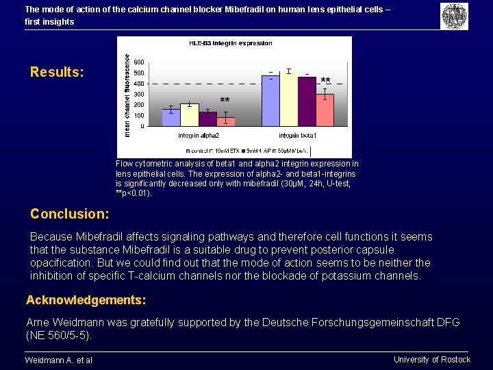
The mode of action of the calcium channel blocker Mibefradil on human lens epithelial cells – first insights Results: ** ** Flow cytometric analysis of beta 1 and alpha 2 integrin expression in lens epithelial cells. The expression of alpha 2 - and beta 1 -integrins is significantly decreased only with mibefradil (30µM, 24 h, U-test, **p<0. 01). Conclusion: Because Mibefradil affects signaling pathways and therefore cell functions it seems that the substance Mibefradil is a suitable drug to prevent posterior capsule opacification. But we could find out that the mode of action seems to be neither the inhibition of specific T-calcium channels nor the blockade of potassium channels. Acknowledgements: Arne Weidmann was gratefully supported by the Deutsche Forschungsgemeinschaft DFG (NE 560/5 -5). Weidmann A. et al University of Rostock