The milan system for reporting salivary gland cytopathology


,")


 Pleomorphic adenoma showing myoepithelial cells and very delicate, pale-staining matrix")


 This aspirate of a pleomorphic adenoma has a predominance of")


 showing classic cytomorphologic features consisting of background")


- Slides: 43
The milan system for reporting salivary gland cytopathology REZA ZIAOLHAGH MD/AP-CP RAZAVI HOSPITAL
Banign neoplasms
Benign Neoplasms
Salivary more gland neoplasms are rare, commonly occur in the parotid gland 80% of salivary gland neoplasms in the parotid gland are benign In adults, pleomorphic adenomas (50%)and Warthin tumor are the most common benign tumors. mucoepidermoid carcinoma is the most common malignant tumor FNA can distinguish with high specificity (97– 98%) a non-neoplastic lesion from a neoplasm
FNA can diagnose the most common benign tumors of the salivary gland, PA, and WT with high specificity (>98%). FNA is generally less effective in providing a specific diagnosis for certain other epithelial neoplasms. Because of the morphologic overlap and diversity among the many different types of salivary gland tumors differentiation between a benign and low-grade malignant neoplasm based on purely cellular and cytoarchitectural features can be challenging in an FNA specimen
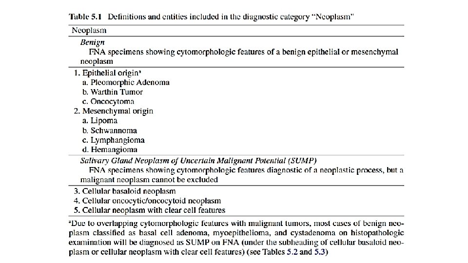
Neoplasm: Benign: when an FNA specimen shows characteristic cytomorphologic features of a specific benign epithelial or mesenchymal neoplasm of the salivary gland. The most common being PA and WT. Salivary Gland Neoplasm of Uncertain Malignant Potential (SUMP): FNA specimens where the cytomorphologic features are diagnostic of a neoplastic process, but the cytologic findings cannot effectively distinguish between a benign and malignant neoplasm. Most malignant tumors included in this diagnostic category will be low-grade carcinomas
Pleomorphic Adenoma a a benign biphasic neoplasm variable admixture of ductal epithelial cells, myoepithelial cells, and mesenchymal matrix.
Pleomorphic Adenoma Cytologic Criteria 1. Distinctive chondromyxoid matrix: in Romanowsky stains (Diff. Quik®, Giemsa), as a bright magenta matrix with a distinct fibrillary/feathery quality; in Papanicolaou grey to translucent green 2. Myoepithelial cells: polygonal, plasmacytoid, round, spindle, or clear, bland nuclear features, often the predominant cell type. 3. Ductal epithelial cells: Bland nuclear features, small groups recapitulating ductal structures
Neoplasm: Benign. Pleomorphic adenoma. Intense metachromatic fibrillary matrix with myoepithelial cells embedded within
Neoplasm: SUMP. FNA of pleomorphic adenoma showing a highly cellular, matrix-poor tumor with a predominance of plasmacytoid myoepithelial cells
Neoplasm: Benign. (a, b) Pleomorphic adenoma showing myoepithelial cells and very delicate, pale-staining matrix
Pleomorphic Adenoma The hallmark and most distinguishing feature of PA is the presence of chondromyxoid matrix, best appreciated using Romanowsky stains (Diff Quik®, Giemsa). other“matrix-producing tumors” includes adenoid cystic carcinoma, basal cell adenoma/adenocarcinoma , and epithelialmyoepithelial carcinoma
Pleomorphic Adenoma FNA specimens with classic features of PA can be readily diagnosed as “Neoplasm: Benign. ” When classic features of PA are not present or when additional “atypical” features are identified, then the FNA should be diagnosed as “Neoplasm: SUMP. ” highly cellular aspirates that have scant to absent matrix would be classified as “Neoplasm: SUMP. ” The differential diagnosis include the solid variant of Ad. CC, myoepithelioma, or other basal cell neoplasms.
Neoplasm: SUMP. FNA of pleomorphic adenoma having adenoid cystic carcinoma-like areas
Neoplasm: SUMP. FNA of pleomorphic adenoma showing squamous metaplasia
Neoplasm: SUMP. (a, b) This aspirate of a pleomorphic adenoma has a predominance of spindled myoepithelial cells mimicking a salivary gland neoplasm of mesenchymal origin
Neoplasm: SUMP. This case of pleomorphic adenoma shows marked nuclear atypia of the myoepithelial cells; in such cases, malignant transformation needs to be excluded
Warthin Tumor the second most common neoplasm of the parotid gland. The majority occur in the 7 th to 9 th decades of life usually have a significant history of smoking. present with a doughy painless mass that may fluctuate in size.
Warthin Tumor Cytologic Tripartite Criteria appearance with dirty proteinaceous background, lymphocytes, and sheets of oncocytes Oncocytes: Abundant homogeneous granular cytoplasm with welldefined borders. Epithelial cell nuclei: Centrally placed and round, with prominent nucleolus Lymphocytes: Mixed population dominated by small mature-appearing cells.
Neoplasm: Benign. FNA of Warthin tumor (WT) showing classic cytomorphologic features consisting of background lymphocytes and groups of oncocytic epithelial cells
Neoplasm: Benign. This classic aspirate of Warthin tumor consists of oncocytic cells with abundant granular cytoplasm and well-defined borders in a background of lymphocytes
Warthin Tumor WT occurs almost exclusively in the parotid gland the tripartite appearance is essentially diagnostic. Aspirates with classic features should be diagnosed as “Neoplasm: Benign. ” Distinction should nodes, lymphoepithelial cyst. be made sialadenitis from (LESA), intraparotid lymph oncocytoma, and Intraparotid lymph nodes and LESA lack the oncocytic epithelium and dirty cyst debris characteristic of a WT.
Warthin Tumor Oncocytomas consist of epithelial cells only and lack the dirty cystic background and lymphocytes of WT. Lymphoepithelial cysts or HIV-associated benign lymphoepithelial lesions contain a mixed lymphoid population among which are distributed rare glandular or more commonly squamous cells. but the sheets of oncocytic epithelium, characteristic of WT are not seen
Oncocytoma Nearly 90% of oncocytomas occur within the major salivary glands, they comprise only 1% of parotid gland neoplasms. Most cases occur in the 6 th to 8 th decades of life.
Oncocytoma Cytologic Criteria 1. Irregular sheets and clusters of large polygonal cells with abundant homogeneous granular cytoplasm 2. Oncocytes: Well-defined cytoplasmic borders 3. Nuclei: Enlarged, round, and distinct nucleolus 4. Background: Clean or contains red blood cells 5. Nuclear pleomorphism and mitotic figures absent
Oncocytoma The differential diagnosis of oncocytoma includes : WT, diffuse oncocytosis, and acinic cell carcinoma (ACC) Aspirates of oncocytoma and oncocytosis are virtually identical; however, oncocytoma presents clinically as a distinct circumscribed mass WT contains occasional groups of oncocytes, but differs from oncocytoma by also having a dirty proteinaceous background a mixed lymphoid population
Oncocytoma ACC contains a mixture of polygonal cells with delicate vacuolated cytoplasm. In contrast, oncocytomas lack the cytoplasmic vacuoles of ACC; . ICC for DOG-1 and SOX 10 is positive in ACC, but negative in oncocytomas Oncocytic carcinoma is very rare and is clinically invasive; clinical correlation is needed.
Neoplasm: Benign. FNAs of oncocytoma showing various patterns of a monotonous population of oncocytic cells with abundant granular cytoplasm and well-defined borders arranged in cohesive groups
Lipoma uncommon neoplasms of the salivary glands, 0. 5% of salivary gland tumors ¾ in the parotid gland often present as a palpable soft nodule.
Lipoma Cytologic 1. 2. 3. 4. Criteria: Lacelike sheets and clusters of very low nuclear-cytoplasmic (N: C) ratio cells with optically clear cytoplasm Individual cells: Single large clear vacuole occupying the entire cytoplasmic volume Nuclei: Small, hyperchromatic, and displaced to the margin of the cell Background: May contain droplets of lipid (best seen on Romanowsky stains)
Lipoma It may be difficult to distinguish fatty change of the salivary gland from lipoma by FNA. While fatty change will contain normal acinar and ductal elements, lipomas are composed purely of adipose tissue, although rare examples of lipomas with entrapped normal serous acini and ducts have been reported. Clinical correlation is useful.
Neoplasm: Benign. FNA of lipoma showing lacelike group of mature adipocytes with abundant clear cytoplasm and small dark nuclei
Neoplasm: Benign. This FNA contains a group of adipocytes from a lipoma characterized by large cells with abundant clear cytoplasm. The small dark nuclei are often displaced to the edge of the cell
Schwannoma the most common benign neural tumor of the salivary glands. FNA of a schwannoma is often associated with pain of a radicular type.
Schwannoma Cytologic 1. 2. 3. 4. 5. 6. Criteria: Scant to moderately cellular aspirate Spindle-shaped cells Form cohesive groups and clusters, sometimes with palisading Cytoplasm: Pale and ill-defined Nuclei: Small, dark, bland, and elongate/spindled, curved, or even Sshaped Nucleoli: Small or absent Background: Myxoid material
Schwannoma The most common differential diagnosis is with PA and myoepithelioma. Some cases of PA and schwannoma can be very difficult to distinguish. Ancillary studies are helpful. Schwannomas are strongly and diffusely positive for S 100 and SOX 10, and negative for keratin and myoepithelial markers. Other differential diagnostic considerations include sarcomas, which are very rare. The possibility of a sarcoma should be considered when aspirates are cellular and display significant nuclear atypia often with scattered mitoses and apoptosis
Neoplasm: Benign. This aspirate of schwannoma shows a group of bland spindle cells with wispy cytoplasm. The cytoplasmic borders are indistinct. Nuclei are spindle-shaped and display bends or curves
Lymphangioma rare with most occurring in children. present as slowly growing fluctuant masses. Most arise in the parotid gland. Cytologic Criteria: 1. Hypocellular smears with watery background 2. Occasional red blood cells 3. Scattered mature-appearing lymphocytes 4. Rare background clusters of nonneoplastic salivary gland acinar tissue may be present
Lymphangioma Aspirates obtained from salivary gland lymphangiomas are frequently Non-Diagnostic, and are composed of watery fluid containing scattered mature-appearing lymphocytes and occasional groups of non-neoplastic acinar cells. Endothelial cells are generally absent from aspirated material. The diagnosis usually requires careful clinical and radiologic correlation.
Hemangioma the most common benign mesenchymal tumor of the salivary gland most examples arising in the parotid gland. the majority of hemangiomas occurs in the first decade of life, especially the first year of life. The latter are of the so-called juvenile type and may regress spontaneously. Juvenile hemangiomas may be highly cellular.
Hemangioma Cytologic Criteria 1. Aspirate dominated by red blood cells 2. Few groups of bland spindle-shaped to polygonal endothelial cells, which may form elongated cord-like structures 3. Individual cells: Oval to spindle-shaped nuclei 4. Nuclei: Small, bland, and lack nucleoli 5. Scattered histiocytes may be present.
Neoplasm: Benign. Smears obtained from hemangiomas are characteristically bloody, but may contain small aggregates of bland spindle-shaped endothelial cells.