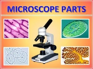
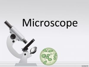
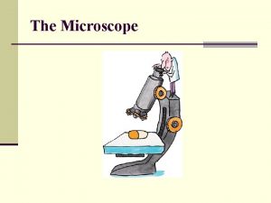
The Microscope The Parts of a Microscope Ocular


- Slides: 25

The Microscope

The Parts of a Microscope

Ocular Lens Body Tube Nose Piece Arm Objective Lenses Stage Clips Diaphragm Stage Coarse Adj. Fine Adjustment Light Source Base Skip to Magnification Section

Body Tube • The body tube holds the objective lenses and the ocular lens at the proper distance Diagram

Nose Piece • The Nose Piece holds the objective lenses and can be turned to increase the magnification Diagram

Objective Lenses • The Objective Lenses increase magnification (usually from 10 x to 40 x) Diagram

Stage Clips • These 2 clips hold the slide/specimen in place on the stage. Diagram

Diaphragm • The Diaphragm controls the amount of light on the slide/specimen Turn to let more light in or to make dimmer. Diagram

Light Source • Projects light upwards through the diaphragm, the specimen and the lenses • Some have lights, others have mirrors where you must move the mirror to reflect light Diagram

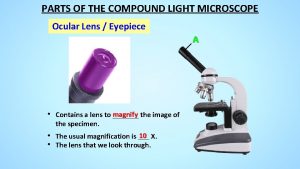
Ocular Lens/Eyepiece • Magnifies the specimen image Diagram

Arm • Used to support the microscope when carried. Holds the body tube, nose piece and objective lenses Diagram

Stage • Supports the slide/specimen Diagram

Coarse Adjustment Knob • Moves the stage up and down (quickly) for focusing your image Diagram

Fine Adjustment Knob • This knob moves the stage SLIGHTLY to sharpen the image Diagram

Base • Supports the microscope Diagram

Magnification

What’s my power? To calculate the power of magnification, multiply the power of the ocular lens by the power of the objective. What are the powers of magnification for each of the objectives we have on our microscopes? Fill in the table on your worksheet.

Comparing Powers of Magnification We can see better details with higher the powers of magnification, but we cannot see as much of the image. Which of these images would be viewed at a higher power of magnification?

Caring for a Microscope • Clean only with a soft cloth/tissue • Make sure it’s on a flat surface • Don’t bang it • Carry it with 2 HANDS…one on the arm and the other on the base

Carry a Microscope Correctly

Using a Microscope • Start on the lowest magnification • Don’t use the coarse adjustment knob on high magnification…you’ll break the slide!!! • Place slide on stage and lock clips • Adjust light source (if it’s a mirror…don’t stand in front of it!) • Use fine adjustment to focus

Let’s give it a try. . . 1 – Turn on the microscope and then rotate the nosepiece to click the red-banded objective into place. 2 – Place a slide on the stage and secure it using the stage clips. Use the coarse adjustment knob (large knob) to get it the image into view and then use the fine adjustment knob (small knob) to make it clearer. 3 – Once you have the image in view, rotate the nosepiece to view it under different powers. Draw what you see on your worksheet! Be careful with the largest objective! Sometimes there is not enough room and you will not be able to use it! 4 – When you are done, turn off the microscope and clean up or put away the slides you used.

How to make a wet-mount slide … 1 – Get a clean slide and coverslip from your teacher. 2 – Place ONE drop of water in the middle of the slide. Don’t use too much or the water will run off the edge and make a mess! 3 – Place the edge of the cover slip on one side of the water drop. 4 - Slowly lower the cover slip on top of the drop. Cover Slip VIDEO Lower slowly 5 – Place the slide on the stage and view it first with the red-banded objective. Once you see the image, you can rotate the nosepiece to view the slide with the different objectives. You do not need to use the stage clips when viewing wet-mount slides!

References • • • http: //www. cerebromente. org. br/n 17/history/neurons 1_i. htm Google Images http: //science. howstuffworks. com/light-microscope 1. htm

This powerpoint was kindly donated to www. worldofteaching. com http: //www. worldofteaching. com is home to over a thousand powerpoints submitted by teachers. This is a completely free site and requires no registration. Please visit and I hope it will help in your teaching.
 Parts of a compound light microscope
 Labeling a compound microscope
 The body tube
 Function of coarse adjustment knob
 Compound microscope draw tube function
 Candida uveitis
 Eye accessory structures
 Epitelio conjuntiva ocular
 Mapa ocular
 Vestibulo ocular reflex
 Pseudoexfoliation glaucoma
 Ocular deviation
 Origen aparente del nervio optico
 Salud ocular his
 Myasthenia gravis.
 Prime astaxanthin
 Vestibulo ocular reflex
 Icd 10 code for allergic reaction to peanuts
 Ocular adnexa
 Aseo ocular
 Esterman visual field test dvla
 Importancia de la profilaxis ocular del recién nacido
 Enroth sign
 Vagabundeo ocular
 Ocular emergency definition
 Ocular migraine
