The Microscope Microscope History Microscopes are instruments which


 • How it works: • • Light Source: Electrons")
 • How it works: Ø it passes a narrow")


 • Consists of 2 lens • Light Source:")
 • Light Source: electrons (passed through specimen) • Resolution:")
 • Light Source: electrons (passed through specimen) • Resolution:")
- Slides: 25
The Microscope
Microscope History • Microscopes are instruments which produce a magnified image to help us to examine small objects and their fine details which our eye cannot see. • Microscopes range from simple devices such as a magnifying glass up to high end compound or electron microscopes.
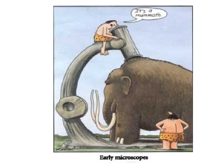
Early Microscopes Simple Microscope Compound Microscope
Microscopes used today Compound Microscope
There are similarities in all microscopes: 1. Magnify a small object 2. Separate fine details in order to achieve a high resolution
Types of Microscopes 1. 2. 3. 4. 5. Simple Dissecting Compound Scanning Electron Microscope (SEM) Transmission Electron Microscope (TEM).
Using a Microscope to Explore the Cell • Resolution or Resolving power – The ability to distinguish between two objects that are close together High resolution Low resolution
1. Simple Microscope • Similar to a magnifying glass and has only one lens • A lens enlarges an image and bends the light toward your eye.
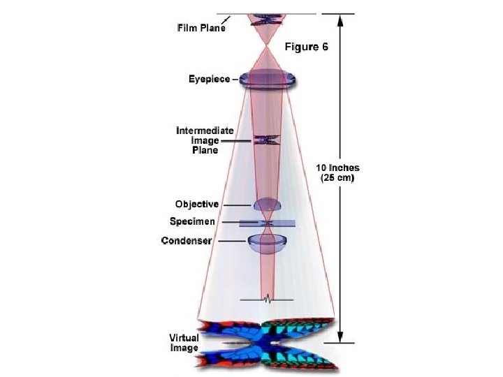
2. Compound light microscope • Light source: Lamp • Resolution: limited to about 0. 2 µm (micrometers) • Magnification: 2000 X • 2 D image produced • Image seen: In eyepiece • Specimen Preparation: Stain cells on a slide • Type of specimens can be used: Both
Total Magnification • To get the total magnification: Eyepiece Lens X Objective Lens = Total Magnification
Low Power = 4 x Medium Power = 10 x High Power = 40 x
3. Electron Microscope • Used to see objects that are smaller than 2µm • There are 2 types of electron microscopes: A. Transmission Electron B. Scanning Electron
A. Transmission Electron Microscope (TEM) • How it works: • • Light Source: Electrons pass through Resolution: 0. 0002 µm Magnification: 500, 000 x Type of image: 2 D Where image is seen: On a screen/ monitor Specimen preparation: Very complex Types that can be used: Dead
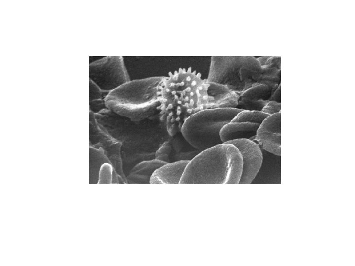
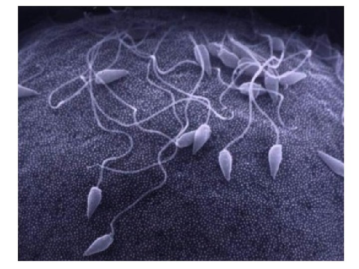
B. Scanning Electron Microscope (SEM) • How it works: Ø it passes a narrow beam of electrons over the surface of a specimen. Ø The specimens must be coated with a very thin film of metal, usually gold. As electrons bounce off the specimen a television screen picks up the image. • It produces a 3 -D image.
• How it works: • Light Source: Electrons pass over surface of specimen • Resolution: 0. 005 µm • Magnification: 300, 000 x • Type of image: 3 D • Where image is seen: On a screen/ monitor • Specimen preparation: Sprayed with gold • Types that can be used: Dead or alive
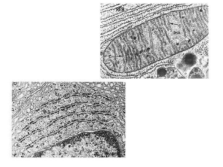
Mitochondria in human liver cell
TEM • Magnification: 500, 000 x • Resolution: 0. 0002 µm SEM • Magnification: 300, 000 x • Resolution: 0. 005 µm *** SEM have lower resolutions than TEM, but they have the advantage of providing 3 -D images Some specially designed SEM’s allow you to observe living specimens
1. Compound Light Microscope (pg. 20) • Consists of 2 lens • Light Source: light (passes through specimen) • Resolution: 200 nm or 0. 2µm • Magnification: 2000 x • Image produced: 2 D • Where image is seen: Eyepiece • Specimen Prep: Slicing/staining • Type of Specimen: Dead or alive
2. Transmission Electron Microscope (TEM) • Light Source: electrons (passed through specimen) • Resolution: 0. 2 nm or 0. 0002 µm • Magnification: 500, 000 x • Image produced: 2 D • Where image is seen: Projected on screen/monitor = picture! (internal study) • Specimen Prep: very complex – cross sections • Type of Specimen: Dead
3. Scanning Electron Microscope (SEM) • Light Source: electrons (passed through specimen) • Resolution: 5 nm or 0. 005 µm • Magnification: 300, 000 x • Image produced: 3 D • Where image is seen: Projected on tv screen (study surface of specimen) • Specimen Prep: Sprayed with gold coating • Type of Specimen: Dead or alive