The Microscope and the Cell The Microscope Compound


 �sweeps a beam of electrons over a surface specimen causing")
 �electrons are passed through a specimen, less dense areas allow fewer")

 and")


 �Rough (has ribosomes) �site")


, thick, waxy")

- Slides: 23
The Microscope and the Cell
The Microscope �Compound Light Microscope �uses two or more glass lenses to magnify an object �magnify 1500 X
�Scanning Electron Microscope (SEM) �sweeps a beam of electrons over a surface specimen causing electrons to be emitted � 3 -D �magnify 60, 000 X
�Transmission Electron Microscope(TEM) �electrons are passed through a specimen, less dense areas allow fewer electrons to pass � 2 -D �magnify hundreds of thousands of times
Scientists �Robert Hooke: �took a thin piece of cork, put it under the microscope, named what he saw CELLS �Anton van Leewenhoek: �First scientist to see a living cell through a compound microscope
The Cell Theory �All living things are made of cells �Matthias Schleiden (botanist) and Theodore Schwann (zoologist) �The cell is the basic building blocks of all living things �Matthias Schleiden (botanist) and Theodore Schwann (zoologist) �All cells come from pre-existing cells �Rudolph Virchow
Prokaryotic vs Eukaryotic • Organelles: things in a cell that make it work (like organs)
Prokaryotic vs Eukaryotic Prokaryotic Cells Eukaryotic Cells �no membrane bound organelles �found in bacteria only �no membrane bound nucleus �only contains DNA, cytoplasm, ribosomes, cell wall, and mitochondria �have membrane bound organelles �found in plants and animals �have membrane bound nucleus
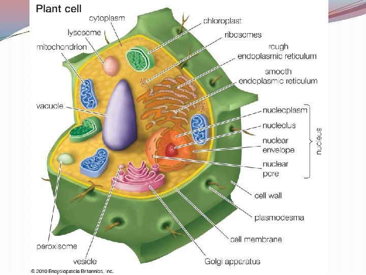
Eukaryotic Plant Cell Organelles Endoplasmic reticulum Microfilaments Cytoplasm Ribosomes Cilia Cell Membrane Mitochondria Flagella Chloroplast Lysosomes Nucleolus Large Vacuole Golgi Apparatus Cytoskeleton Cell Wall Microtubules Nucleus
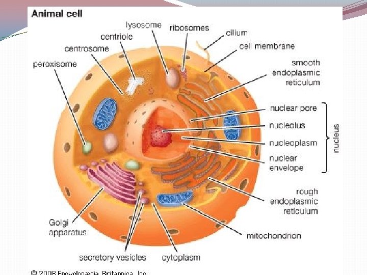
Eukaryotic Animal Cell Organelles Endoplasmic reticulum Microfilaments Cytoplasm Ribosomes Cilia Cell Membrane Mitochondria Flagella Centrioles Lysosomes Nucleolus Small Vacuole Golgi Apparatus Cytoskeleton Microtubules Nucleus
Cell Control �Nucleus: control center of the cell, found in eukaryotic cell, contains DNA �DNA: contains directions for making proteins �Nucleolus: center of the nucleus, makes ribosomes
�Ribosomes: site of protein synthesis �Centrioles: only in animals, helps pull apart the cell during mitosis �Nuclear membrane: keeps materials inside the cell
Cell Assembly �Endoplasmic reticulum: � 2 types: �Smooth (no ribosomes) �Rough (has ribosomes) �site of cellular chemical reactions (protein synthesis) �Golgi Apparatus: POST OFFICE, transports & packages proteins to be sent out into the cell
Storage & Clean-up �Vacuole: storage facility of the cell, larger in plants, holds water and other minerals important to the cell �Lysosomes: contains digestive enzymes, gets rid of worn out organelles , lysosomes can fuse to the vacuole to digest foreign matter in the vacuole, �“suicide sacs”
Structure, Support, & Locomotion �Cytoskeleton: support system for the cell…. . Cyto=cell � 2 components of the cytoskeleton: �microtubules �microfilaments
�Cilia & Flagella: helps the cell move �Cilia: short hair-like structures �Flagella: long whip-like structures
�Cytoplasm: jelly-like substance that suspends the organelles inside the cell, adds fluidity to the cell
Power House �Mitochondria: generates power for the cell, contains ATP, mighty mitochondria, numerous in the cell �Chloroplast: converts sun into energy the plant can use
Highly Folded Membranes • The golgi apparatus and the mitochondria have highly folded inner membranes to increase the surface area to allow for greater absorption of nutrients
Barriers for Protection �Cell Wall: only in plants, made of cellulose (sugar), thick, waxy to prevent water loss, gives plant shape, support, and protection �Plasma/Cell Membrane � Found in plants and animals � Semi-permeable/selectively permeable � “heads” are hydrophilic (phosphate groups) � “tails” are hydrophobic (lipids) � AKA: Phospholipid bilayer
Fluid Mosaic Model �Model of the plasma membrane �Helps demonstrate how the “heads” of the membrane will move up and down in a fluid manner �the membrane moves with the current