The Meninges Dura mater outermost layer Arachnoid mater


 DESCENDING impulses travel to the muscles (motor)")


")


 Figure 13. 7 a Sulcus = groove Gyrus =")


 fluid that protects and supports brain Figure 13. 27 b")


")


.")

- Slides: 33
The Meninges Dura mater - outermost layer Arachnoid mater - no blood vessels, in between layer (resembles a spider web) Pia mater -inner membrane, contains nerves and blood vessels to nourish cells
CSF - cerebrospinal fluid Figure 13. 25 a
Dura mater is being peeled away in this photo.
Subdural Hematoma
CNN Video Showing cognitive tasks during brain surgery as a tumor is removed. Natgeo Brain Surgery Video - removal of tumor http: //video. nationalgeographic. com/vi deo/science/health-human-bodysci/human-body/brain-tumor-sci/
Spinal Cord passes down the vertebral canal, has 31 segments (each with a pair of spinal nerves) Cervical enlargement = supplies nerves to upper limbs (neck) Lumbar enlargement = supplies nerves to the lower limbs (lower back) FUNCTION: conducting nerve impulses, serves as a center for spinal reflexes
ASCENDING impulses travel to the brain (sensory) DESCENDING impulses travel to the muscles (motor)
Spinal reflexes - reflex arcs pass through the spinal cord
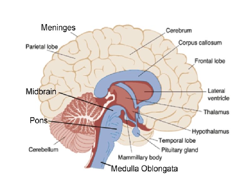
THE BRAIN • ANATOMICAL REGIONS o. Cerebrum o. Cerebellum o. Brain Stem
CEREBELLUM • Balance and coordination
CEREBRUM wrinkly large part of the brain, largest area in humans, higher mental function
Brain Stem regulates visceral functions (autonomic system)
1. Cerebral Hemispheres - left and right side separated by the. . Corpus Callosum - connects the two hemispheres
The Cerebral Hemispheres Figure 13. 7 b, c
3. Convolutions of the Brain - the wrinkles and grooves of the cerebrum Fissures = deep groove Sulcus = shallow groove Gyrus = bump
4. Fissures – separate lobes Longitudinal fissure - separate right and left sides
Lobes of the Brain (general 5. Frontal – reasoning, thinking, language 6. Parietal – touch, pain, relation of body parts (somatosensory) 7. Temporal Lobe – hearing 8. Occipital – vision functions)
LOBES OF THE BRAIN (CEREBRUM) Figure 13. 7 a Sulcus = groove Gyrus = raised bump Fissure = deep groove
9. Cerebral Cortex - thin layer of gray matter that is the outermost portion of cerebrum (the part with all the wrinkles)
Functional and Structural Areas of the Cerebral Cortex Figure 13. 11 a
11. Cerebrospinal Fluid (CSF) fluid that protects and supports brain Figure 13. 27 b
FUNCTIONAL REGIONS • A. MOTOR AREAS • B. SENSORY AREAS • C. ASSOCIATION
12. Motor Areas - controls voluntary movements - the right side of the brain generally controls the left side of the body -also has Broca's Area (speech)
13. Sensory Area - involved in feelings and sensations (visual, auditory, smell, touch, taste)
14. Association Areas - higher levels of thinking, interpreting and analyzing information
BRAIN STEM Consists of three parts: -MIDBRAIN -PONS -MEDULLA OBLONGATA
Cerebellum balance, coordination 5. Midbrain – visual reflexes, eye movements 6. Pons - relay sensory information 7. Medulla – heart, respiration, blood pressure
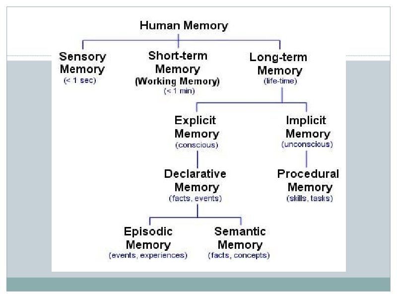
9. HIPPOCAMPUS • Memory is controlled by the HIPPOCAMPUS (“sea horse”; that’s its shape). The hippocampus plays a major role in memories.
How important are your memories? If you were involved in a traumatic event, such as a rape or a terrorist attack, would you take a pill that would make it so that you did not remember the event? http: //psychcentral. com/news/2011/ 05/27/drug-metyrapone-to-erasebad-memories/26532. html