The Limping Child The good bad and do

The Limping Child: The good, bad and do not miss KELLY CROWN, PA-C PEDIATRIC ORTHOPEDIC SURGERY
 of any commercial product(s)")
Disclosures I have no relevant financial relationships with the manufacturer(s) of any commercial product(s) and/or provider(s) of commercial services discussed in this CME activity. I do not intend to discuss an unapproved/investigative use of a commercial product/device in my presentation

Objectives 1. Learn to recognize which pediatric limps are concerning and require urgent/emergent care. Ultimately: When should you be concerned 2. Review the most common and the most concerning etiologies of pediatric limp Learn how clinical, radiographic and lab values can help you differentiate between these various etiologies 3.

What is a Limp? Asymmetric gait Results in decreased time in the Stance Phase When it’s painful = Antalgic Gait Painless: Typically Trendelenburg Gait seen

BIGGEST TAKE AWAY When a child says they have knee pain…. . ALWAYS EXAMINE THE HIPS
 Toddler (1 -3) v Transient Synovitis v Septic Arthritis")
Differential Diagnosis Child (4 -12) Toddler (1 -3) v Transient Synovitis v Septic Arthritis v Toddler’s Fracture v CP v DDH (Hip Dysplasia) v JRA (Juvenile Rheumatoid Arthritis) v Transient Synovitis v Septic Arthritis v Osteomyelitis v Legg-Calve-Perthes v Leg Length Teen (13 -18) v SCFE v Dysplasia v Tarsal Coalition v Osgood-Schlatter Disease Discrepancy v Sever’s Disease v Kohler’s Disease All ages: Tumors, Trauma

Septic Arthritis #1 thing you MUST rule this out in all limping kids — Serious bacterial infection within the joint — Delay in treatment cartilage damage, AVN, osteomyelitis — Hematogenus spread Neonates: Group B Strep, Gram Negative Bacteria — Infants: MSSA, H. Influenza — Children: MSSA, Salmonella — Adolescents: MSSA, Gonorrhea — — Can affect any body part and kids of any age but 50% are under the age of 2 — 0 -2 y/o Hips 50% — 3+ Knees 50%, Hip 20% SURGICAL

Septic Arthritis Symptoms § Refuse to bear weight or limp § Knee or hip pain § Fever § NO injury § Physical Exam Febrile or low grade fever § Toxic/sick looking § § § No improvement with NSAIDs/Motrin Challenge § SEVERE pain with passive ROM. § Positive Log Roll (hips) § Asymmetry in motion Very hesitant to let you examine them § § Neonates less likely to be toxic Hip held in flexion and ER Can have TTP, erythema and when severe, visible effusion

Kocher Criteria Used to identify septic hip arthritis vs. transient synovitis 1. Refusal to bear weight 2. Fever >38. 5°C (101. 3°F) 3. WBC >12, 000 cells/u. L 4. ESR >40 mm/h **CRP >20 mg/L or >2 mg/d. L ** (not part of original criteria, but shown to be most reliable indicator) # Criteria Met 1 2 3 4 Percentage chance of having Septic Arthritis 3% 40% 93% 99% (J. Bone Joint Surg. Am. 1999; 81: 1662 -70)

Septic Arthritis Tests § CBC, CRP, ESR § § Treatment *Kocher Criteria § § Xray: Pelvis AP/Frog View Normal doesn’t mean something isn’t wrong. § Late sign is narrowing, edema and ANV/collapse of the femoral head § Ultrasound if 2+ Kocher Criteria § MRI more sensitive but can delay treatment § Aspiration § >50, 000 cells/mm 3, >75% segmented neutrophils Positive gram stain EITHER SURGERY EMERGENT SURGERY for I&D § Sometimes, repease I&D’s are nessesary § continue to follow the CRP § If it’s increasing, take back to surgery 6 weeks IV antibiotics
 Synovitis * No. 1 Cause of limp/hip pain in children age 2")
Transient (Toxic) Synovitis * No. 1 Cause of limp/hip pain in children age 2 -8 § Self-limited inflammatory joint condition § Most commonly affects the hip § Most commonly complain of “Knee/Thigh Pain” § Males 2: 1 § Diagnosis of Exclusion § § MUST RULE OUT SEPTIC ARTHRITIS No single laboratory or imaging study will be definitive to confirm or exclude transient synovitis.
 Synovitis Symptoms Physical Exam § Limp or refuse to bear weight §")
Transient (Toxic) Synovitis Symptoms Physical Exam § Limp or refuse to bear weight § Afebrile or low grade fever § Knee or hip pain § § REMOTE history of an ILLNESS Not toxic/sick looking § Passive ROM: Asymmetry +/- guarding and pain § 1 to 5 weeks ago § Viral or bacterial § NO injury § Typically worse in the morning § AFEBRILE or low grade fever (99 -101) § Limp improves with NSAIDs § § Hips: decreased Abduction and Internal Rotation (+Impingement) § Knee: lack full flexion +/- lack terminal extension No TTP, No erythema, No visible effusion
 Synovitis Tests § CBC, CRP, *ESR § § Normal Xray: Pelvis AP/Frog")
Transient (Toxic) Synovitis Tests § CBC, CRP, *ESR § § Normal Xray: Pelvis AP/Frog View § § Treatment Normal vs slight widening Ultrasound 0. 43 0. 49 § Self limiting § Symptoms can resolve in a week or as long as 4 -5 weeks § The faster the patient is put on Motrin, TID, the faster their pain/symptoms resolve
 Trip and")
Toddler’s Fractures § § § Low energy injury § i. e. ) Trip and fall over a toy § Classic: going down a slide Age < 3 y/o § Walking toddlers: low likelihood of child abuse § If not walking, suspect abuse STABLE fractures: commonly spiral/oblique fracture of the tibia § Other common locations § 1 st Metatarsal § Tibia +/- Fibula § Femur (higher suspicion for abuse)

Toddler’s Fractures Symptoms § Limp or refuse to bear weight Physical Exam § Afebrile, Nontoxic § Crawling: tibia, fibula fx § BONY TTP and/or pain with rotation § Not walking: tibia or femur fx § § Walk on lateral aspect of foot or on their heel: Metatarsal fx Passive ROM: Asymmetry +/- guarding and pain § Injury § AFEBRILE § Pain with hip ROM: femur § Pain with knee ROM: distal femur or proximal tibia § Pain with ankle ROM: distal tibia/fibula

Toddler’s Fractures Tests § Xray: nondisplaced subtle fracture Treatment § Self limiting § Sometimes a clinical diagnosis § Might not be visible for a week § Late finding is new periosteal reaction § Limp up to 6 weeks § § § Cast vs observation Heal in 2 -4 weeks Typically no long term sequela § Proximal Tibia (Cozen Fracture) slight risk of developing Cozen Deformity (Valgus deformity of the knee 1 -2 years after)

Leg-Calve-Perthes § aka Perthes Avascular Necrosis of the Femoral Head Unknown etiology § Can cause permanent deformity of the femoral head § Earlier treatment, minimizes this risk of permanent deformity § Boys 5: 1 more likely § Age typically 4 -7 § Age <6 better prognosis § Age > 8 worse prognosis §
 § +/- Hip, thigh or Knee § NO")
Leg-Calve-Perthes Symptoms PAINLESS LIMP (first symptom) § +/- Hip, thigh or Knee § NO injury § Afebrile § Symptoms Wax and Wane § Worse during or after activities § Improve with rest and NSAIDs Physical Exam § Antalgic gait § § § Trendelenburg gait late finding once femoral head collapses Decreased hip ROM § Loss of Abduction and Internal Rotation § Hip flexion contracture Limp Length Discrepancy (late finding) § Hip contracture can exacerbate this
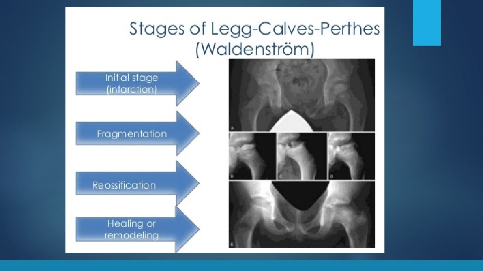

Leg-Calve-Perthes Tests § Xray Treatment **type of treatment recommended is based on patient’s age, stage of disease, and radiographic class** § Normal first 3 -6 mo § Early signs is medial joint space narrowing, sclerosis and flattening MAIN GOALS of the femoral head § Symptomatic relief: NSAIDs § MRI is the gold standard for diagnosis § § Follow with serial xrays Can take 18 mo to 2 years to fully subside Protected weight bearing through the acute phase with crutches +/- abduction brace

SCFE (Slipped Capital Femoral § Femoral head Slips off the Femoral neck § Most common adolescent hip disorder Epiphysis) § “Ice cream falling off the cone” § During rapid growth Males: 13. 4 y/o (12 -16) § Females 12. 2 y/o (10 -14) § Younger: Hypothyroid § **Obesity** § Male (2 to 1. 5 ratio) § Unilateral (40% BL) § SURGICAL EMERGENCY Jorge Muniz, PA-C, Medcomic. com
 §")
SCFE Symptoms § PAINFUL LIMP § § Acute or chronic Hip pain (L>R) § Commonly get referred thigh or knee pain § +/- minor injury § Afebrile Physical Exam Gait § Antalgic gait = Stable SCFE § NWB = Unstable SCFE § Decreased hip ROM § Loss of Internal Rotation § Obligatory ER with hip flexion § Guarding § Limp Length Discrepancy (unstable) § Short and Externally rotated §

SCFE Tests § Treatment Xray Pelvis § AP & Frog-leg Lateral § § Frog-leg best at subtle SCFE MRI if nl Xray: can dx pre-slip § § Physeal edema or widening § Percutaneous Screw Fixation § URGENT § Contralateral Pinning? Correct the deformity after it heals § Minimal displacement: hip arthroscopy with chondroplasty (FAI surgery) § Displaced: Varus Derotational Osteotomy (VDRO)

Case 1: Left leg pain x 13 days. Hasn’t walked in 12 days 7 y/o Female No PMH 2 weeks ago fell from a seated position off a rocking chair. Walked and had no pain after the fall. Next morning woke up with left knee pain and wouldn’t walk 2 days later, had a tactile fever. Taken to local ER, Temp 100. 4. Hip Xray Slight Narrowing. Positive for Strep. A and put on Amoxicillin Day 7 of her ABX develops a rash, back to urgent care. Still not walking! Temp 99. Thought to be a drug rash. Stopped the Amoxicillin and put on Ceftin. § CBC: elevated WBC (13. 6), Anemic, Hgb 10. 5, Hct 31. 1, Platelets 724 PCP instructed the family OK not to take ABX since 7 days was sufficient

Case 1 § Sees PCP the AM before coming to the ortho clinic, order labs § CBC: nl WBC (10), worsening anemia Hgb 9. 1, Hct 28, Platelets 671 § Sed Rate 95 DAY 13: Sees me § HASN’T WALKED IN 12 DAYS. § Parents still say that tactilely she is still febrile (don’t have a thermometer) § No improvement in pain with Motrin or Tylenol. § Any small movement, she cries in pain.

Case 1 § § Vitals § Temp 98. 2 F (took motrin 2 hrs prior) § Unable to get height and weight because she refuses to stand PE § Sitting in a baby stroller § Looks sick § SEVERE pain with even minor left hip ROM § Hip flexion contracture with obligatory ER § Full knee ROM without pain § TTP at Psoas

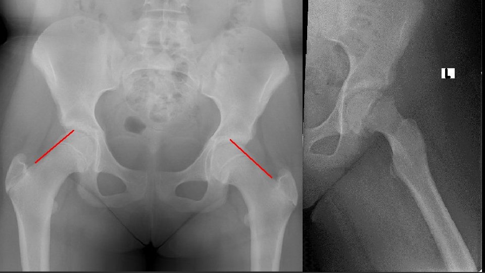
Day 4

Day 13

Case 1 § Admitted to Fairfax ED for STAT MRI with aspiration § Labs § WBC 14. 12 4. 80 - 13. 00 x 10 3/u. L § Hgb 9. 5 11. 5 - 14. 5 g/d. L § Hematocrit 29. 0 § Platelets § CRP § ESR 33. 0 - 43. 0 % 681 6. 3 99 140 - 400 x 10 3/u. L 0. 0 - 0. 8 mg/d. L 0 - 20 mm/Hr

Kocher Criteria Used to identify septic hip arthritis vs. transient synovitis 1. Refusal to bear weight 2. Fever >38. 5°C (101. 3°F) 3. WBC >12, 000 cells/u. L 4. ESR >40 mm/h **CRP >20 mg/L or >2 mg/d. L ** (not part of original criteria, but shown to be most reliable indicator) # Criteria Met 1 2 3 4 Percentage chance of having Septic Arthritis 3% 40% 93% 99% (J. Bone Joint Surg. Am. 1999; 81: 1662 -70)

Case 1 § MRI § § URGENT I&D § § Septic arthritis with Psoas Abscess Cartilage of her hip was gone, underlying osteomyelitis 6 weeks IV ABX

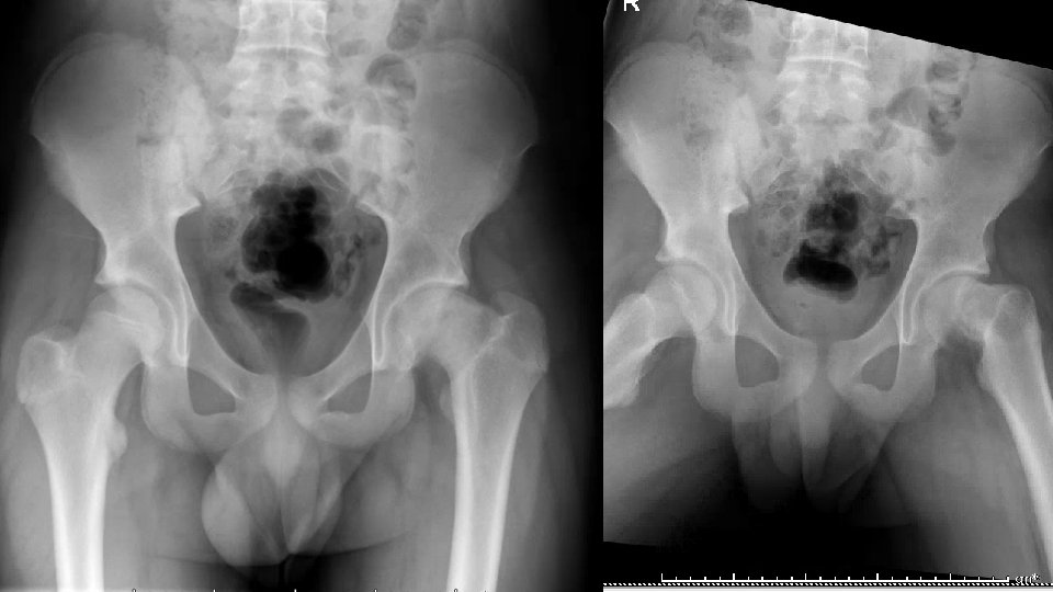
3 months post-op Still has pain and limited hip ROM

1 year post op

Case 2: Left hip pain x 2 weeks 11 y. o. female No PMH § Left hip pain x 2 weeks after a fall § jumping over hurdles at camp and landed on her LLE "awkwardly. " § Pain= sharp, non-radiating, worse with movement § Able to walk PE: § Vitals § 5’ 3” 130 lbs § Afebrile § AAO x 3, NAD § Slightly antalgic gait but FWB § TTP at the groin § Pain and Limited IR

Case 2 § URGENT Percutaneous Screw Fixation § 7 weeks post-op § Pain free § FWB § Only lacked 10 degrees IR § Full abduction § CLEARED FOR ALL ACTIVITIES

Case 3: 1 year of left knee pain 16 y/o M with no PMH here for 2 nd opinion § Left knee pain x 1 year after a twisting injury during soccer § Didn’t get treatment for 2 months § Was seen by an outside ortho group for concern for meniscus tear § MRI showed small lateral meniscus tear § Tx included rest, NSAIDs, corticosteroid injection and months of physical therapy § Physical Therapist noted that he had minimal hip ROM § Has had no hip pain x 2 months

Case 3 Vitals § 5’ 7” 187 lbs § Afebrile PE § IR of the Right was 40, Left was 0 and with mild pain § Obligate ER of the left § Full knee ROM, negative Mc. Murry § Gait: Significant Trendelenburg and External Foot progression of the left

Case 3 TREATMENT Option 1: § Percutaneous Screw Fixation § POSITVE: would stop further Slippage, minor surgery, WBAT after § NEGATIVE: wouldn’t correct the deformity, likely need a second surgery Option 2: § VDRO (Valgus Derotational Osteotomy) § POSITIVE: Would correct the deformity and regain his motion § NEGATIVE: Major surgery, would be NWB for 4 -6 week and not able to run for 12 weeks

They choose option 2 10 weeks post-op -now FWB -knee pain -recently started PT to help with his hip ROM and strength now that the osteotomy is healed

BIGGEST TAKE AWAY When a child says they have knee pain…. . ALWAYS EXAMINE THE HIPS

Thank you! Questions?
- Slides: 43