The lacrimal system Raghad Al Haj Hasan Raghad


 • Congenital NLD obstruction is the most common cause of")
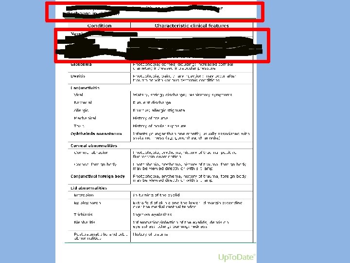
 Clinical features : • Chronic or intermittent tearing. • Debris")


 • Produced by proximal and distal obstructions of")


- Slides: 37
The lacrimal system - Raghad Al Haj Hasan - Raghad Al Qazaqi
Reflex tearing
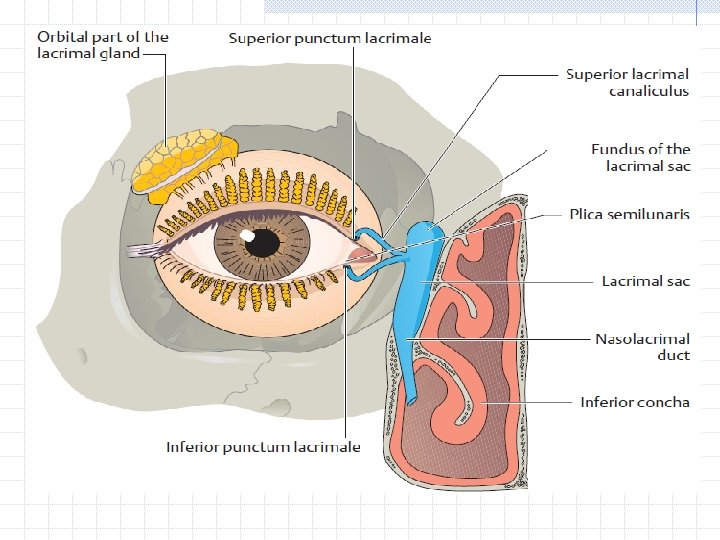
Anatomy & physiology of the lacrimal system Lacrimal gland with two components Accessory lacrimal glands Conjuctival sac Upper & lower puncti Upper & lower canaliculi Lacrimal sac NLD
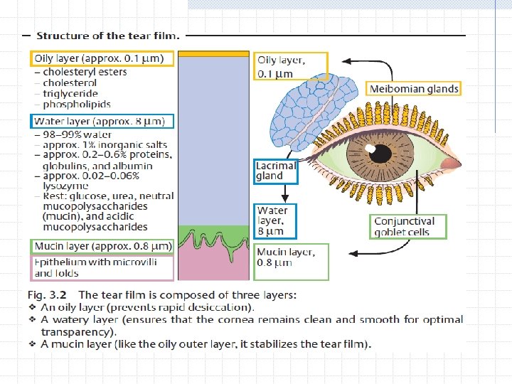
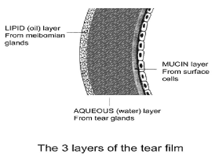
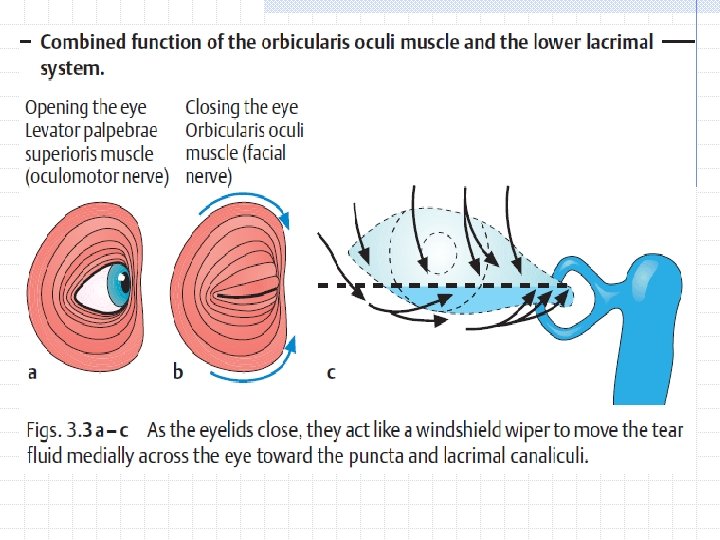
Physiology ü Tear film : * Refraction - air-tear interference for sharp & clear vision Importance of good apposition Importance of smooth ocular surfaces Importance and function of the facial pump Importance of the tear layer integrity and components Normal production rate 1. 2µl/min
Abnormalities of lacrimal system Tear composition abnormalities Drainage system abnormalities
Tear composition abnormalities Abnormal aqueous production Abnormal mucus production Abnormal meibomian secretion
Aqueous insufficiency Deficiency of lacrimal secretions occur with age and results in Kerato. Conjuctivitis Sicca (KCS ) Primary Sjogren syndrome : dry eye and mouth, is an autoimmune disease Secondary Sjogren : when associated with connective tissue disease with Rheumatoid Arthritis as the commonest.
Symptom & Signs Non-specific symptoms as FB sensation , tiredness , grittiness , burning/itching , heaviness photophobia and ocular fatigue, problem in wearing contact lenses … These symptoms are worse at the end of the day and when exposed to dry or windy atmosphere.
Signs In mild cases may show staining of the cornea with fluorescence (punctate erosion > punctate staining ) In severe cases Tags of mucus attached to the corneal surface ( filamentary keratitis ) Very severe cases may show structural corneal changes.
Filamentary keratitis
Management Tear supplementations Humidify the surrounding atmosphere Occlude the punci to preserve tears. Prognosis depend on the severity and the underlying cause. - Severe > perforation of cornea
Inadequate mucus production Destruction of goblet cells can occur in many conditions. Causes : Cicatricial conjuctival conditions Chemical burns Trachoma Vit. A deficiency (Xerophthalmia ) e. g of cicatricial conj. Disorders is Steven_Johnson syndrome.
Management Artificial Tear Vit A supplement for Xerophthalmia
Abnormal or inadequate meibomian gland secretion It will cause tear film instability Giving symptoms of dry eye Treatment is the same.
Malposition of the eyelid margin Causes : Ectropion Entropion Facial palsy Proptosis All of these will cause unstable preocular tear film > improper lid to cornea Relationship …
Management Artificial Tear & lubricants Temporary ptosis induction by Botullinum Toxin injection in the levator muscle Lateral tarsorrhaphy when permanent cause
Tear drainage disorders When tear production exceeds the drainage the tear will overflow on the cheeks Can be caused by : * Ocular surface irritation as in FB or infection causing (Lacrimation ) * Occlusion pf part of the drainage system (Epiphora)
Naso. Lacrimal Duct Obstruction Congenital Acquired
• The nasolacrimal apparatus appears in the 3 -5 week and gradually forms a cord of epithelium that extends from the eyelids to the nose. • Canalization of the cord begins at the punctum in the eyelid during the third month of intrauterine life and extends toward the nose. • Canalization is usually complete by birth. Cord canalized
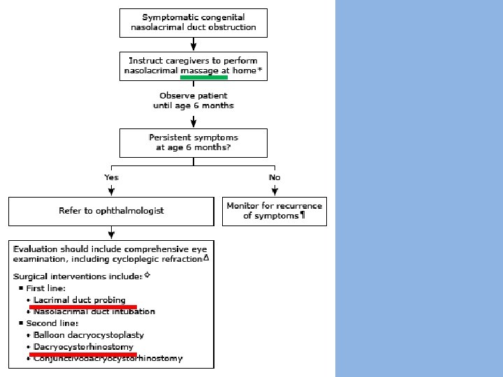
Dactryostenosis (nasolacrimal duct obstruction) • Congenital NLD obstruction is the most common cause of persistent tearing and ocular discharge in infants and young children. • Acquired caused by: Infection, Trauma , Tumor , Radiation • Most common cause of congenital obstruction is incomplete canalization at the distal end (closest to the nose). • Spontaneous resolution occurs by 6 months of age in 90% of infants. • obstructions persisting beyond the age of 12 months are unlikely to resolve spontaneously ! • Exclude other ocular causes of lacrimation as blepharitis *Suspect it’s an infantile glaucoma* When (abnormal tearing with photophobia, blepharospasm, and/or large or asymmetric corneal diameters)
Dactryostenosis (nasolacrimal duct obstruction) Clinical features : • Chronic or intermittent tearing. • Debris on the eyelashes ("mattering"). • Mild redness of the lower eyelid (irritation from overflow tearing and chronic rubbing of the eyes). • Overflow of tears when the child is in an environment that stimulates a greater production of tears (eg, wind or cold) or reduced outflow of tears (swelling of nasal mucosa during an upper respiratory infection). Physical examination : • Increase in the size of the tear meniscus. • Palpation of the lacrimal sac cause reflux of tears and/or mucoid discharge onto the eye through the puncta. Diagnosis : history and physical examination alone.
Punctal occlusion can be seen using slit lamp. Syringing and saline injection Dacrocystogram
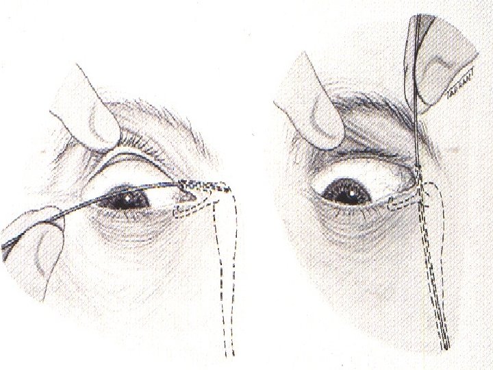
A small probe is introduced into the punctum and advanced through the lacrimal drainage system until it abuts the obstruction.
DACRYO-CYSTO-CELE (dacryoceles/ amniotoceles/ nasolacrimal duct cysts) • Produced by proximal and distal obstructions of the NLD. • Proximal obstruction is a one-way valve (permits tears to enter, but not to reflux out of the canaliculi of the lacrimal drainage system). • Noted at or shortly after birth. • Bluish swelling of the skin overlying the lacrimal sac and superior displacement of the medial canthal tendon are the typical findings • Dx : clinically (further work-up is not necessary). • Rx : Decompression of a dacryocystocele into the fornices with digital massage and/or probing of the common canaliculus. • Some ophthalmologists may observe for a short time to look for spontaneous resolution.
• Note the bluish swelling of the skin overlying the lacrimal sac and the upward displacement of the medial canthal tendon.
Acute dacryocystitis • The most common cause : Staph aureus. • Symptomes: erythema, swelling, warmth, tenderness of the lacrimal sac, and/or purulent discharge. Fever and Subtle early symptoms of infection include mild erythema over the lacrimal sac, poor feeding. . • A rare complication of isolated congenital NLD obstruction but occurs commonly with dacryocystoceles. • A medical emergency, complications (preseptal or orbital cellulitis, sepsis, meningitis, or brain abscess). • Blood cultures and cultures of material obtained during drainage guide definitive antimicrobial therapy • Empiric systemic antibiotic should be provided with suspected acute dacryocystitis pending culture results.
• Chronic dacryocystitis — bacterial overgrowth in the stagnant tear pool of the lacrimal sac. • characterized by mucopurulent drainage from the puncta without other signs of infection. • Nasal obstruction − Distension of the mucosal lining of the NLD from entrapment of tears can create mucoceles that extend into the nose and cause obstruction. • infants are obligate nose breathers, hence, nasal obstruction from mucoceles can cause respiratory distress !
Acquired NLD obstruction Causes : Infection Trauma Tumour Radiation
Thank You