The Knee Anatomy The Bones The knee is

The Knee Anatomy

The Bones

• The knee is comprised of 3 bones – Femur – Patella – Tibia • The Fibula has no bearing on the knee

The Femur

12

15 segments of the Femur • • • • Head Fovea Capitis Neck Greater Trochanter Intertrochanteric Line Lesser Trochanter Gluteal Tuberosity Linea aspera Pectineal Line Body Medial/Lateral Epicondyle Medial/Lateral condyle Patellar Grove Intercondylar Fossa

Patella – Body’s largest sesamoid bone

4

4 Segments of the Patella • • Base – superior pole Apex – inferior pole Medial Articulating Facet Lateral Articulating Facet

Tibia

6

6 segments of the Tibia • • • Medial Condyle Lateral Condyle Intercondylar Eminance Tibial Tuberosity Shaft Medial Malleolus

Muscles

Refer to Handout • The knee uses muscles from both the upper leg and lower leg – Hamstring to flex the knee – Gastrocnemius to the flex knee

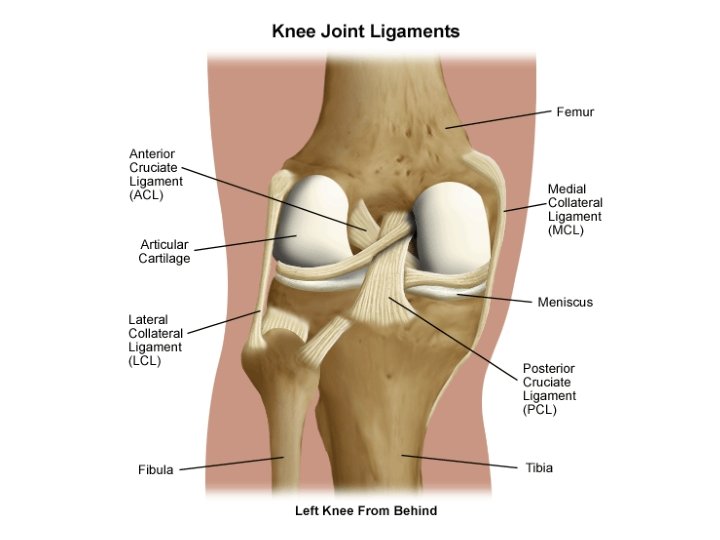
Ligaments
 – Purpose: Keep the Tibia from translating posteriorly")
• Posterior Cruciate Ligament (PCL) – Purpose: Keep the Tibia from translating posteriorly (bkwrds) • 120% to 150% wider than ACL; is Primary stabilizer of knee • Medial Collateral Ligament (MCL) – Purpose: Prevent excessive Valgus (lateral) force • Anterior Cruciate Ligament (ACL) – Purpose: Stabilizing against – • • Tibia from translating forward Internal rotation of tibia on femur External rotation of tibia on femur Hyperextension of tibiofemoral joint • Lateral Collateral Ligament (LCL) – Purpose: Prevent excessive Varus (medial) force

Origin / Insertion of Ligs • PCL – O: 2 bands; Posterior aspect of tibia I: Medial Femoral condyle • MCL – O: Medial condyle of Femur I: Neck of the Tibia • ACL – O: Anteriormedial Tibial condyle I: Lateral Femoral condyle • LCL – O: Lateral condyle of Femur I: Head of Fibula

Anterior View

Origin /Insertion: • Quadriceps Tendon - O: Quadriceps becomes tendon I: Superior aspect of Patella • Patellar Tendon - O: Anterior Inferior Patella I: Tibial Tuberosity.

Patella and Patellar Lig

Mensicii

Meniscii Purpose: Act as “shock absorbers” for the knee, and provide structural integrity. • Purpose: Limit extremes of flexion and extension in knee • Medial Meniscus – more of a crescent shape, wider posteriorly than anteriorly • Lateral Meniscus – more of a “c” shape
")
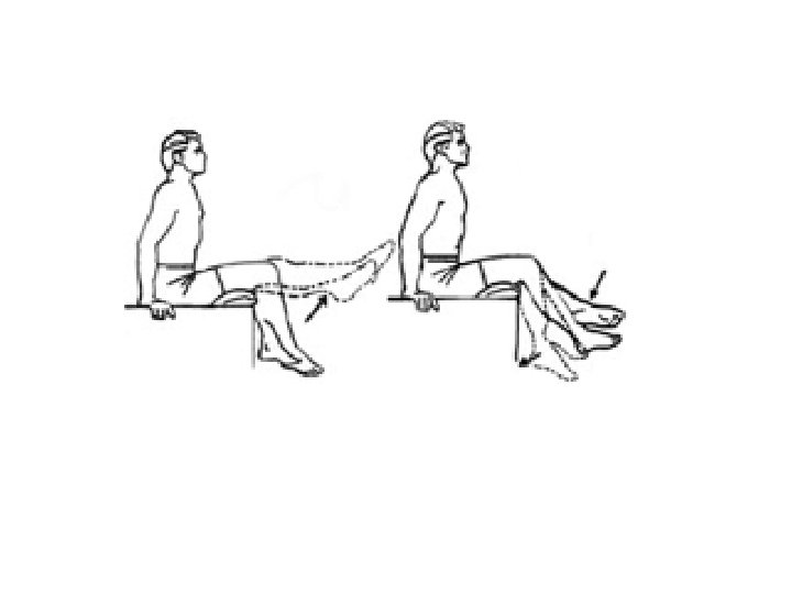
ROM • Flexion & Extension • Internal & External Rotation (from the hip)

Knee Joint

The End…
- Slides: 27