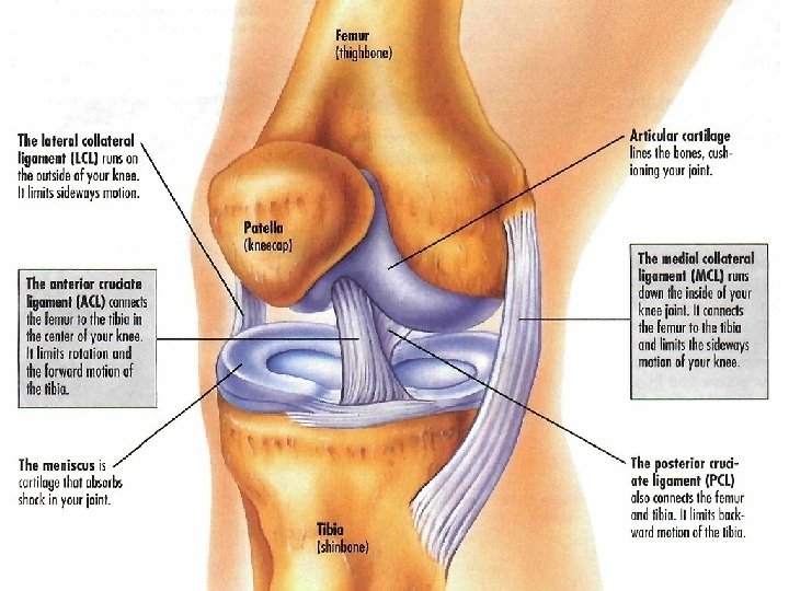
The Knee Anatomy Femur Tibia Patella Fibula Knee

The Knee - Anatomy • Femur • Tibia • Patella • Fibula

Knee Factoids - The knee joint consists of two articulations: one between the femur and tibia (tibiofemoral joint), and one between the femur and patella (patellofemoral joint). - The knee is a modified hinge joint, which permits flexion and extension as well as slight internal and external rotation. - At birth, the patella is just formed from cartilage, and this will ossify (change to bone) between ages 3 -5.
 • Dislocation • Anterior Cruciate")
Common knee ailments • Fracture (Patella is most common) • Dislocation • Anterior Cruciate Ligament (ACL) Injuries • Posterior Cruciate Ligament Injuries • Collateral Ligament Injuries • Meniscal Tears • Tendon Tears

ACL Injuri es

Osgood Schlatter Development of an avulsion fracture at the tibial tubercle.

Osgood Schlatter: Etiology Forceful contraction of quadriceps femoris tendon onto immature tibial tubercle due to sports, pronation, etc. Children in rapid growth period are predisposed.

Osgood Schlatter: S & S’s 1. 2. 3. 4. Pain, tenderness, swelling at tibial tuberosity Swelling may or may not be present Climbing stairs, running, kneeling Possibly enlarged tibial tuberosity

Osgood Schlatter: Treatment Physiotherapy modalities: Adjust: Tibia Patella Femur

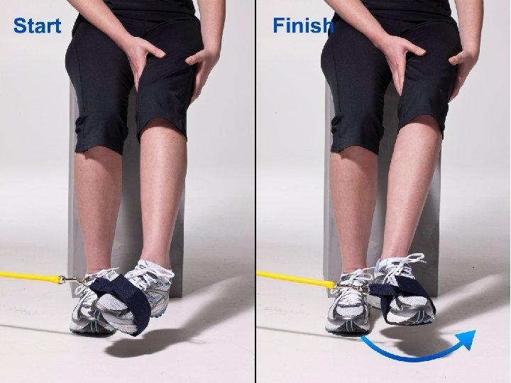
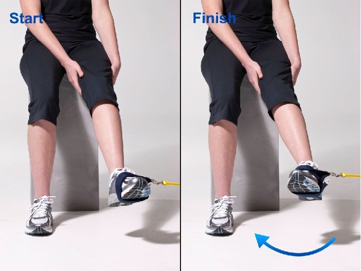
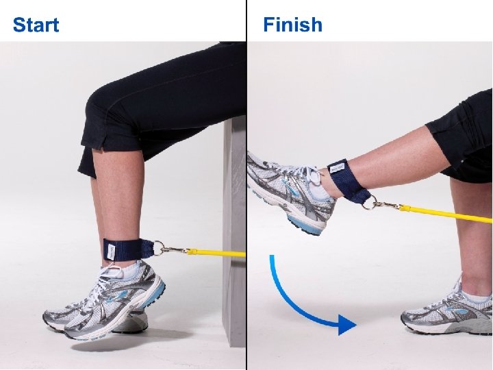
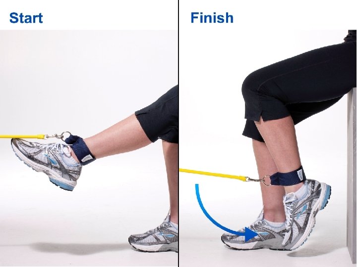
Osgood Schlatter: Treatment Rehabiltiation: Knee Series using the Elastic bands
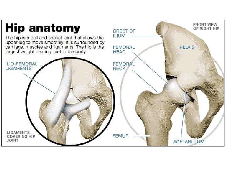

Hip Joint • Very stable anatomy. Deep socket, thick joint capsule, strong spiral ligaments, several powerful stabilizing muscle groups • Joint is rarely injured. Most injuries are secondary to biomechanical dysfunctions.

Hip joint Imbalance Syndromes • Iliotibial band syndrome • Tensor fascia lata strain • Trochanteric bursitis • Hip joint capsulitis • Hip flexor muscle strain • Hip adductor muscle strain • Anterior pelvic tilt
 Syndrome")
Iliotibial Band (ITB) Syndrome

Trochanteric Bursitis

Hip Flexor Muscle Strain
- Slides: 20