The Integumentary System Skin and its appendages hair
 By Dr.")


 -")


- Slides: 18
The Integumentary System Skin, and its appendages (hair and various skin-related glands) By Dr. Esam Omar
The Integumentary System • The Integumentary System includes the skin and its appendages (hair and various skin-related glands) • Skin is the protective covering of the whole body that forms about 2. 3 m 2 and 16 % of body weight (so skin is the largest and heaviest organ in the body).
Functions of Skin and subcutaneous layer • Protection of underlying tissues and organs against shocks, abrasion, and chemical attack; • Excretion of salts, water, and organic wastes by integumentary glands; • Maintenance of normal body temperature though either insulation or evaporative cooling, as need; • Synthesis of vitamin D 3 • Storage of nutrients: lipids are stored in adipocytes in the dermis and in adipose tissue in the subcutaneous layer; • Detection of touch, pressure, pain, and temperature stimuli and relay of that information to the nervous system.
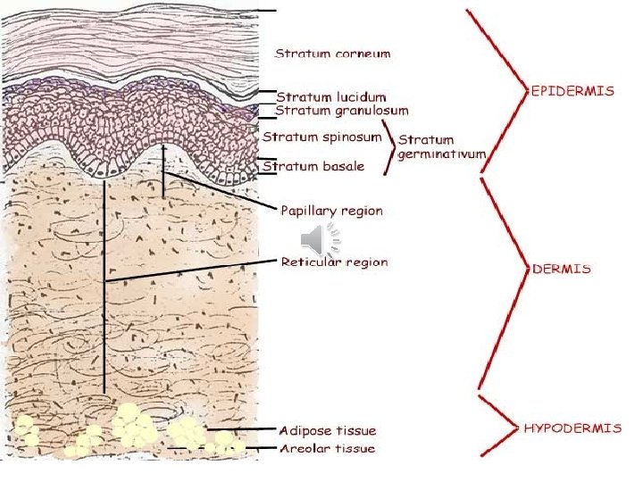
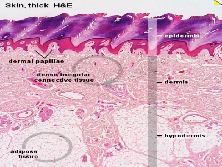
Histological Structure of Skin • Skin: • Epidermis: keratinized stratified squamous epithelium forms an outer waterproof shield. It is avascular and receives its nutrition by diffusion from the vascular superficial papillary layer of dermis. - Dermis: a tough, leathery layer of dense fibroelastic connective tissue. It is vascular. - Epidermis is fixed to dermis by hemidesmosomes, basement membrane and dermal papillae. • Subcutaneous layer (hypodermis): primarily adipose tissue (a thermoinsulator and a mechanical shock absorber). Often not considered a part of the integument.
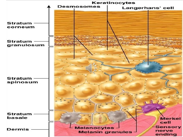
Epidremal cells Keratinocyte s Forms 85% of epidermis Non keratinocytes • Melanocytes • Langerhans’ cells • Merkels’ cells 5
Keratinocytes: produce keratin (a tough, fibrous, waterproof protein but gives skin its resiliency and strength). • Keratinization: the normal formation of keratin by the stratified squamous epithelia of skin. As the epithelial cells mature, they fill with granules filled with keratin and at the same time give up vital organelles; ultimately become lifeless sheets of keratin. In human, an entirely new epidermis forms every 7 -8 weeks.
Epidermal Cell Layers • Stratum basale – – A single layer of columnar cells The cells in this layer considered to be stem cells L/M: basal oval nuclei with basophilic cytoplasm Desmosomes are found between cells • Stratum spinosum (Prickle cell layer) – 4 to 8 layers of basophilic polygonal cells (the cells are angular) with central nuclei – The cells are rich in keratin filaments – Desmosomes are found
Epidermal Cell Layers • Stratum granulosum - 2 -3 layers of flat cells rich in basophilic granules (keratohyaline granules, lamellar granules, phospholipid granules and mucopolysaccharide granules). - These granules form a cement intercellular space that forms skin barrier. - Moreover, lamellar granules produce a lipid-rich impermeable layer around cells for prevention of water loss. • Stratum lucidum - Formed of acidophilic flat non-nucleated cells rich in immature keratin filaments. - A translucent layer present only in thick skin. • Stratum corneum - Formed of acidophilic horny scales rich in mature keratin filaments.
Non-keratinocytes • They include: Merkel’s cells, Langerhans cells and melanocytes.
Merkel’s cells • Present in between the cells of the stratum basale • The nuclei are indented and the cytoplasm contains small dense granules • Free nerve endings form expanded disk at the base of cells called Merkel’s disc • They are mechanoreceptors
Langerhans cells - They are originating from the bone marrow (mesodermal in origin) - Mainly located in between the cells of the stratum spinosum - LM: star-shaped cells with dense nucleus, pale cytoplasm and slender processes - EM: the cytoplasm contains Golgi body, many lysosomes and vermiform granules (membranebound granules have racquet-like appearance with unknown function) - No desmosomes, no intermediate filaments - They are antigen-presenting cells
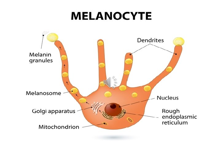
Melanocytes • They are originating from the neural crest • LM: they have rounded cell bodies, rounded nucleus with prominent nucleolus and long processes that extend between the surrounding cells • EM: the cytoplasm contains Golgi, r. ER, mitochondria, intermediate filaments and melanosome granules • The cells contain tyrosinase enz that converts tyrosine into dopa then into dopaquinone which converted into melanin. Melanosomes then move into the tips of melanocytes processes to be injected into the keratinocytes
Thick skin Thin skin Palms and soles Whole body except palms and soles Thicker Thinner + - Thinner Thicker More in number, higher and narrower Fewer, lower and broader Hair and hair follicles - + Sebaceous glands - + Eccrine weat glands + + Apocrine sweat glands - + Sites Epidrmis Stratum lucidum Dermis Dermal papillae
Thin skin