The Integumentary System An Introduction to the Integumentary


 and cell types (on right)")


 n n Epidermal Growth Factor (EGF) ¨ Powerful peptide growth")


 thick skin and (b) thin skin (which one makes")

 = below the skin “Subcutaneous”")


 n In the")


 glands n n n Entire body except palms and soles Produce sebum")
 glands n n n Entire skin surface except nipples and part of")

![n Hydration ¨ Results from immersion in hypotonic solution (e. g. , freshwater [osmosis])](https://slidetodoc.com/presentation_image/eda1e8a2dcb212479296a1857e37f5b2/image-38.jpg "n Hydration ¨ Results from immersion in hypotonic solution (e. g. , freshwater [osmosis])")

 Second-degree (epidermis and dermis, with blistering) Third-degree (full thickness,")


- Slides: 44
The Integumentary System
An Introduction to the Integumentary System n The Integument ¨ Is the largest system of the body n 16% of body weight n 1. 5 to 2 m 2 in area n The integument is made up of two parts 1. Cutaneous membrane (skin) 2. Accessory structures (hair, nails, exocrone glands)
The Integumentary System Integument is skin n Skin and its appendages make up the integumentary system n Two distinct regions of skin n ¨ Epidermis ¨ Dermis ¨ Hypodermis/Subcutaneous Layer
How does this System help other systems? ¨ Cardiovascular n Blood vessels in the dermis ¨ Nervous n system Sensory receptors for pain, touch, and temperature
Functions of skin n Protection ¨ Cushions and insulates and is waterproof ¨ Protects from chemicals, heat, cold, bacteria ¨ Screens UV by producing Melanin n n n Synthesizes vitamin D with UV Regulates body heat Prevents unnecessary water loss Sensory reception (nerve endings) Excretion of salts, water and chemicals Storage of lipids
Epidermis n n Keratinized stratified squamous epithelium Four types of cells Keratinocytes – deepest, produce keratin (tough fibrous protein) ¨ Melanocytes - make dark skin pigment melanin ¨ Merkel cells – associated with sensory nerve endings ¨ Langerhans cells – macrophage-like dendritic cells ¨ n Layers (from deep to superficial) ¨ ¨ ¨ Stratum basale or germinativum – single row of cells attached to dermis; youngest cells; forms fingerprints Stratum spinosum – spinyness is artifactual; tonofilaments (bundles of protein) resist tension Stratum granulosum – layers of flattened keratinocytes producing keratin (hair and nails made of it also) Stratum lucidum (only on palms and soles) Stratum corneum – horny layer (cells dead, many layers thick). Thickest on palms and soles of feet. (see figure on next slide)
Epithelium: layers (on left) and cell types (on right)
5 -1 Epidermis n Stratum germinativum or stratum basale ¨ Forms a strong bond between epidermis and dermis ¨ Forms epidermal ridges (e. g. , fingerprints) ¨ Has many basal cells or germinative cells Thick skin Epidermal ridge
n Specialized Cells of Stratum Basale ¨ Merkel cells n Found in hairless skin n Respond to touch (trigger nervous system) ¨ Melanocytes n Contain the pigment melanin n Scattered throughout stratum basale
n Stratum Spinosum — the “spiny layer” ¨ Produced by division of stratum basale ¨ Cells shrink until cytoskeletons stick out (spiny) ¨ Continue to divide, increasing thickness of epithelium ¨ Contain dendritic (Langerhans) cells, active in immune response
n Stratum Granulosum — the “grainy layer” ¨ Stops n n dividing, starts producing Keratin ¨ A tough, fibrous protein ¨ Makes up hair and nails Keratohyalin ¨ Dense granules ¨ Cross-link keratin fibers
n Stratum Lucidum — the “clear layer” n Found only in thick skin n Covers stratum granulosum
n Stratum Corneum — the “horn layer” n Exposed surface of skin n 15 to 30 layers of keratinized cells n Water resistant n Shed and replaced every 2 weeks
n Keratinization ¨ The formation of a layer of dead, protective cells filled with keratin ¨ Occurs on all exposed skin surfaces except eyes ¨ Skin ¨ It life cycle takes 15– 30 days for a cell to move from stratum basale to stratum corneum
Epidermal Growth Factor (EGF) n n Epidermal Growth Factor (EGF) ¨ Powerful peptide growth factor ¨ Produced by glands (salivary and duodenum) ¨ Used in laboratories to grow skin grafts Functions of EGF ¨ Promotes division of germinative cells ¨ Accelerates keratin production ¨ Stimulates epidermal repair ¨ Stimulates glandular secretion
Remember… n Four basic types of tissue ¨Epithelium – epidermis just discussed ¨Connective tissue - dermis ¨Muscle tissue ¨Nervous tissue
Dermis n n n Strong, flexible connective tissue: your “hide” Cells: fibroblasts, macrophages, mast cells, WBCs Fiber types: collagen, elastic, reticular Rich supply of nerves and vessels Critical role in temperature regulation (the vessels) Two layers (see next slides) ¨ Papillary – areolar connective tissue; includes dermal papillae ¨ Reticular – “reticulum” (network) of collagen and reticular fibers
*Dermis layers *Dermal papillae * *
Epidermis and dermis of (a) thick skin and (b) thin skin (which one makes the difference? )
Fingerprints, palmprints, footprints n n Dermal papillae lie atop dermal ridges Elevate the overlying epidermis into epidermal ridges Are “sweat films” because of sweat pores Genetically determined Flexion creases n Deep dermis, from continual folding Fibers n n Collagen: strength and resilience Elastic fibers: stretch-recoil ¨ n Striae: stretch marks Tension lines (or lines of cleavage) ¨ The direction the bundles of fibers are directed The dermis is the receptive site for the pigment of tattoos
Hypodermis or Subcutaneous Layer n n n “Hypodermis” (Gk) = below the skin “Subcutaneous” (Latin) = below the skin Also called “superficial fascia” “fascia” (Latin) =band; in anatomy: sheet of connective tissue Fatty tissue which stores fat and anchors skin (areolar tissue and adipose cells) Different patterns of accumulation (male/female)
Skin color n n Three skin pigments ¨ Melanin: the most important ¨ Carotene: from carrots and yellow veggies ¨ Hemoglobin: the pink of light skin Melanin in granules passes from melanocytes (same number in all races) to keratinocytes in stratum basale ¨ Digested by lysosomes ¨ Variations in color ¨ Protection from UV light vs vitamin D?
Figure 5 -5 Melanocytes in stratum basale Melanin pigment Basement membrane Melanocytes LM 600
Figure 5 -5 Melanocytes Melanosome Keratinocyte Melanin pigment Melanocyte Basement membrane
n Capillaries and Skin Color ¨ Oxygenated red blood contributes to skin color n Blood vessels dilate from heat, skin reddens n Blood flow decreases, skin pales ¨ Cyanosis n Bluish skin tint n Caused by severe reduction in blood flow or oxygenation
n Illness and Skin Color ¨ Jaundice n n ¨ Pituitary tumor n ¨ Excess MSH Addison’s disease n n ¨ Buildup of bile produced by liver Yellow color A disease of the pituitary gland Skin darkening Vitiligo n n Loss of melanocytes Loss of color
n Vitamin D 3 ¨ Epidermal cells produce (vitamin D 3) n In the presence of UV radiation ¨ Liver and kidneys convert vitamin D 3 into calcitriol n Aids absorption of calcium and phosphorus ¨ Insufficient n vitamin D 3 Can cause rickets
n Function of Melanocytes ¨ Melanin protects skin from sun damage ¨ Ultraviolet n (UV) radiation Causes DNA mutations and burns that lead to cancer and wrinkles ¨ Skin color depends on melanin production, not number of melanocytes
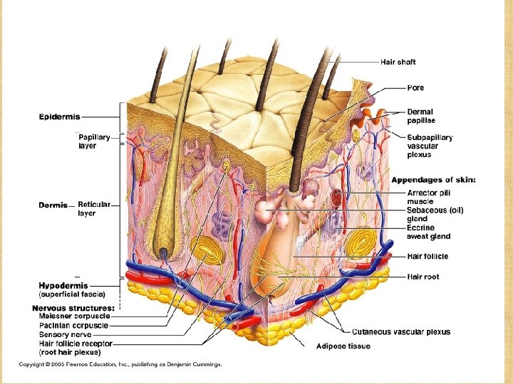
Accessory Structures or Skin appendages Derived from epidermis but extend into dermis n Include n ¨ Hair and hair follicles ¨ Sebaceous (oil) glands ¨ Sweat (sudoiferous) glands ¨ Nails
Hair and hair follicles: complex Derived from epidermis and dermis Everywhere but palms, soles, nipples, parts of genitalia * *“arrector pili” is smooth muscle Hair bulb: epithelial cells surrounding papilla Hair papilla is connective tissue________
n Types of hair ¨ Vellus: fine, short hairs ¨ Intermediate hairs ¨ Terminal: longer, courser n Hair growth: averages 2 mm/week ¨ Active: growing ¨ Resting phase then n hair shed Hair loss ¨ Thinning – age related ¨ Male pattern baldness n Hair color ¨ Amount of melanin for black or brown; distinct form of melanin for red ¨ White: decreased melanin and air bubbles in the medulla ¨ Genetically determined though influenced by hormones and environment
n Functions of hair ¨ Warmth – less in man than other mammals ¨ Sense light touch of the skin ¨ Protection - scalp n Parts ¨ Root imbedded in skin ¨ Shaft projecting above skin surface Make up of hair – hard keratin n Three concentric layers n ¨ Medulla (core) ¨ Cortex (surrounds medulla) ¨ Cuticle (single layers, overlapping)
Nails Of hard keratin n Corresponds to hooves and claws n Grows from nail matrix n
Sebaceous (oil) glands n n n Entire body except palms and soles Produce sebum by holocrine secretion Oils and lubricates
Sweat (sudoriferous) glands n n n Entire skin surface except nipples and part of external genitalia Prevent overheating 500 cc to 12 l/day! (is mostly water) Humans most efficient (only mammals have) Produced in response to stress as well as heat
Types of sweat glands n Eccrine or merocrine ¨ Most numerous ¨ True sweat: 99% water, some salts, traces of waste ¨ Open through pores n Apocrine ¨ Axillary, anal and genital areas only ¨ Ducts open into hair follices ¨ The organic molecules in it decompose with time - odor n Modified apocrine glands ¨ Ceruminous – secrete earwax ¨ Mammary – secrete milk
n Hydration ¨ Results from immersion in hypotonic solution (e. g. , freshwater [osmosis]) ¨ Causes swelling of epithelial cells, evident on the palms and soles
Disorders of the integumentary system n Burns ¨ Threat to life n Catastrophic loss of body fluids n Dehydration and fatal circulatory shock n Infection ¨ Types n First degree – epidermis: redness (e. g. sunburn) n Second degree – epidermis and upper dermis: blister n Third degree - full thickness Infections n Skin cancer n
Burns First-degree (epidermis only; redness) Second-degree (epidermis and dermis, with blistering) Third-degree (full thickness, destroying epidermis, often part of hypodermis)
Critical burns n n n Over 10% of the body has thirddegree burns 25 % of the body has seconddegree burns Third-degree burns on face, hands, or feet Estimate by “rule of 9’s”
Tumors of the skin Benign, e. g. warts n Cancer – associated with UV exposure (also skin aging) n ¨ Aktinic keratosis - premalignant ¨ Basal cell - cells of stratum basale ¨ Squamous cell - keratinocytes ¨ Melanoma – melanocytes: most dangerous; recognition: A - Asymmetry n B - Border irregularity n C - Colors n D - Diameter larger than 6 mm n
Skin Cancer Sqaumous cell carcinoma Basal cell carcinoma Melanoma
Figure 5 -6 Skin Cancers Basal cell carcinoma Melanoma