The influence of breathing on Cerebrospinal Fluid Flow

The influence of breathing on Cerebrospinal Fluid Flow Vegard Vinje Postdoctoral Fellow Simula Research Laboratory 31. August 2019
 surrounds the brain and the spinal cord")
The cerebrospinal fluid (CSF) surrounds the brain and the spinal cord

CSF flows from the choroid plexus to the arachnoid granulations

CSF flows from the choroid plexus to the arachnoid granulations, but not exclusively

CSF may enter paravascular spaces

CSF may enter paravascular spaces, and water may be filtrated over the capillary wall

In addition, cardiac and respiratory effects add pulsatility to CSF flow

CSF flow through the brain has been proposed as a mechanism for waste removal Rasmussen et al. 2018

Glymphatic flow has been shown to increase during sleep Rasmussen et al. 2018

Measuring CSF/ISF flow with MRI is a difficult task

Measuring CSF/ISF flow with MRI is a difficult task

Figure: Cardiac gated PCMRI of two different flow fields, one consisting of only the cardiac (green) and the other of cardiac and respiratory components (black). Yildiz et al. 2017

Real Time MRI shows great promise to identify both cardiac and respiratory components CSF flow in the aqueduct in two different patients. From Dreha. Kulaczewski et al. 2015 CSF flow in the foramen magnum during a breathing protocol. From Yildiz et al. 2017.

Respiration may affect CSF flow despite inducing a smaller pressure gradient than the cardiac component

In 9 i. NPH patients, pressure gradients were computed from simultaneous ICP measurements

The Fourier transform of the pressure difference reveal cardiac and respiratory components

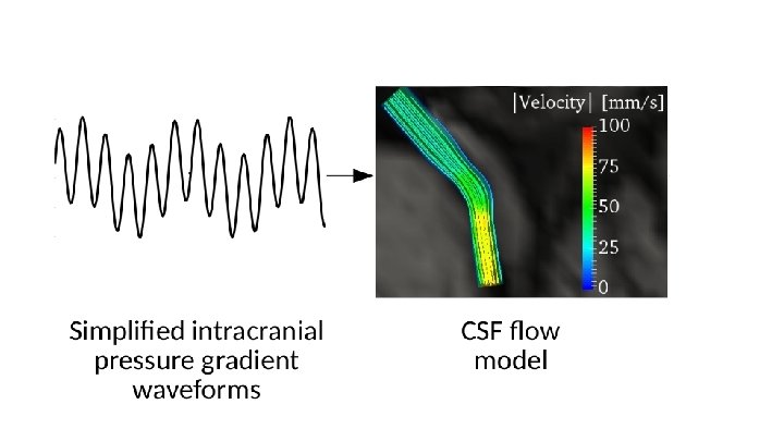
We extracted respiratory and cardiac components from the Fourier transform to construct a simplified pressure gradient

We extracted respiratory and cardiac components from the Fourier transform to construct a simplified pressure gradient d. ICP(t) = a 0 sin(2�tf 0) + a 1 sin(2�tf 1)

Simplified pressure curve shows a fairly good fit with raw data


Pressure gradients did not change consistently in the sleeping state
 = Δp(t) v=0 p(t) = 0")
p(t) = Δp(t) v=0 p(t) = 0

Respiration may affect CSF flow despite inducing a smaller pressure gradient than the cardiac component Figure: Example of a patient’s simplified pressure gradient and the corresponding computed flow rate

Respiration may affect CSF flow despite inducing a smaller pressure gradient than the cardiac component Cohort Average Cardiac: 1. 46 mm. Hg/m Respiratory: 0. 52 mm. Hg/m Cardiac: 0. 31 m. L/sec Respiratory: 0. 35 m. L/sec Figure: Example of a patient’s simplified pressure gradient and the corresponding computed flow rate

Respiration may affect CSF flow despite inducing a smaller pressure gradient than the cardiac component Cohort Average Cardiac: 1. 46 mm. Hg/m Respiratory: 0. 52 mm. Hg/m Static: ~0. 005 mm. Hg/m Cardiac: 0. 31 m. L/sec Respiratory: 0. 35 m. L/sec Figure: Example of a patient’s simplified pressure gradient and the corresponding computed flow rate Static: ~0. 006 m. L/sec

The long respiratory wave carry a greater volume than the shorter cardiac wave

Respiratory rates were found at 15 breaths per minute, however breath rates can be consciously controlled

In ancient eastern cultures, breathing control in yoga has been used for health benefits When you practice one breath a minute, then you become Pavan Guru — you become the light and knowledge of the prana, and then you know the Universe, the Universe knows you. -Yogi Bhajan 7/26/96

To conclude. . .

Pressure gradients in the brain are dominated by cardiac pulsations while CSF flow volumes are dominated by respiration
- Slides: 33