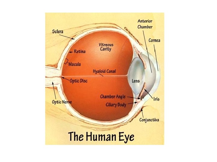
The Human Eye Anatomy and Function Vitreous Humor


 and away (artery) from")

- Slides: 17
The Human Eye Anatomy and Function
Vitreous Humor • A jelly-like substance that composes much of the interior of the eyeball • Makes up about 80% of the eyeball in the human eye
Sclera • Tough layer around the outside of the eyeball • Commonly referred to as the “white of the eye” • Maintains the shape of the eyeball
Conjunctiva • Thin, clear, moist membrane that coats the inner surfaces of the eyelids and the outer surface of the eye
Cornea • Strong, clear bump at the front of the eye • Refracts light that enters the eye through the pupil onto the lens (which focuses the light onto the retina)
Pupil • Hole through which light enters the eye • Formed by the iris • Size increases or decreases as size of iris increases or decreases
Iris • The colored part of the eye – Blue, green, brown, hazel, or grey • Regulates the size of the pupil • Regulates the amount of light let into the eye
Lens • Focuses light onto the retina
Ciliary Body • Ring shaped muscle attached to the iris • Controls the shape of the lens by contracting and relaxing
Choroid • Layer of the eyeball located between the retina and the sclera • Absorbs unused radiation
Retina • Located at the back of the human eye • The “screen” on which an image is formed • Collects information contained in that image and transmit it to the brain • Contains Rods and Cones – Rods—sees black and white – Cones—sees color
Fovea • Shallow pit in the retina at the back of each eye • Contains a large number of cones • Responsible for acute vision
Optic Disk • Location on the retina at which the optic nerve leaves the eye
Optic Nerve • Route by which information is sent from the eye for processing by the brain • Leaves the posterior surface of each eye • Each optic nerve contains appx. One million fibers carrying info. From the rods and cones • Goes to the occipital lobe of the brain
Central Retinal Artery and Vein • Carries blood to (vein) and away (artery) from the eyeball
How the eye works: