The Human Brain Master Watermark Image http williamcalvin

The Human Brain Master Watermark Image: http: //williamcalvin. com/Brain. For. All. Seasons/img/bonobo. LH-human. LH-via. TWD. gif

Cerebrum -The largest division of the brain. It is divided into two hemispheres, each of which is divided into four lobes. Cerebrum Cerebellum http: //williamcalvin. com/Brain. For. All. Seasons/img/bonobo. LH-human. LH-via. TWD. gif

Cerebral Cortex - The outermost layer of gray matter making up the superficial aspect of the cerebrum. Cerebral Cortex http: //www. bioon. com/book/biology/whole/image/1/1 -6. tif. jpg

Cerebral Features: • Gyri – Elevated ridges “winding” around the brain. • Sulci – Small grooves dividing the gyri – Central Sulcus – Divides the Frontal Lobe from the Parietal Lobe • Fissures – Deep grooves, generally dividing large regions/lobes of the brain – Longitudinal Fissure – Divides the two Cerebral Hemispheres – Transverse Fissure – Separates the Cerebrum from the Cerebellum – Sylvian/Lateral Fissure – Divides the Temporal Lobe from the Frontal and Parietal Lobes
 Sulci (groove) Fissure (deep groove) http: //williamcalvin. com/Brain. For. All. Seasons/img/bonobo. LH-human.")
Gyri (ridge) Sulci (groove) Fissure (deep groove) http: //williamcalvin. com/Brain. For. All. Seasons/img/bonobo. LH-human. LH-via. TWD. gif

Specific Sulci/Fissures: Central Sulcus Longitudinal Fissure Sylvian/Lateral Fissure Transverse Fissure http: //www. bioon. com/book/biology/whole/image/1/1 -8. tif. jpg http: //www. dalbsoutss. eq. edu. au/Sheepbrains_Me/human_brain. gif
 • • Frontal Parietal Occipital Temporal http: //www. bioon.")
Lobes of the Brain (4) • • Frontal Parietal Occipital Temporal http: //www. bioon. com/book/biology/whole/image/1/1 -8. tif. jpg * Note: Occasionally, the Insula is considered the fifth lobe. It is located deep to the Temporal Lobe.

Lobes of the Brain - Frontal • The Frontal Lobe of the brain is located deep to the Frontal Bone of the skull. • It plays an integral role in the following functions/actions: - Memory Formation - Emotions - Decision Making/Reasoning - Personality Modified from: http: //www. bioon. com/book/biology/whole/image/1/1 -8. tif. jpg

Lobes of the Brain - Parietal Lobe • The Parietal Lobe of the brain is located deep to the Parietal Bone of the skull. • It plays a major role in the following functions/actions: - Senses and integrates sensation(s) - Spatial awareness and perception (Proprioception - Awareness of body/ body parts in space and in relation to each other) Modified from: http: //www. bioon. com/book/biology/whole/image/1/1 -8. tif. jpg

Lobes of the Brain – Occipital Lobe • The Occipital Lobe of the Brain is located deep to the Occipital Bone of the Skull. • Its primary function is the processing, integration, interpretation, etc. of VISION and visual stimuli. Modified from: http: //www. bioon. com/book/biology/whole/image/1/1 -8. tif. jpg

Primary Visual Cortex Visual Association Area Modified from: http: //www. bioon. com/book/biology/whole/image/1/1 -8. tif. jpg Regions

Lobes of the Brain – Temporal Lobe • The Temporal Lobes are located on the sides of the brain, deep to the Temporal Bones of the skull. • They play an integral role in the following functions: - Hearing - Organization/Comprehension of language - Information Retrieval (Memory and Memory Formation) Modified from: http: //www. bioon. com/book/biology/whole/image/1/1 -8. tif. jpg

Brain Stem The brain stem connects the brain to the spinal cord. With in the brain stem is the midbrain, the pons and the medulla oblongata. The midbrain relays visual and auditory information. The pons relays information between the cerebellum and the cereberal hemispheres. The medulla oblongata is a relay center AND control for heart rate, respiratory rate and maintaining homeostasis.
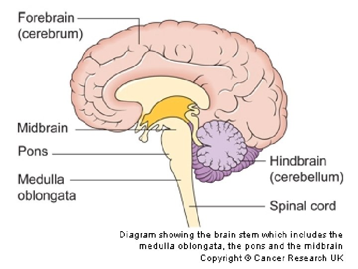

Thalamus and Hypothalamus The thalamus and hypothalamus are the main relay centers. They get the information from the spinal cord and direct the information to the correct lobe of the brain. They also help maintain homeostasis and govern emotion and memory.

Cerebellum The cerebellum lies at the back of the brain under the cerebral hemispheres. The cerebellum helps control muscle action, coordination, body position and balance.

Copyright: Gary Larson Q: Assuming this comical situation was factually accurate, what Cortical Region of the brain would these doctors be stimulating?

B. Lobes and Structures of the Brain A. G. F. C. E. D. http: //williamcalvin. com/Brain. For. All. Seasons/img/bonobo. LH-human. LH-via. TWD. gif

Lobes and Structures of the Brain A. Central Sulcus B. Frontal Lobe C. Sylvian/Lateral Fissure D. Temporal Lobe A. (groove) G. B. F. E. Transverse Fissure F. Occipital Lobe G. Parietal Lobe C. (groove) D. E. (groove) http: //williamcalvin. com/Brain. For. All. Seasons/img/bonobo. LH-human. LH-via. TWD. gif

Further Investigation Phineas Gage: Phineas Gage was a railroad worker in the 19 th century living in Cavendish, Vermont. One of his jobs was to set off explosive charges in large rock in order to break them into smaller pieces. On one of these instances, the detonation occurred prior to his expectations, resulting in a 42 inch long, 1. 2 inch wide, metal rod to be blown right up through his skull and out the top. The rod entered his skull below his left cheek bone and exited after passing through the anterior frontal lobe of his brain. Frontal
,")
Remarkably, Gage never lost consciousness, or quickly regained it (there is still some debate), suffered little to no pain, and was awake and alert when he reached a doctor approximately 45 minutes later. He had a normal pulse and normal vision, and following a short period of rest, returned to work several days later. However, he was not unaffected by this accident. http: //www. sruweb. com/~walsh/gage 5. jpg Learn more about Phineas Gage: http: //en. wikipedia. org/wiki/Phineas_Gage Frontal

Resources Images: • • • http: //www. dalbsoutss. eq. edu. au/Sheepbrains_Me/human_brain. gif http: //www. bioon. com/book/biology/whole/image/1/1 -8. tif. jpg http: //www. bioon. com/book/biology/whole/image/1/1 -6. tif. jpg http: //williamcalvin. com/Brain. For. All. Seasons/img/bonobo. LH-human. LH-via. TWD. gif http: //www. math. tu-dresden. de/~belov/brain/motorcor 2. gif Larson, Gary. The Far Side. Phineas Gage: • • http: //www. sruweb. com/~walsh/gage 5. jpg http: //soma. npa. uiuc. edu/courses/bio 303/Image 7. jpg http: //en. wikipedia. org/wiki/Phineas_Gage http: //scienceeducation. nih. gov/nih. HTML/ose/snapshots/multimedia/ritn/Gage/Broken_brain 1. html
- Slides: 22