The hierarchy of life At the most basic

The hierarchy of life • At the most basic level, life is made of atoms molecules macromolecules organelles cells may be unicellular (e. g. bacteria) or multicellular (plants, animals, etc. ) • Multicellular life has higher levels of organization above cells • For humans cells make up tissues organ systems organisms (full human)

Levels of Structural Organization Smooth muscle cell Molecules Cellular level Cells are made up of molecules Atoms Chemical level Atoms combine to form molecules Smooth muscle tissue Tissue level Tissues consist of similar types of cells Epithelial tissue Smooth muscle Blood tissue vessel (organ) Connective tissue Organ level Organs are made up of different types of tissues Cardio- Organismal level vascular Human organisms system are made up of many organ systems Organ system level Organ systems consist of different organs that work together closely Figure 1. 1

The two forms of cells Eukaryotic means Before nucleus: These cells do NOT have a nucleus Or organelles Eukaryotic means true nucleus: These cells will have a nucleus and organelles like ER, GA, Mitochondria etc

Prokaryote Vs. Eukaryote • • • No nucleus DNA in a nucleoid Cytosol No organelles other than ribosomes Small size Primitive i. e. Bacteria & Archaea • Has nucleus and nuclear envelope • Cytosol • Membrane-bound organelles with specialized structure/function • Much larger in size • More complex • i. e. plant/animal cell
 which is responsible for production of proteins")
The Nucleus Contains the genetic material (DNA) which is responsible for production of proteins in the cells Nuclear envelop: – Consists of a double membrane that bounds the nucleus – Contains nuclear pores that allow for exchange of material with the rest of the cell – Encloses the jellylike fluid called the nucleoplasm – Nucleoplasm is rich with proteins, RNA and genetic material
 Nucleoli: One or more small dark staining bodies; sites of")
The Nucleus (cont. ) Nucleoli: One or more small dark staining bodies; sites of ribosome synthesis & assembly Ribosomes migrate into the cytoplasm through nuclear pores to serve as the site of protein synthesis Chromatin: Chromatin is the combination of DNA + proteins in resting cells. As cells prepare for dividing, chromatin threads become condensed forming dense rod-like segmented threads (chromosomes).

Plasma membrane • Border between cell environment and outside • Fluid mosaic model: A tail-to-tail phospholipid bilayer with cholesterol (in animal cells) + proteins + glycoproteins placed in various configurations ( = mosaic). • Tail-to-tail = polar heads (hydrophilic) facing water, nonpolar tails (hydrophobic) away from water.
 free to move in a")
Plasma membrane • Phospholipids align together (no chemical linking) free to move in a lateral fashion (fluid). • Glycoproteins in plasma membrane function as structural proteins, enzymes, transport proteins, ion channels, receptors, etc. • Sugars and glycoproteins are sticky give plasma membrane a fuzzy appearance and used for cell to cell recognition.

Role of Cholesterol in the Plasma membrane • • • Cholesterol works a buffer for membrane fluidity. It allows our cell membrane to remain stable and at normal level of fluidity At High Temperature phospholipids will move faster and the membrane will be More fluid, in the case Cholesterol will reduce membrane fluidity to bring it back to normal levels At low Temperature phospholipids will move slower and the membrane will be Less fluid, in the case Cholesterol will increase membrane fluidity to bring it back to normal levels

Cytoplasm • Cellular constituents located between nuclear envelop and plasma membrane. • Most of the cell is basically cytoplasm manufacturing area of the cell (where cells produce and metabolize substances and molecules) • The cytoplasm consists of: – Cytosol: (water + dissolved nutrients and other elements), – Cellular organelles: mitochondria, endoplasmic reticulum, Golgi apparatus, lysosomes, peroxisomes, …etc. – Cytoplasmic inclusions: stored fat in fat cells (adipocytes), glycogen in liver cells (hepatocytes), pigments in skin cells (keratinocytes), et.

Mitochondria • Mitochondrial wall is a double membrane: equal to two plasma membranes, placed side by side. • Double membrane: outer and inner membrane • Cristae: folds of inner membrane; contains enzymes for ATP production; increased surface area to ATP made • Matrix: fluid-filled inner compartment Contains enzymes dissolved in the fluid inside the mitochondria and anchored to inner membrane • Has own genetic material that is maternally inherited • Major function: “Powerhouse of the cell”: Produces ATP by cellular respiration.
;")
Chloroplasts • • Function: site of photosynthesis Double membrane Thylakoid disks in stacks (grana); stroma (fluid) Contains chlorophylls (pigments) for capturing sunlight energy
 • Made of proteins")
Ribosomes • Small bi-lobed structures (small subunit + large subunit) • Made of proteins and r. RNA • Sites for protein synthesis (m. RNA translation) • Free floating or attached to outer surface of ER (rough endoplasmic reticulum) 1. 2. Free ribosomes: float in cytosol, produce proteins used within cell Bound ribosomes: attached to ER, make proteins for export from cell
 • Accounts for ½ of cell’s membrane system • Rough (RER)")
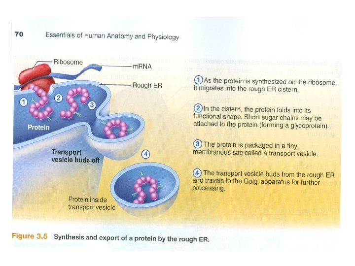
Endoplasmic reticulum (ER) • Accounts for ½ of cell’s membrane system • Rough (RER) & Smooth (SER) types • ER switches between RER and SER • RER: ribosomes attached to ER surface; participate in proteins synthesis synthesized protein buds off RER in transport vesicles attaches to receiving phase of GA( Golgi apparatus) • RER is the cell membrane factory. • Abundant in cells making and exporting proteins and digestive enzymes (e. g. pancreatic cells releasing insulin)
 • SER( smooth ): Plays no role in protein synthesis •")
Endoplasmic reticulum (ER) • SER( smooth ): Plays no role in protein synthesis • Participates in lipid metabolism (cholesterol and fat synthesis and breakdown) • Detoxification of drugs and pesticides (liver cells are very rich in SER) • Body cells producing steroid hormones such as male testes producing testosterone

Golgi Apparatus • Stack of flattened membranous sacs with many vesicles fusing in and budding off. synthesis & packaging of materials (small molecules) for transport (in vesicles); produce lysosomes Series of flattened membrane sacs (cisternae) Cis face: receives vesicles Trans face: ships vesicles • Modifies and package incoming proteins for final shipping to target location (folding, glycosylation, etc) • Vesicles from GA move in 1 (one) of 3 pathways – Pathway 1: trafficking of mature proteins to final destination (cytoplasm, cell membrane, secretory proteins) – Pathway 2: Empty vesicles bud out of GA fuse with pm enable cells to increase in size – Pathway 3: Vesicles containing digestive enzymes become lysosomes, fuse with ingested substances to degrade them (e. g. pathogens)

Figure 3. 6 Role of the Golgi apparatus in packaging the products of the rough ER. Rough ER Cisterns Proteins in cisterns Membrane Transport vesicle Lysosome fuses with ingested substances. Golgi vesicle containing digestive enzymes becomes a lysosome. Pathway 3 Golgi apparatus Pathway 2 Pathway 1 Golgi vesicle containing proteins to be secreted becomes a secretory vesicle. Secretory vesicles Proteins Secretion by exocytosis Golgi vesicle containing membrane components fuses with the plasma membrane and is incorporated into it. Plasma membrane Extracellular fluid Interaction of ribosomes, ER, and GA in protein synthesis, packaging and export

Lysosomes • Membranous bags filled with digestive enzymes • Degrade damaged or no-longer-needed molecules for recycling purposes (e. g. protein amino acids exit for reuse in protein synthesis • Abundant in certain types of cells that are know for their ability to degrade macromolecules (pathogen-degrade phagocytes, macrophages, etc. )
 • Membrane-bound vesicles")
Vacuoles • Function: storage of materials (food, water, minerals, pigments, poisons) • Membrane-bound vesicles • Eg. food vacuoles, contractile vacuoles • Plants: large central vacuole -- stores water, ions
 that use molecular oxygen (O 2)")
Peroxisomes • Membranous bags of oxidative enzymes (oxidases) that use molecular oxygen (O 2) to detoxify poisons, alcohol, & other harmful substances) • Free radicals are normal by-products of cellular metabolism; should not accumulate as they are very reactive chemicals that can damage proteins and DNA • Peroxisomes convert free radicals into H 2 O 2; excess H 2 O 2 is converted into H 2 O by the catalase enzyme • Numerous in liver and kidney cells (major organs of drug and poison detoxification) • Peroxisomes do not arise from GA( golgi apparatus). They replicate and arise from old ones by pinching (budding).

Cytoskeleton • A network of fibers & filaments that form internal frameworks within cells • Give cells shape, support organelles & furnishes the machinery to transport substances inside cells and for cells to move (e. g. cilia, flagella, etc. ) • 3 different types: – Microfilaments: mostly involved in cell mobility and in changing shape of cell (e. g. ACTIN & MYOSIN) – Intermediate filaments: form desmosomes, act like internal anchors that resist pulling forces on the cell. – Microtubules: give cell its overall shape, help in distributing & locking organelles in place inside cells. Very important during cell division
")
Figure 3. 7 Cytoskeletal elements support the cell and help to generate movement. (a) Microfilaments (b) Intermediate filaments (c) Microtubules Tubulin subunits Fibrous subunits Actin subunit 7 nm Microfilaments form the blue batlike network. 10 nm Intermediate filaments form the purple network surrounding the pink nucleus. 25 nm Microtubules appear as gold networks surrounding the cells’ pink nuclei.
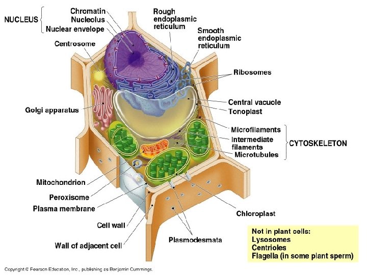

Parts of plant & animal cell p 108 -109

Endosymbiont theory • Mitochondria & chloroplasts share similar origin • Prokaryotic cells engulfed by ancestors of eukaryotic cells • Evidence: – Double-membrane structure – Have own ribosomes & DNA – Reproduce independently within cell
- Slides: 26