The Heart Path of Blood through heart Heart

The Heart Path of Blood through heart Heart structures and internal/external anatomy
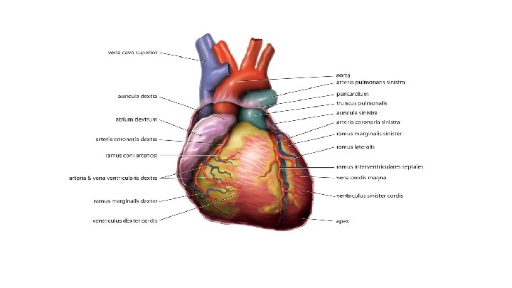

Heart Parts Heart is located just left of sternum and size of fist. 4 chambered ● ● ● Myocardium - heart muscle Pericardium - sac surrounding the heart Cardiac muscle - contracts by itself Atria - thin walled and pump blood into ventricles Ventricles - thick muscle wall, pump blood out of heart Valves - ensure 1 direction flow of blood
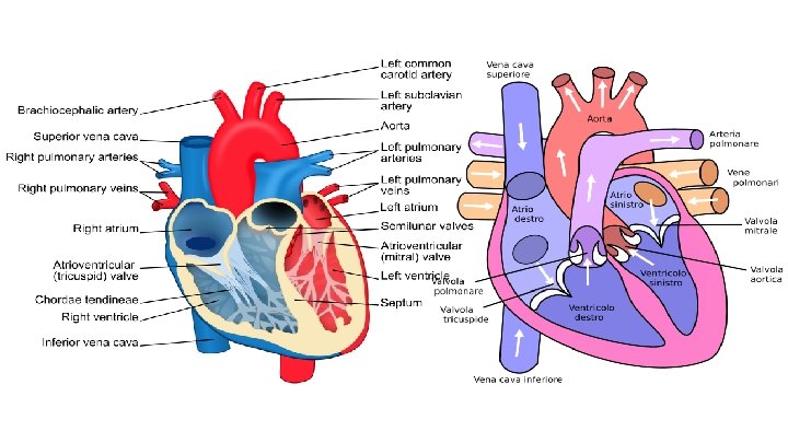

Heart to Lungs 1. Blood enters the heart via the inferior and superior vena cava (veins) 2. Blood is oxygen-poor and pressure is low. It enters the RIGHT side of the heart into the right atrium 3. The right atrium contracts, and blood passes through the tricuspid valve and into the right VENTRICLE 4. The heart contracts again, pushing blood through the semilunar valve (pulmonic valve) towards the lungs. The blood leaves via the left/right pulmonary arteries

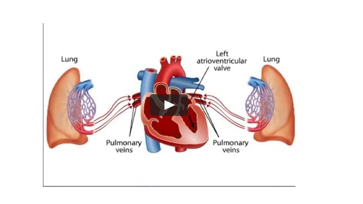
Lungs to heart 1. Blood leaving the lungs is now oxygen-rich and reenters the heart via the pulmonary veins, passing into the LEFT atrium. 2. The left atrium contracts, and blood pushes past the mitral valve into the LEFT ventricle. 3. The heart contraction pushes blood through the other semilunar valve (aortic valve) into the aorta, which connects to arteries that will lead to other parts of the body.

Why does the heart have Valves? To prevent backflow of blood into previous chambers.

What causes the LUB DUB sound? Valves open and close at the same time. The tricuspid and mitral valves will close together, and semilunar valves close together. This prevents any back flow. There also chordae tendinae that help pull the valves open and prevent them from folding improperly. LUB - tricuspid and mitral valves will close DUB - semilunar valves close together. - The DUB is louder and stronger because the ventricles have to push harder to increase blood pressure and push blood to rest of body

How does the heart receive the signal to beat? 1. Atria signalled to contract via SA Node (Sino Atrial Node = PACEMAKER) - located in Right Atrium - causes atria to contract 2. Electrical Signal transmitted through Perkinji fibers to the AV node (Atrio Ventricular node) 3. AV node receiving the signal causes Ventricles to contract

How does the heart receive nutrients? The heart does not get nutrients/oxygen from the blood that enters it. A coronary artery, and other arteries help supply the heart with blood.

ECG - Electrocardiograph
 QRS complex")
P wave → SA node triggers atria (they are about to contract) QRS complex - ventricles about to contract T wave - ventricular muscles recover
 - WHY? - Often a drug overdose")
ECG abnormalities Irregular heart beat (ventricular fibrillation) - WHY? - Often a drug overdose Most common cause of death of healthy individuals Can shock heart into a normal rhythm (defibrillation)

Heart Murmur - When valves don’t shut properly Pressure is backed-up and blood travels backwards in heart Leads to - - Decreased blood pressure (affects all organs/tissues) Swelling in tissues Fluid build up on lungs May be born with it (congenital) or caused by illness Many heart murmurs that are congenital correct themselves (baby’s 1 st cry closes many open circuits/clears airway/starts organ function)

Blood Pressure Systolic blood pressure - Pressure exerted when ventricle contract and send blood OUT of heart Highest ~ 120 mm Hg (average) if you are healthier/active/younger this may be lower Diastolic blood pressure - Pressure exerted on blood by artery when heart relaxing During filling of ventricles ~ 80 mm Hg average. This can be slightly lower.
 ● ● ● Normal less than")
High and low blood pressure Hypertension (high BP) ● ● ● Normal less than 120 /80 mm Hg (Systolic/diastolic) Prehypertension (120 -129 /80 -90 mm Hg (Systolic/diastolic) High BP anything over 130/90 mm Hg Hypotension (low BP) ● Less than 90/60 mm Hg CAUSES High blood volume CAUSES Constricted blood vessels Low blood volume - Blood loss Increased cardiac output (eg. exercise, drugs) Wide blood vessels - Lots of solute in blood so water increases - Plaque in arteries, hardening of arteries Reduced cardiac output
 ● ● Broken blood vessels Damaged")
Problems associated with. . . Hypertension (high BP) ● ● Broken blood vessels Damaged tissue Heart attack Stroke Hypotension (low BP) ● ● Kidney malfunction Tissue starvation Death of tissue Dizziness
- Slides: 18