The Heart Overview of the Heart double pump

The Heart

Overview of the Heart • double pump • about the size of a fist - cone shaped • weight less than 1 lb.
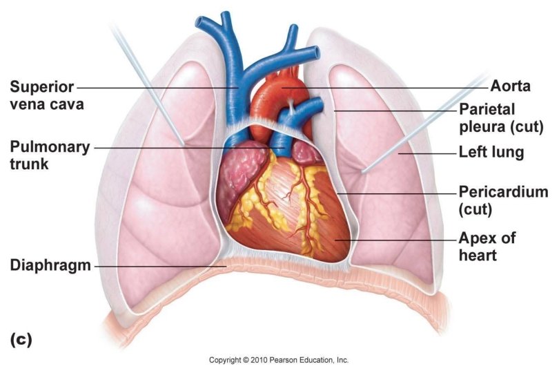

• located mid-thoracic cavity between the lungs • lies at an angle under the sternum • angles to the left

• covered with double walled membrane pericardium § outer layer is tough § inner is a serous § potential space between pericardial cavity with serous fluid to reduce friction

3 Layers of Heart Wall 1. pericardium – § outer 2. myocardium – § cardiac muscle § bulk of the heart § contracts 3. endocardium – § lines chambers


Heart Chambers

Heart Chambers 2 1 4 3 • 4 chambers • as in all birds and mammals

R and L Atria • receive blood • small and thin walled Left Atrium Right Atrium

R and L Ventricles • RV pumps blood to the lungs • LV pumps blood to the body • large and thick walled because has hardest job to do


• septum § divides the heart into R and L sides

Great Vessels

Great Vessels • superior vena cava § blood from superior body to heart • inferior vena cava § blood from below heart RA

• pulmonary veins § 4 total § from lungs LA

• pulmonary trunk § to lungs RV

• aorta § largest artery § to all organs LV

Heart Valves

Atrioventricular Valves = AV valves • between atrium and ventricle • right is tricuspid • left is the bicuspid or mitral
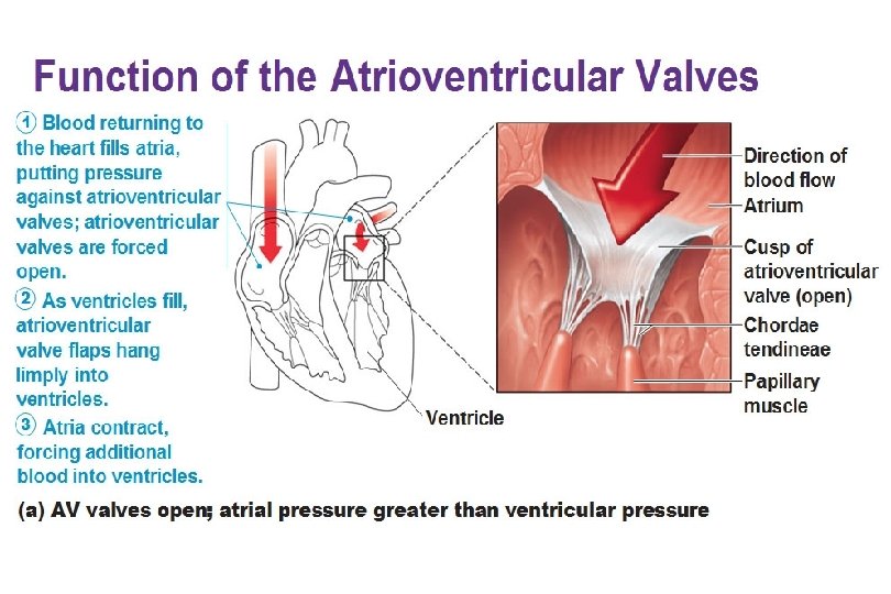
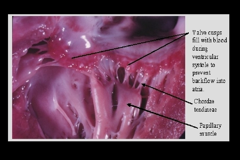

• papillary muscles attached to chordae tendineae • contraction pulls valves open • when V begins to contract, blood is forced against valve and it shuts

Semilunar Valves • Aortic Semilunar Valve § between ventricle and aorta • Pulmonary Semilunar Valve § between ventricle and pulmonary trunk
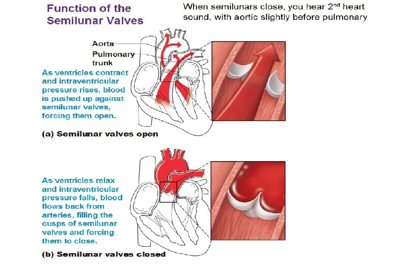

Semilunar Valves • force of blood pushes them open • attempted backflow after contraction closes them

Left Atrium Right Atrium Left Ventricle Right Ventricle

Superior Vena Cava Inferior Vena Cava

Pulmonary Trunk R Pulmonary Artery L Pulmonary Artery

Right Pulmonary Veins Left Pulmonary Veins

Aorta

Superior Vena Cava Aorta Pulmonary Trunk L Pulmonary Artery R Pulmonary Artery Left Pulmonary Veins Right Pulmonary Veins Inferior Vena Cava Aorta

Pulmonary Semilunar Valve Tricuspid Valve Aortic Semilunar Valve Bicuspid Valve

Septum Chordae Tendineae Apex

Circulation

Pulmonary Circulation • right side of heart • from body to heart to lungs for oxygen

Pulmonary Circulation From body ↓ Superior and inferior vena cava ↓ Right atrium ↓ Tricuspid valve ↓ Right ventricle ↓ Pulmonary Semilunar valve ↓ Pulmonary trunk ↓ R and L pulmonary arteries ↓ Capillary beds of lungs (gas exchange) ↓ Pulmonary veins

System Circulation • left side of heart • supplies body with oxygenated blood

Systemic Circulation From lungs ↓ Pulmonary Veins ↓ Left atrium ↓ Bicuspid valve ↓ Left ventricle ↓ Aortic Semilunar valve ↓ Aorta ↓ Arteries ↓ Capillary beds of body (gas exchange) ↓ Superior and Inferior Vena Cava

Coronary Arteries

Coronary Arteries • supply the heart muscle with oxygen rich blood

Atherosclerosis • these are clogged with plaque • contract in spasms called angina or get clots stuck in them • one cause of heart attack

• cardiac muscles that die are not replaced dead cells = infarct • myocardial infarction or MI is proper term for a heart attack

• coronary bypass surgery is most common surgery in USA

Conduction System

Conduction System • coordinated activity depends on gap junctions and “in house” conduction system
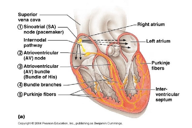

Steps: 1. sinoatrial node in RA near superior vena cava acts as “pace maker” by initiating each wave of nerve activity 2. wave moves to atrioventricular node in septum between atria
 in upper ventricular region of septum")
3. to the AV bundle (bundle of His) in upper ventricular region of septum 4. to the Pyrkinje fibers which branch down septum to ventricles
 of ventricles to push blood up and out")
5. contraction starts a bottom (apex) of ventricles to push blood up and out arteries

Fibrillation • uncoordinated contractions • A fibs - rapid flutters and weakness

• V fibs - fatal unless heart is defibrillated shocked back into normal

Electrocardiogram • ECG • measures electrical events of the heart

Heart Excitation Related to ECG

Electrocardiography

Cardiac Cycle

Cardiac Cycle • all the events associated with the flow of blood through the heart in 1 beat • Diastole - period of cardiac ventricle filling • Systole period of cardiac ventricle pumping (contracts)

Heartsounds • LUB-dup § closing of valves § LUB - AV valves closing, longer and louder § dup - Semilunar valves closing, shorter and sharper

Murmurs • abnormal sounds are called murmurs and are usually caused by leaky of narrowed valves
- Slides: 59