
The Heart Dr Isazadehfar Location of the Heart
- Slides: 28
The Heart Dr. Isazadehfar
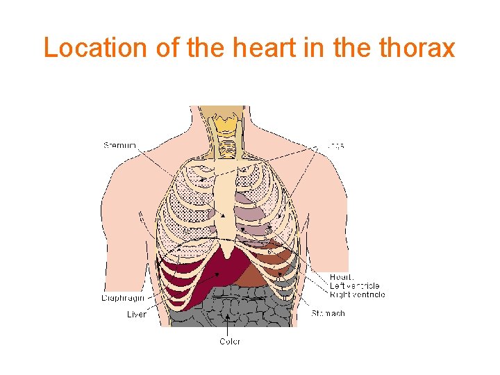
Location of the Heart • The heart is located between the lungs behind the sternum and above the diaphragm. • It is surrounded by the pericardium. • Its size is about that of a fist, and its weight is about 250 -300 g. • Its center is located about 1. 5 cm to the left of the midsagittal plane.
Location of the heart in the thorax
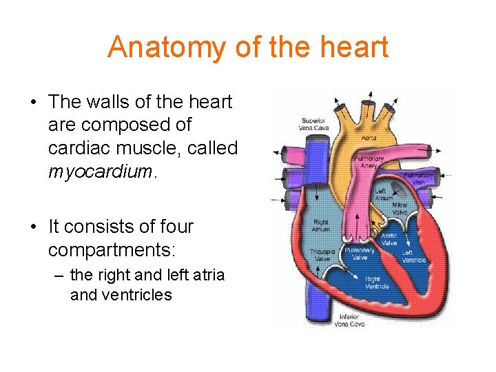
Anatomy of the heart • The walls of the heart are composed of cardiac muscle, called myocardium. • It consists of four compartments: – the right and left atria and ventricles
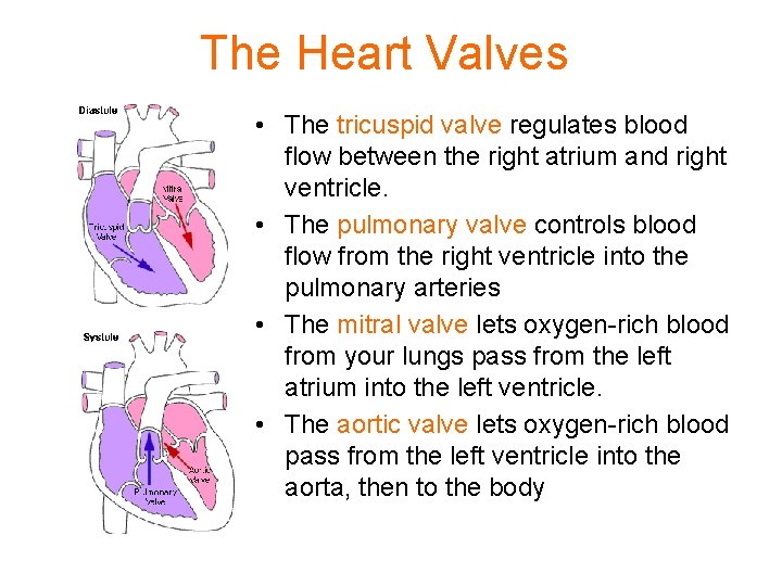
The Heart Valves • The tricuspid valve regulates blood flow between the right atrium and right ventricle. • The pulmonary valve controls blood flow from the right ventricle into the pulmonary arteries • The mitral valve lets oxygen-rich blood from your lungs pass from the left atrium into the left ventricle. • The aortic valve lets oxygen-rich blood pass from the left ventricle into the aorta, then to the body
Blood circulation via heart • The blood returns from the systemic circulation to the right atrium and from there goes through the tricuspid valve to the right ventricle. • It is ejected from the right ventricle through the pulmonary valve to the lungs. • Oxygenated blood returns from the lungs to the left atrium, and from there through the mitral valve to the left ventricle. • Finally blood is pumped through the aortic valve to the aorta and the systemic circulation. .
The Heartbeat • A heartbeat is a two-part pumping action that takes about a second. As blood collects in the upper chambers, the heart's natural pacemaker (the SA node) sends out an electrical signal that causes the atria to contract. This contraction pushes blood through the tricuspid and mitral valves into the resting lower chambers. This part of the two-part pumping phase (the longer of the two) is called the diastole.
The Heartbeat • The second part of the pumping phase begins when the ventricles are full of blood. The electrical signals from the SA node travel along a pathway of cells to the ventricles, causing them to contract. This is called systole. • As the tricuspid and mitral valves shut tight to prevent a back flow of blood, the pulmonary and aortic valves are pushed open. While blood is pushed from the right ventricle into the lungs to pick up oxygen, oxygen-rich blood flows from the left ventricle to the heart and other parts of the body.
The Heartbeat • After blood moves into the pulmonary artery and the aorta, the ventricles relax, and the pulmonary and aortic valves close. • The lower pressure in the ventricles causes the tricuspid and mitral valves to open, and the cycle begins again. • This series of contractions is repeated over and over again, increasing during times of exertion and decreasing while you are at rest.
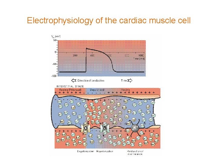
Electrical activation of the heart • In the heart muscle cell, or myocyte, electric activation takes place by means of the same mechanism as in the nerve cell, i. e. , from the inflow of Na ions across the cell membrane. • The amplitude of the action potential is also similar, being 100 m. V for both nerve and muscle.
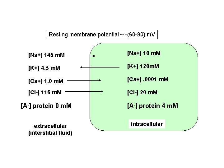
Resting membrane potential ~ -(60 -80) m. V [Na+] 145 m. M [Na+] 10 m. M [K+] 4. 5 m. M [K+] 120 m. M [Ca+] 1. 0 m. M [Ca+]. 0001 m. M [Cl-] 116 m. M [Cl-] 20 m. M [A-] protein 0 m. M extracellular (interstitial fluid) [A-] protein 4 m. M intracellular
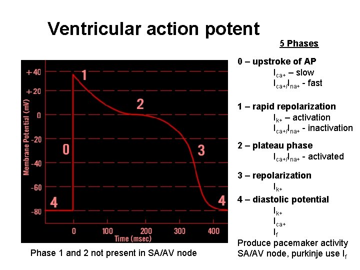
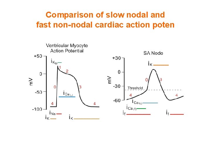
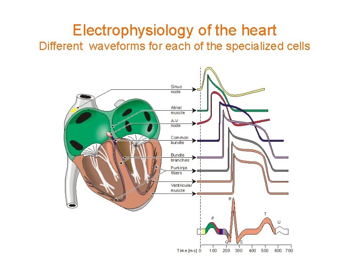
Ventricular action potent 5 Phases 0 – upstroke of AP Ica+ – slow Ica+/Ina+ - fast 1 – rapid repolarization Ik+ – activation Ica+/Ina+ - inactivation 2 – plateau phase Ica+/Ina+ - activated Phase 1 and 2 not present in SA/AV node 3 – repolarization Ik+ 4 – diastolic potential Ik+ Ica+ If Produce pacemaker activity SA/AV node, purkinje use If
Comparison of slow nodal and fast non-nodal cardiac action poten
Electrophysiology of the cardiac muscle cell
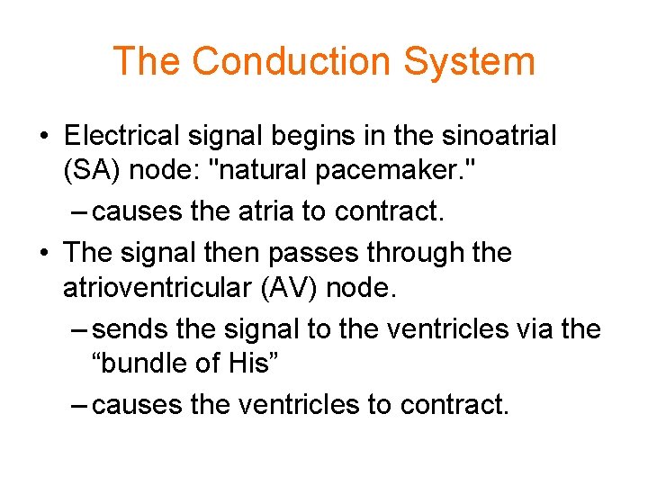
The Conduction System • Electrical signal begins in the sinoatrial (SA) node: "natural pacemaker. " – causes the atria to contract. • The signal then passes through the atrioventricular (AV) node. – sends the signal to the ventricles via the “bundle of His” – causes the ventricles to contract.
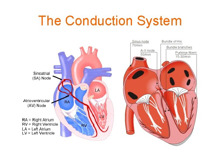
The Conduction System
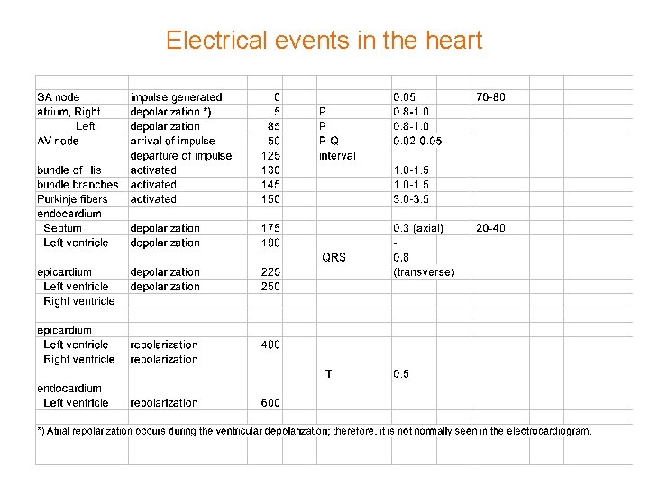
Conduction on the Heart • The sinoatrial node in humans is in the shape of a crescent and is about 15 mm long and 5 mm wide. • The SA nodal cells are self-excitatory, pacemaker cells. • They generate an action potential at the rate of about 70 per minute. • From the sinus node, activation propagates throughout the atria, but cannot propagate directly across the boundary between atria and ventricles. • The atrioventricular node (AV node) is located at the boundary between the atria and ventricles; it has an intrinsic frequency of about 50 pulses/min. • If the AV node is triggered with a higher pulse frequency, it follows this higher frequency. In a normal heart, the AV node provides the only conducting path from the atria to the ventricles.
• Propagation from the AV node to the ventricles is provided by a specialized conduction system. Proximally, this system is composed of a common bundle, called the • bundle of His • More distally, it separates into two bundle branches propagating along each side of the septum, constituting the right and left bundle branches. (The left bundle subsequently divides into an anterior and posterior branch. ) • Even more distally the bundles ramify into Purkinje fibers that diverge to the inner sides of the ventricular walls. • Propagation along the conduction system takes place at a relatively high speed once it is within the ventricular region, but prior to this (through the AV node) the velocity is extremely slow.
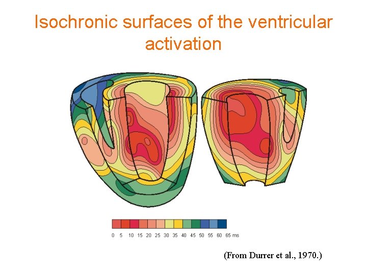
Propagation on ventricular wall • From the inner side of the ventricular wall, the many activation sites cause the formation of a wavefront which propagates through the ventricular mass toward the outer wall. • This process results from cell-to-cell activation. • After each ventricular muscle region has depolarized, repolarization occurs.
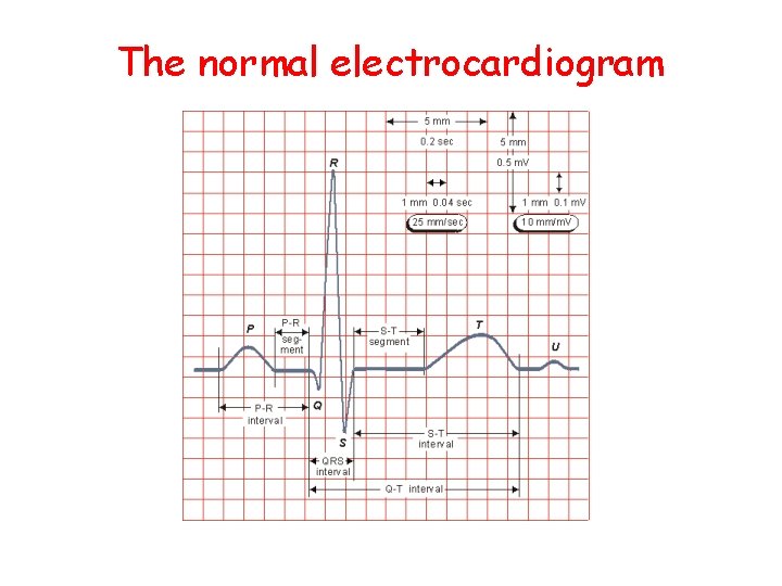
The normal electrocardiogram
Electrical events in the heart
Electrophysiology of the heart Different waveforms for each of the specialized cells
Isochronic surfaces of the ventricular activation (From Durrer et al. , 1970. )
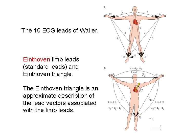
The 10 ECG leads of Waller. Einthoven limb leads (standard leads) and Einthoven triangle. The Einthoven triangle is an approximate description of the lead vectors associated with the limb leads.
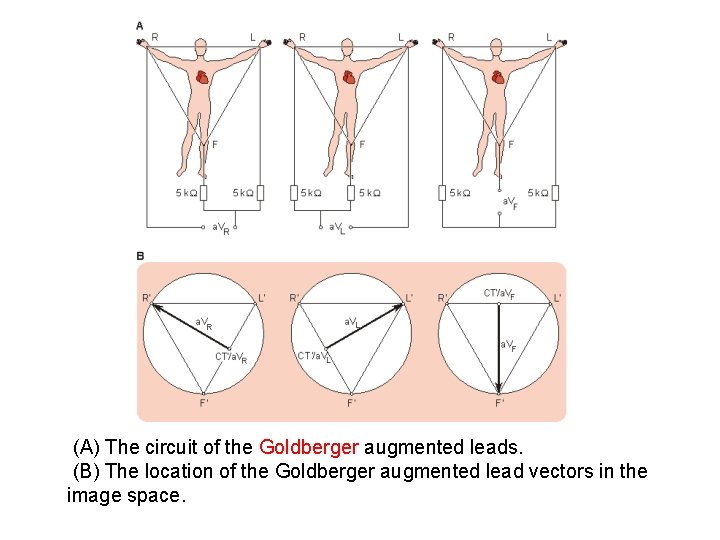
(A) The circuit of the Goldberger augmented leads. (B) The location of the Goldberger augmented lead vectors in the image space.
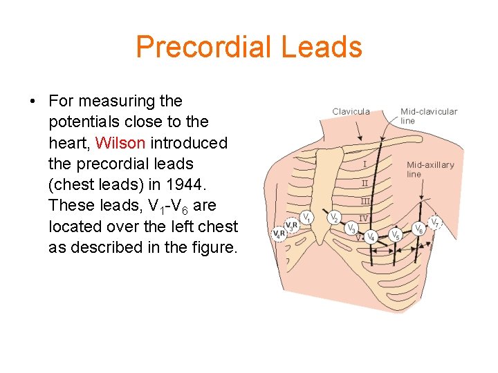
Precordial Leads • For measuring the potentials close to the heart, Wilson introduced the precordial leads (chest leads) in 1944. These leads, V 1 -V 6 are located over the left chest as described in the figure.
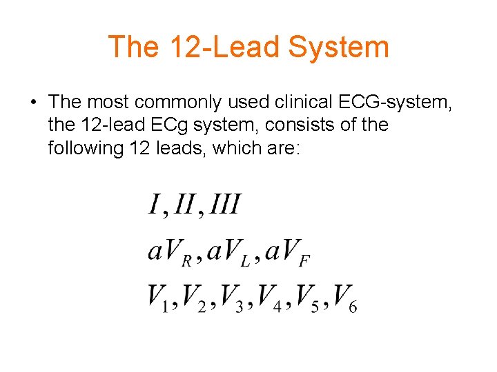
The 12 -Lead System • The most commonly used clinical ECG-system, the 12 -lead ECg system, consists of the following 12 leads, which are:
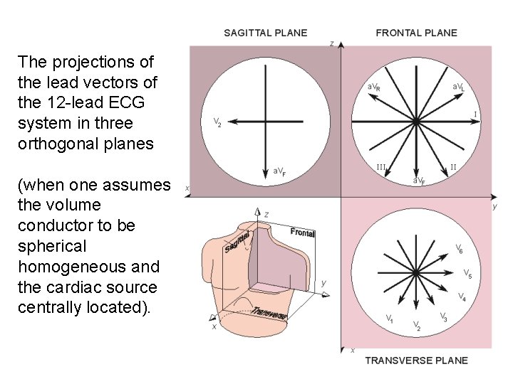
The projections of the lead vectors of the 12 -lead ECG system in three orthogonal planes (when one assumes the volume conductor to be spherical homogeneous and the cardiac source centrally located).