The Heart By Dr Anyanwu GE INTRODUCTION It

The Heart By Dr Anyanwu GE

INTRODUCTION • It is a conical hollow muscular organ responsible for blood circulation. • it is located in the middle of the mediastinum • it is enclosed within the pericardium • Greek name: cardia • Latin : Cor • The heart lies obliquely behind the sternum • 1/3 lies to the right while 2/3 lies to the left

Heart’s position in thorax

Heart’s position in thorax • In mediastinum – behind sternum and pointing left, lying on the diaphragm Feel your heart beat at apex (this is of a person lying down)

FEATURES • Its about the size owners clenched fist • 1/3 lies to the right while 2/3 lies to the left • Heart dimension 12 x 9 cm • Weight : 300 g males, 250 g females

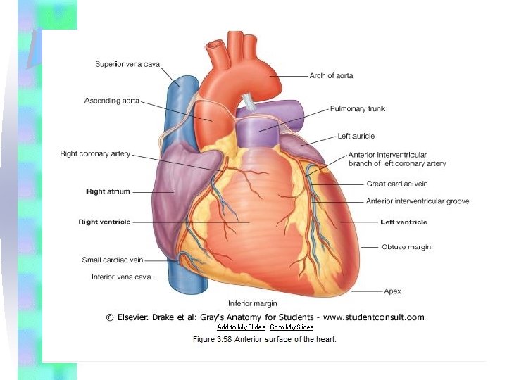
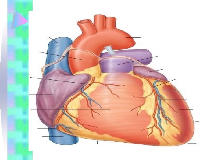
Features contd • It is an organ with four chambers—right, and left atria, right and left ventricles, IT HAS 3 SURFACES • Anterior or sternocostal surface, an • Inferior or diaphragmatic surface, • A base or posterior surface, • It also has an Apex.
 Normal male")
CXR (chest x ray) Normal male
")
Chest x rays Normal female Lateral (male)
 blood from")
• The right side of the heart receives poorly oxygenated (venous) blood from the body through the SVC and IVC and pumps it through the pulmonary trunk and arteries to the lungs for oxygenation. • The left side of the heart receives welloxygenated (arterial) blood from the lungs through the pulmonary veins and pumps it into the aorta for distribution to the body.

The heart=a muscular double pump with 2 functions Overview • The right side receives oxygen-poor blood from the body and tissues and then pumps it to the lungs to pick up oxygen and dispel carbon dioxide • Its left side receives oxygenated blood returning from the lungs and pumps this blood throughout the body to supply oxygen and nutrients to the body tissues

• Sulci and AV grooves: Externally, the atria are demarcated from the ventricles by the coronary sulcus (atrioventricular groove), and the right and left ventricles are demarcated from each other by anterior and posterior interventricular sulci (grooves)

The apex of the heart Is formed by the inferolateral part of the left ventricle. • At the junction of the inferior and left borders of the heart • Lies posterior to the left 5 th intercostal space in adults, usually approximately 9 cm (a hand's breadth) from the median plane. • Remains motionless throughout the cardiac cycle. • Is where the sounds of mitral valve closure are maximal (apex beat); the apex underlies the site where the heartbeat may be auscultated on the thoracic wall.
 of")
• The base of the heart • The posterior surface (or base) of the heart consists • almost entirely of the left atrium, receiving the four • pulmonary veins. • Is formed mainly by the left atrium, with a lesser contribution by the right atrium.

• Faces posteriorly toward the bodies of vertebrae T 6 -T 9 and is separated from them by the pericardium, oblique pericardial sinus, esophagus, and aorta. • Extends superiorly to the bifurcation of the pulmonary trunk and inferiorly to the coronary sulcus. • Receives the pulmonary veins on the right and left sides of its left atrial portion and SVC AND IVC ends of its right atrial portion.

The Anterior/Sternocostal surface Consists of the • right atrium, • the vertical atrioventricular groove • the right ventricle, • a narrow strip of the left ventricle • tip of the left auricular appendage

THE INFERIOR, OR DIAPHRAGMATIC SURFACE • Consists of the • Right atrium • The anteroposterior atrioventricular groove • The ventricular surface made up of 1/3 right ventricle • 2/3 left ventricle separated by the posterior interventricular branch of the • right coronary artery.

• The heart appears trapezoidal in both anterior and posterior views. This make the heart to have borders The four borders of the heart are the: Superior, right, inferior and left borders,
, formed by the right atrium and extending")
• Right border • (slightly convex), formed by the right atrium and extending between the SVC and the IVC. • it extends from the lower border of the right third costal cartilage to the lower border of the right sixth costal cartilage, just beyond the • right margin of the sternum and describing a slight • convex curve between these points.
, formed mainly by the right ventricle and slightly by the")
Inferior border (nearly horizontal), formed mainly by the right ventricle and slightly by the left ventricle. • It passes from the right sixth costal cartilage to the apex, which is normally in the left fifth intercostal space about 9 cm (3 x in) from the midline Left border (oblique, nearly vertical), formed mainly by the left ventricle and slightly by the left auricle. • From the apex the left border extends upwards to the lower border of the left • second costal cartilage about 2 cm from the sternal margin.

• • Superior border, formed by the right and left atria and auricles in an anterior view; the ascending aorta and pulmonary trunk emerge from this border and the SVC enters its right side. Posterior to the aorta and pulmonary trunk and anterior to the SVC, this border forms the inferior boundary of the transverse pericardial sinus.

Other features of the heart • Some general descriptions of cardiac orientation refer to right, left, inferior (acute), and obtuse margins: • the right and left margins are the same as the right and left pulmonary surfaces of the heart; • the inferior margin is defined as the sharp edge between the anterior and diaphragmatic surfaces of the heart and it is formed mostly by the right ventricle and a small portion of the left ventricle near the apex. • The obtuse margin separates the anterior and left pulmonary surfaces • it is round and extends from the left auricle to the cardiac apex and is formed mostly by the left ventricle and superiorly by a small portion of the left auricle

• • • Other features of the heart Some general descriptions of cardiac orientation refer to right, left, inferior (acute), and obtuse margins: the right and left margins are the same as the right and left pulmonary surfaces of the heart; the inferior margin is defined as the sharp edge between the anterior and diaphragmatic surfaces of the heart and it is formed mostly by the right ventricle and a small portion of the left ventricle near the apex. The obtuse margin separates the anterior and left pulmonary surfaces it is round and extends from the left auricle to the cardiac apex and is formed mostly by the left ventricle and superiorly by a small portion of the left auricle

Fibrous skeleton • This is a pair of conjoined fibrous rings which has the form of the figure 8 • The 'figure 8' lies on its side, very nearly in the sagittal plane. • This fibrous frame gives the points of attachments for the muscles of the heart. • The muscle fibres encircle the chambers of the heart in a series of whorls • and spirals. • The atria lie to the right and the ventricles to the left of the fibrous skeleton and there is no muscular continuity between the two.

RIGHT ATRIUM • • • The right atrium forms the right border of the heart and receives venous blood from the SVC, IVC, and coronary sinus This elongated chamber lies between the superior and inferior venae cavae, Inferior vena cava is located at its lower end at the upper end the SVC left of the superior vena cava Is the right auricle. The right auricle is an ear-like conical muscular pouch that projects from this chamber. it increases the capacity of the atrium.

• • The auricle overlies the commencement of the aorta and the upper part of the right atrioventricular groove and, with the left auricle, it clasps the infundibulum (upper end) of the right ventricle. A groove called the Sulcus Terminalis runs from angle between the superior vena cava and the right auricle. This groove is projected inside the hollow of the left atrium as a ridge called the Crista terminalis.

• • • The smooth and rough parts of the atrial wall are separated externally by the shallow vertical groove, the sulcus terminalis or terminal groove and internally by a vertical ridge, the crista terminalis or terminal crest The interior of the right atrium is smooth to the right of the crista terminalis. The rough part of the wall is due to the presence of Pectinate muscles which appear as series of horizontal ridges. This rough area represents the true auricular chamber of the embryonic heart.

• The remainder of the atrial cavity, smooth walled, is produced by incorporation of the right horn of the sinus venosus • The opening of the coronary sinus, a short venous trunk receiving most of the cardiac veins, is between the right AV orifice and the IVC orifice The interatrial septum forms the posterior wall of the right atrium above the opening of the coronary sinus. • The left atrium is attached behind the right atrium, but at a higher level; it does not extend down to the diaphragmatic surface.

• • • A shallow saucer-shaped depression called the fossa ovalis lies toward the lower part of this wall. The fossa ovalis represents the primary septum of the fetal heart. A crescentic upper margin of the fossa ovalis is called the limbus,

• The limbus indicates the lower edge of the secondary septum of the fetal heart. • Failure of fusion of the two septa gives rise to a persistent foramen ovale
 RV is a triangular chamber. it receives blood")
• • RIGHT VENTRICLE (RV) RV is a triangular chamber. it receives blood from the RA. It forms the largest part of the anterior surface of the heart, a small part of the diaphragmatic surface, and almost the entire inferior border of the heart RV lies to the left of the right atrium The atrioventricular groove between the two is vertical over the front of the heart and anteroposterior on the inferior surface.

• RIGHT VENTRICLE • This AV, usually filled with fat groove lodges right coronary artery. • RV narrows as it passes Superiorly forming an arterial cone called the conus arteriosus or Infundibulum. • The infundibulum leads into the pulmonary trunk •

The interior of the cavity • Walls of the interior of the cavity of RV are much thicker than those of the atrium. • Walls of RV • The interior of RV forms 3 walls; anterior, inferior and • septal walls. • Inferior wall is also known as the floor and also forms the interventricular wall of both ventricles.

• Orifices • The interior of the RV has 2 orifices • The right AV or tricuspid orifice guarded by the Tricuspid valve • The pulmonary orifice guarded by the Pulmonary valve

Parts of RV • The interior of RV has two parts • The inflowing part • The out flowing part • The two parts are separated by a muscular ridge called the supraventricular crest or Infundibuloventric ular crest. • This crest is located between the tricuspid and pulmonary orifices

The inflowing part • This the rough part of the interior of RV. • Rough appearance is due to the presence of series of muscular ridges and bundles called the trabeculae carneae. • This part of the RV develops from the proximal portion of the bulbus cordis of the heart tube. • The inflow part of the ventricle receives blood from the right atrium through the right AV or tricuspid orifice.

Right AV or tricuspid orifice. • • • This orifice is located posterior to the body of the sternum at the level of the 4 th and 5 th intercostal spaces. The right AV orifice is surrounded by one of the fibrous rings of the fibrous skeleton of the heart The fibrous ring keeps the caliber of the orifice constant (large enough to admit the tips of three fingers). It also enables the orifice to resist the dilation that might otherwise result from blood being forced through it at varying pressures.

• • • The out flowing part The outflow part/ tract is the upper conical part of the RV. This part of the cavity connects RV to the pulmonary trunk. It is the region of the conus arteriosus (infundibulum ). It develops from the mid portion of the bulbus cordis of the heart tube

The Trabeculae carneae • • These are the muscular ridges in the walls of the inflowing part of the RV. They either attached to the ventricular walls throughout their length, forming Ridges Or attached at both ends, forming Bridges. A few trabeculae carneae (papillary muscles) have only one end attached to the ventricular surface forming Pillars.

• The Pillars are also known as the Papillary muscles. • The other end of the Papillary muscles are attached to the free edges of the cusps of the tricuspid valve through tendon-like fibrous cords called the chordae tendineae.
, forms a")
Septomarginal Trabecula • A single specialized trabeculum, the septomarginal trabecula (moderator band), forms a bridge between the lower portion of the interventricular septum and the base of the anterior papillary muscle. The septomarginal trabeculum carries a portion of the cardiac conduction system, right bundle of the atrioventricular bundle, to the anterior wall of the right ventricle.

Papillary muscles • They are 3 in number. • They are named relative to their point of origin on the ventricular surface. • They are the Anterior, Posterior, and septal papillary muscles.

• • • The anterior papillary muscle is the largest and most constant papillary muscle, and arises from the anterior wall of the ventricle; The posterior papillary muscle may consist of one, two, or three structures, with some chordae tendineae arising directly from the ventricular wall; The septal papillary muscle is the most inconsistent papillary muscle, being either small or absent, with chordae tendineae emerging directly from the septal wall.

Tricuspid valve • • • The tricuspid valve guards the right atrioventricular orifice. It has three cusps and admits the tips of three fingers The right atrioventricular orifice is closed during ventricular contraction by the tricuspid valve. The base of each cusp is secured to the fibrous ring that surrounds the atrioventricular orifice. This fibrous ring helps to maintain the shape of the opening. The cusps are continuous with each other near their bases at sites termed commissures.

• The naming of the three cusps, the anterior, septal, and posterior cusps, is based on their relative position in the right ventricle. • Posterior cusp lies against the inferior wall or floor of the ventricle ( is • called posterior because it is behind the anterior cusp but would be better called the inferior cusp) • Anterior and Septal cusps lie on the corresponding sides of the ventricular wall. • The free margins of the cusps are attached to the chordae tendineae, which arise from the tips of the papillary muscles. • During filling of the right ventricle, the tricuspid valve is open, and the three cusps project into the right ventricle.

Function of AV valves

Pulmonary valve • The pulmonary valve guards the opening of the outflow tract (opening into the pulmonary trunk) • This is at the apex of the infundibulum.

• • • It consists of three semilunar cusps with free edges projecting upward into the lumen of the pulmonary trunk, The free superior edge of each cusp has a middle, thickened portion the nodule of the semilunar cusp, and a thin lateral portion, the lunule of the semilunar cusp The cusps are named the left, right and anterior semilunar cusps, relative to their fetal position Each cusp forms a pocketlike sinus; pulmonary sinuses -a dilation in the wall of the initial portion of the pulmonary trunk. After ventricular contraction, the recoil of blood fills these pulmonary sinuses and forces the cusps closed. This prevents blood in the pulmonary trunk from refilling the right ventricle
")
Function of semilunar valves (Aortic and pulmonic valves)

The interventricular septum • This is composed 2 parts : the muscular • and membranous parts, • it is a strong, obliquely placed partition between the right and left ventricles • It forming part of the walls of each. • Upper part of septum is thin and membranous and lower part is thick and muscular • Because of the much • The muscular part of the IVS, which forms the majority of the septum • it is 2 to 3 times as thick as the wall of the rest part of the right ventricle • It bulges into the cavity of the right ventricle.
 of the")
The left Atrium • The left atrium forms the posterior surface (base) of the heart • it lies behind the right atrium • It has a quadrangular shaped chamber • It has an auricular appendage which projected anteriorly to overlap the infundibulum of the right ventricle • it forms the left 2/3 of the base of the heart • it receives oxygenated blood from the lungs through the 4 pulmonary veins that opened into it

• It pumps blood to the left ventricle • The left atrioventricular orifice is guarded by the bicuspid valve also called mitral valve • Posterior surface of the LA forms the anterior wall of the oblique sinus of pericardium • The fibrous pericardium separates this surface from the oesophagus. • The anterior wall of LA is the interatrial septum; its common wall with RA. • The posterior half, or inflow portion, receives the four pulmonary veins

• • • THE INTERIOR Most of the cavity of the left atrium is smooth-walled except in the auricle The auricular part has rough muscular ridges due to the presence of pectinate muscles The rough area of LA indicate part of the chamber formed from original auricular chamber of the embryonic heart. All the smooth-walled portion is derived by incorporation of the embryonic pulmonary veins into the atrial cavity.

LEFT VENTRICLE • LV receives blood from the LV and pumps it out through the aorta • It forms the apex of the heart • The walls of this cavity are three times as thick as those • of the right ventricle

Relative thickness of the ventricular walls

• • • NTERIOR OF THE LV In a cross section the cavity of LV appears circular b/c the interventricular septum bulges into the cavity of the RV A conical cavity that is longer than that of the right ventricle The chamber has two orifices which are; The Atrioventricular/ bicuspid/mitral orifice, guarded by the atrioventricular/ bicuspid/mitral valve The Aortic orifice guarded by the aortic valve The interior is made up of two parts: The Upper smooth part The lower rough part

• • • The upper smooth part is the outflow tract. it is the smooth-walled, nonmuscular, superoanterior region known as the aortic vestibule, This part leads to the aortic orifice and aortic valve. The aortic vestibule lies posterior to the infundibulum of the right ventricle it develops from the mid portion of the bulbus cordis It lies btw the membranous part of the interventricular septum and the anterior cusp of the bicuspid valve

• The lower rough part has several muscular ridges of Trabeculae carneae. This region develops from primitive ventricle of the heart tube. . • The trabeculae carneae in the walls are well developed, finer and more numerous than those of the right ventricle •

• The aortic valve also has right, posterior, and left cusps • The aortic orifice lies at the right posterosuperior part of the chamber. • It is surrounded by a fibrous ring to which the right posterior, • and left cusps of the aortic valve are attached • The ascending aorta begins at the aortic orifice.

• • • There are two papillary muscles PM, anterior and posterior muscles which are larger than those in the RV. The anterior Papillary muz is larger the posterior. Both PMs are connected by chordae tendineae to each valve cusp The posterior cusp receives the chordae on both its margin and its ventricular surface While the Anterior cusp is only attached to the chordae tendinae only along its margins. This is b/c blood is squirted across both surfaces of the anterior cusp (down through the mitral orifice and up to the aortic) (NB; The anterior cusp lies between the mitral and the aortic orifices).

• • • THE BICUSPID MITRAL The mitral valve has two cusps, anterior and posterior It admits the tips of two fingers The cusps are named anterior and posterior The anterior cusp lies between the mitral and the aortic orifices. The bases of the cusps are attached to the margins of the fibrous atrioventricular ring. The mitral cusps are smaller in area and thicker than those of the tricuspid valve and consequently are not ballooned back so much into the atrium during ventricular systole. The anterior cusp of the mitral valve is thicker and more rigid than the posterior cusp.

Aortic valve • • • Aortic opening is guarded by the aortic valve This valve is similar in structure to the pulmonary valve. It consists of three semilunar cusps with the free edge of each projecting upward into the lumen of the ascending aorta Between the semilunar cusps and the wall of the ascending aorta are pocket-like sinuses. These are named : the right, left, and posterior aortic sinuses. The right and left coronary arteries originate from the right and left aortic sinuses. Because of this, the posterior aortic sinus and cusp are sometimes referred to as the noncoronary sinus and cusp. .
")
Function of semilunar valves (Aortic and pulmonic valves)

• The functioning of the aortic valve is similar to that of the pulmonary valve with one important additional process: as blood recoils after ventricular contraction and fills the aortic sinuses, it is automatically forced into the coronary arteries because these vessels originate from the right and left aortic sinuses

Arterial Supply of Heart. • The coronary arteries, the first branches of the aorta • The right and left coronary arteries arise from the corresponding aortic sinuses at the proximal part of the ascending aorta, just superior to the aortic valve, and pass around opposite sides of the pulmonary trunk.
 A: Right Coronary Artery;")
Blood supply to the heart (there’s a lot of variation) A: Right Coronary Artery; B: Left Main Coronary Artery; C: Left Anterior Descending (LAD, or Left Anterior Interventricular); D: Left Circumflex Coronary Artery; G: Marginal Artery; H: Great Cardiac Vein; I: Coronary sinus, Anterior Cardiac Veins.
 • • It arises from the right aortic sinus")
The right coronary artery (RCA) • • It arises from the right aortic sinus of the ascending aorta and passes to the right side of the pulmonary trunk, it runs downwards in the rt anterior coronary sulcus Near its origin, the RCA usually gives off an ascending sinuatrial nodal branch, which supplies the SA node. The RCA then descends in the coronary sulcus and gives off the right marginal branch, which supplies the right border of the heart as it runs toward (but does not reach) the apex of the heart. After giving off this branch, the RCA turns to the left and continues in the rt posterior coronary sulcus to the posterior aspect of the heart. At the posterior aspect of the crux of the heart—the junction of the interatrial and interventricular (IV) septa between the four heart chambers —the RCA gives rise to the atrioventricular nodal branch, which supplies the AV node.

Coronary Circulation: Arterial Supply Figure 18. 7 a
 A: Right Coronary Artery;")
Blood supply to the heart (there’s a lot of variation) A: Right Coronary Artery; B: Left Main Coronary Artery; C: Left Anterior Descending (LAD, or Left Anterior Interventricular); D: Left Circumflex Coronary Artery; G: Marginal Artery; H: Great Cardiac Vein; I: Coronary sinus, Anterior Cardiac Veins.

• The RCA continues posteriorl an gives rise to the large posterior interventricular branch, which descends in the posterior IV groove toward the apex of the heart. • This branch supplies adjacent areas of both ventricles and sends perforating interventricular septal branches into the IV septum • The terminal (left ventricular) branch of the RCA then continues for a short distance in the coronary sulcus. • Typically, the RCA supplies • The right atrium. • Most of right ventricle. • Part of the left ventricle (the diaphragmatic surface). • Part of the IV septum, usually the posterior third. • The SA node (in approximately 60% of people). • The AV node (in approximately 80% of people).

Coronary Circulation: Arterial Supply Figure 18. 7 a

Anterior view L main coronary artery arises from the left side of the aorta and has 2 branches: LAD and circumflex R coronary artery emerges from right side of aorta

Note that the usual name for “anterior interventricular artery” is the LAD (left anterior descending)
 • It arises from the left aortic sinus of")
The left coronary artery (LCA) • It arises from the left aortic sinus of the ascending aorta • It passes between the left auricle and the left side of the pulmonary trunk • It runs in the coronary sulcus • In approximately 40% of people, the SA nodal branch arises from the circumflex branch of the LCA and ascends on the posterior surface of the • left atrium to the SA node. • As it enters the coronary sulcus, at the superior end of the anterior IV groove, the LCA divides into two branches, the anterior IV branch ( LAD, “left anterior descending” artery) and the circumflex branch

Coronary Circulation: Arterial Supply Figure 18. 7 a
 A: Right Coronary Artery;")
Blood supply to the heart (there’s a lot of variation) A: Right Coronary Artery; B: Left Main Coronary Artery; C: Left Anterior Descending (LAD, or Left Anterior Interventricular); D: Left Circumflex Coronary Artery; G: Marginal Artery; H: Great Cardiac Vein; I: Coronary sinus, Anterior Cardiac Veins.

• The anterior IV branch passes along the IV groove to the apex of the heart. Here it turns around the inferior border of the heart and commonly anastomoses with the posterior IV branch of the right coronary artery • The anterior IV branch supplies adjacent parts of both ventricles and, via IV septal branches, the anterior two thirds of the IVS • In many people, the anterior IV branch gives rise to a lateral branch (diagonal artery), which descends on the anterior surface of the heart • The smaller circumflex branch of the LCA follows the coronary sulcus around the left border of the heart to the posterior surface of the heart. • The left marginal branch of the circumflex branch follows the left margin of the heart and supplies the left ventricle.

• Most commonly, the circumflex branch of the LCA terminates in the coronary sulcus on the posterior aspect of the heart before reaching the crux of the heart • But in approximately one third of hearts it continues to supply a branch that runs in or adjacent to the posterior IV groove • Typically, the LCA supplies • The left atrium. • Most of the left ventricle. • Part of the right ventricle. • Most of the IVS (usually its anterior two thirds),

• • Variations of the Coronary Arteries. Dominance of the coronary arterial system is defined by which artery gives rise to the posterior interventricular (IV) branch (posterior descending artery). In the most common right dominant pattern, present in approximately 67% of people codominance in approximately 18% of people, in which branches of both the right and left coronary arteries reach the crux of the heart and give rise to branches that course in or near the posterior IV groove. In approximately 15% of hearts, the LCA is dominant in that the posterior IV branch is a branch of the circumflex artery A few people have only one coronary artery In other people, the circumflex branch arises from the right aortic sinus Approximately 4% of people have an accessory coronary artery.

Coronary Circulation: Venous Supply Figure 18. 7 b

Coronary lymphatics • The lymphatic vessels of the heart follow the coronary arteries and drain mainly into: • Brachiocephalic nodes, anterior to the brachiocephalic veins; and • Tracheobronchial nodes, at the inferior end of the trachea.

Cardiac conduction system • The musculature of the atria and ventricles is capable of contracting spontaneously. The cardiac conduction system initiates and coordinates contraction. The conduction system consists of nodes and networks of specialized myocardial cells organized into four basic components: • the sinu-atrial node; • the atrioventricular bundle with its right and left bundle branches; • the subendocardial plexus of conduction cells (the Purkinje fibers).

FROM HERE THE JOURNEY BEGINS THANKS
- Slides: 84