
The Hand Lymphatics Week 6 TA Review Things





- Slides: 71
The Hand & Lymphatics Week 6 TA Review
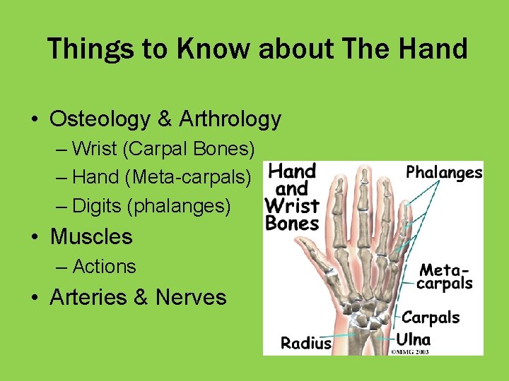
Things to Know about The Hand • Osteology & Arthrology – Wrist (Carpal Bones) – Hand (Meta-carpals) – Digits (phalanges) • Muscles – Actions • Arteries & Nerves
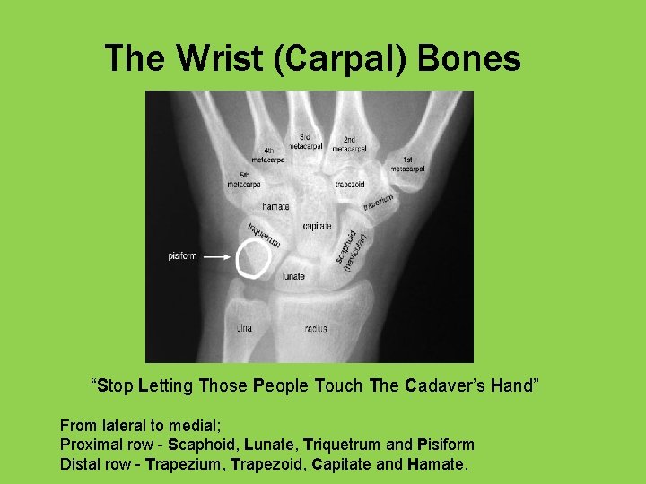
The Wrist (Carpal) Bones “Stop Letting Those People Touch The Cadaver’s Hand” From lateral to medial; Proximal row - Scaphoid, Lunate, Triquetrum and Pisiform Distal row - Trapezium, Trapezoid, Capitate and Hamate.
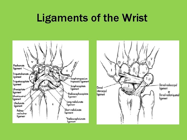
Arthrology of the Wrist • Radiocarpal Joint • Intercarpal Joint • Carpometacarpal Joints
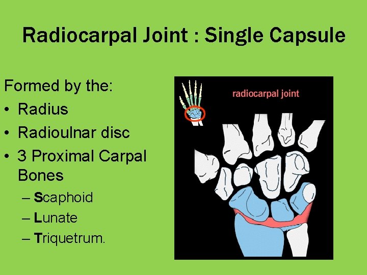
Radiocarpal Joint : Single Capsule Formed by the: • Radius • Radioulnar disc • 3 Proximal Carpal Bones – Scaphoid – Lunate – Triquetrum.
Intercarpal Joint • Articulations b/w the individual carpal bones. • Plane synovial joints. • The small movement b/w carpal bones at these joints contributes to total wrist mobility
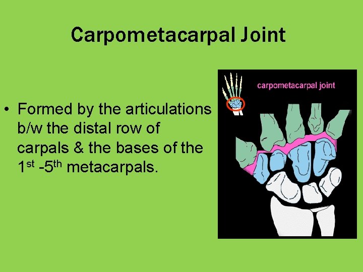
Carpometacarpal Joint • Formed by the articulations b/w the distal row of carpals & the bases of the 1 st -5 th metacarpals.
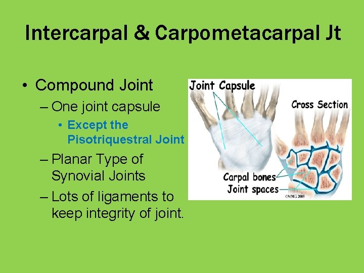
Intercarpal & Carpometacarpal Jt • Compound Joint – One joint capsule • Except the Pisotriquestral Joint – Planar Type of Synovial Joints – Lots of ligaments to keep integrity of joint.
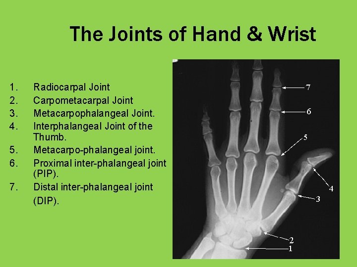
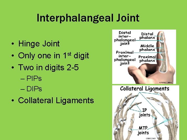
The Joints of Hand & Wrist 1. 2. 3. 4. 5. 6. 7. Radiocarpal Joint Carpometacarpal Joint Metacarpophalangeal Joint. Interphalangeal Joint of the Thumb. Metacarpo-phalangeal joint. Proximal inter-phalangeal joint (PIP). Distal inter-phalangeal joint (DIP).
Ligaments of the Wrist
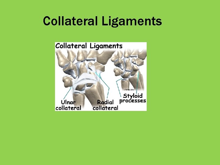
Collateral Ligaments
Arthrology of the Hand • Carpometacarpal Joints • Metacarpophalangeal Joints • Interphalangeal Joints
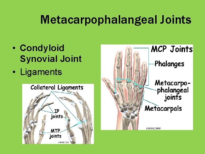
Metacarpophalangeal Joints • Condyloid Synovial Joint • Ligaments
Interphalangeal Joint • Hinge Joint • Only one in 1 st digit • Two in digits 2 -5 – PIPs – DIPs • Collateral Ligaments
Actions of Wrist and Hand • • • Flexion Extension AB-duction AD-duction Opposition (Hand)
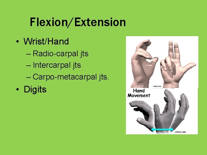
Flexion/Extension • Wrist/Hand – Radio-carpal jts – Intercarpal jts – Carpo-metacarpal jts. • Digits
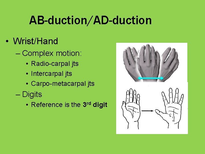
AB-duction/AD-duction • Wrist/Hand – Complex motion: • Radio-carpal jts • Intercarpal jts • Carpo-metacarpal jts – Digits • Reference is the 3 rd digit
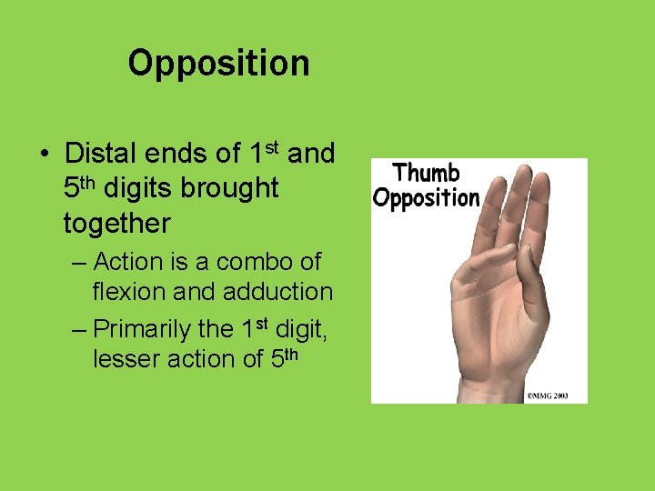
Opposition • Distal ends of 1 st and 5 th digits brought together – Action is a combo of flexion and adduction – Primarily the 1 st digit, lesser action of 5 th
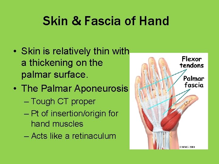
Skin & Fascia of Hand • Skin is relatively thin with a thickening on the palmar surface. • The Palmar Aponeurosis – Tough CT proper – Pt of insertion/origin for hand muscles – Acts like a retinaculum
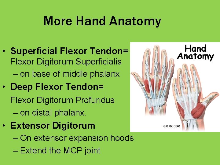
More Hand Anatomy • Superficial Flexor Tendon= Flexor Digitorum Superficialis – on base of middle phalanx • Deep Flexor Tendon= Flexor Digitorum Profundus – on distal phalanx. • Extensor Digitorum – On extensor expansion hoods – Extend the MCP joint
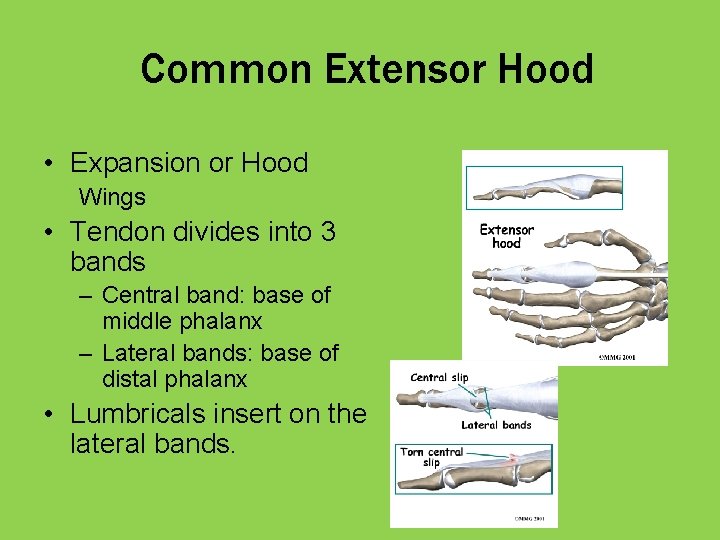
Common Extensor Hood • Expansion or Hood Wings • Tendon divides into 3 bands – Central band: base of middle phalanx – Lateral bands: base of distal phalanx • Lumbricals insert on the lateral bands.
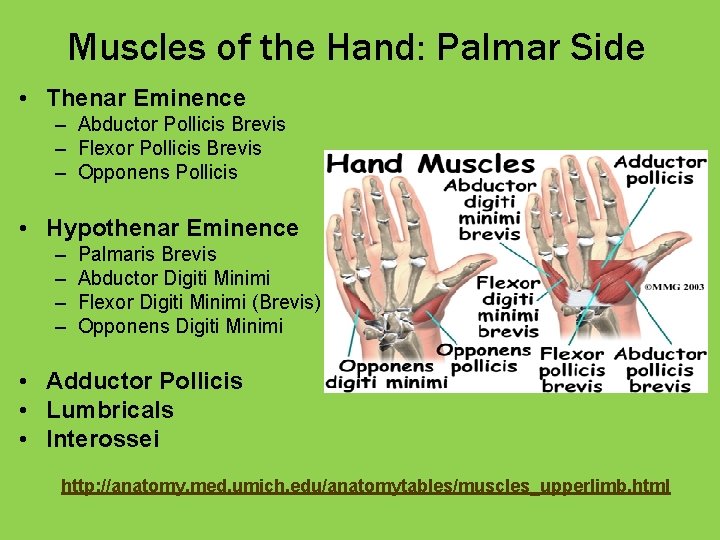
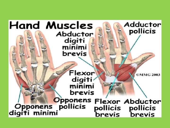
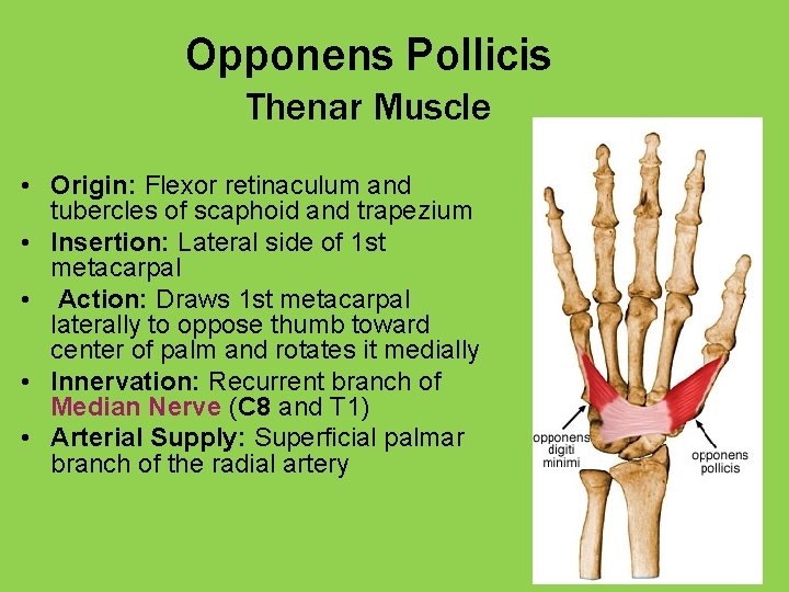
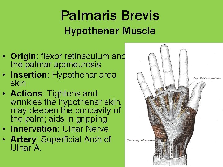
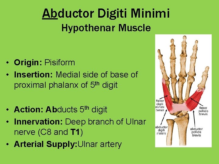
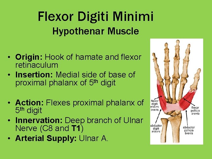
Muscles of the Hand: Palmar Side • Thenar Eminence – Abductor Pollicis Brevis – Flexor Pollicis Brevis – Opponens Pollicis • Hypothenar Eminence – – Palmaris Brevis Abductor Digiti Minimi Flexor Digiti Minimi (Brevis) Opponens Digiti Minimi • Adductor Pollicis • Lumbricals • Interossei http: //anatomy. med. umich. edu/anatomytables/muscles_upperlimb. html
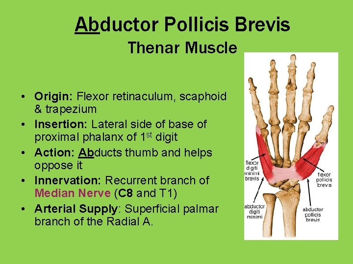
Abductor Pollicis Brevis Thenar Muscle • Origin: Flexor retinaculum, scaphoid & trapezium • Insertion: Lateral side of base of proximal phalanx of 1 st digit • Action: Abducts thumb and helps oppose it • Innervation: Recurrent branch of Median Nerve (C 8 and T 1) • Arterial Supply: Superficial palmar branch of the Radial A.
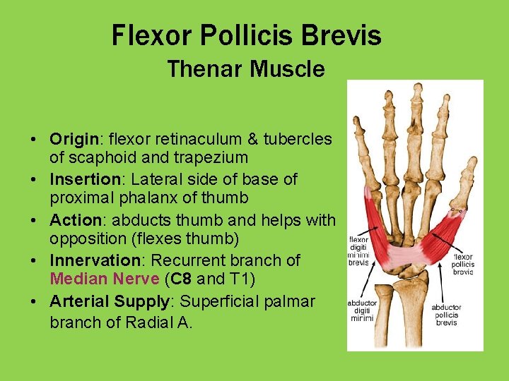
Flexor Pollicis Brevis Thenar Muscle • Origin: flexor retinaculum & tubercles of scaphoid and trapezium • Insertion: Lateral side of base of proximal phalanx of thumb • Action: abducts thumb and helps with opposition (flexes thumb) • Innervation: Recurrent branch of Median Nerve (C 8 and T 1) • Arterial Supply: Superficial palmar branch of Radial A.
Opponens Pollicis Thenar Muscle • Origin: Flexor retinaculum and tubercles of scaphoid and trapezium • Insertion: Lateral side of 1 st metacarpal • Action: Draws 1 st metacarpal laterally to oppose thumb toward center of palm and rotates it medially • Innervation: Recurrent branch of Median Nerve (C 8 and T 1) • Arterial Supply: Superficial palmar branch of the radial artery
Palmaris Brevis Hypothenar Muscle • Origin: flexor retinaculum and the palmar aponeurosis • Insertion: Hypothenar area skin • Actions: Tightens and wrinkles the hypothenar skin, may deepen the concavity of the palm; aids in gripping • Innervation: Ulnar Nerve • Artery: Superficial Arch of Ulnar A.
Abductor Digiti Minimi Hypothenar Muscle • Origin: Pisiform • Insertion: Medial side of base of proximal phalanx of 5 th digit • Action: Abducts 5 th digit • Innervation: Deep branch of Ulnar nerve (C 8 and T 1) • Arterial Supply: Ulnar artery
Flexor Digiti Minimi Hypothenar Muscle • Origin: Hook of hamate and flexor retinaculum • Insertion: Medial side of base of proximal phalanx of 5 th digit • Action: Flexes proximal phalanx of 5 th digit • Innervation: Deep branch of Ulnar Nerve (C 8 and T 1) • Arterial Supply: Ulnar A.
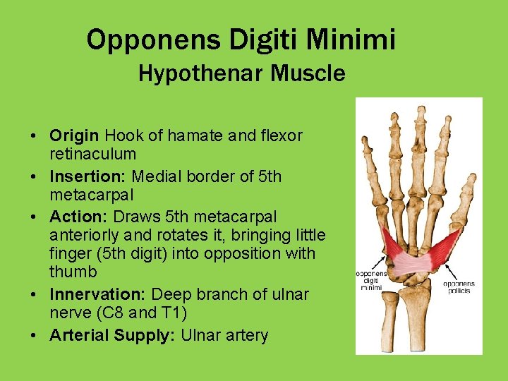
Opponens Digiti Minimi Hypothenar Muscle • Origin Hook of hamate and flexor retinaculum • Insertion: Medial border of 5 th metacarpal • Action: Draws 5 th metacarpal anteriorly and rotates it, bringing little finger (5 th digit) into opposition with thumb • Innervation: Deep branch of ulnar nerve (C 8 and T 1) • Arterial Supply: Ulnar artery
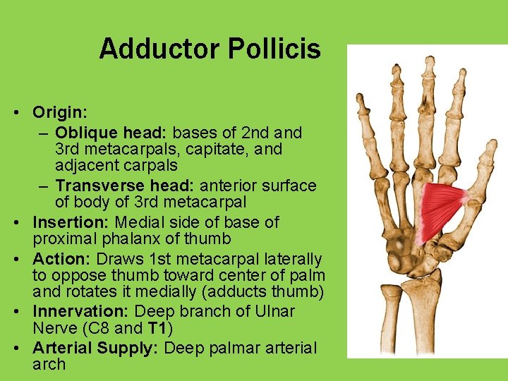
Adductor Pollicis • Origin: – Oblique head: bases of 2 nd and 3 rd metacarpals, capitate, and adjacent carpals – Transverse head: anterior surface of body of 3 rd metacarpal • Insertion: Medial side of base of proximal phalanx of thumb • Action: Draws 1 st metacarpal laterally to oppose thumb toward center of palm and rotates it medially (adducts thumb) • Innervation: Deep branch of Ulnar Nerve (C 8 and T 1) • Arterial Supply: Deep palmar arterial arch
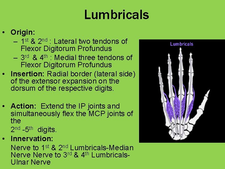
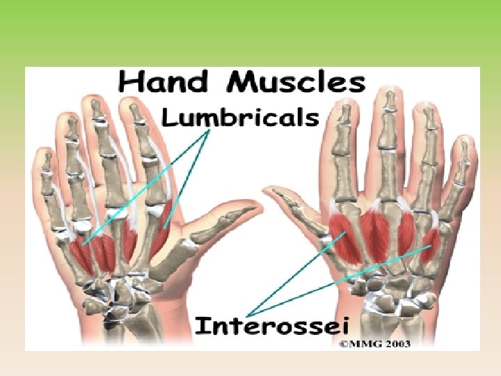
Lumbricals • Origin: – 1 st & 2 nd : Lateral two tendons of Flexor Digitorum Profundus – 3 rd & 4 th : Medial three tendons of Flexor Digitorum Profundus • Insertion: Radial border (lateral side) of the extensor expansion on the dorsum of the respective digits. • Action: Extend the IP joints and simultaneously flex the MCP joints of the 2 nd -5 th digits. • Innervation: Nerve to 1 st & 2 nd Lumbricals-Median Nerve to 3 rd & 4 th Lumbricals. Ulnar Nerve
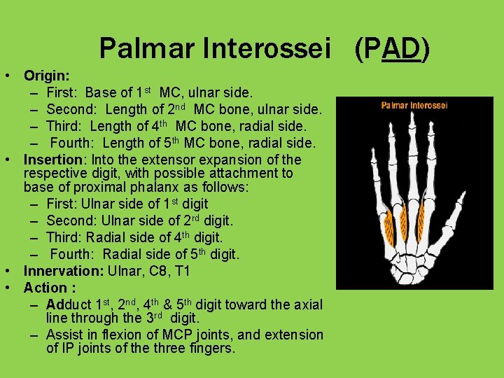
Palmar Interossei (PAD) • Origin: – First: Base of 1 st MC, ulnar side. – Second: Length of 2 nd MC bone, ulnar side. – Third: Length of 4 th MC bone, radial side. – Fourth: Length of 5 th MC bone, radial side. • Insertion: Into the extensor expansion of the respective digit, with possible attachment to base of proximal phalanx as follows: – First: Ulnar side of 1 st digit – Second: Ulnar side of 2 rd digit. – Third: Radial side of 4 th digit. – Fourth: Radial side of 5 th digit. • Innervation: Ulnar, C 8, T 1 • Action : – Adduct 1 st, 2 nd, 4 th & 5 th digit toward the axial line through the 3 rd digit. – Assist in flexion of MCP joints, and extension of IP joints of the three fingers.
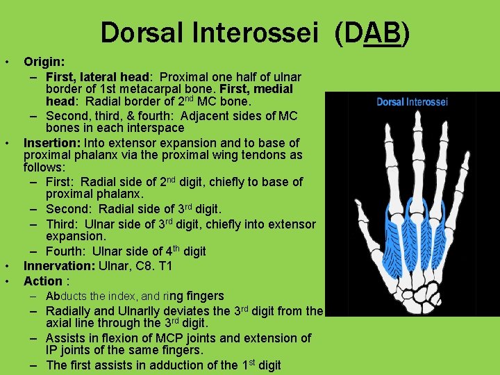
Dorsal Interossei (DAB) • • Origin: – First, lateral head: Proximal one half of ulnar border of 1 st metacarpal bone. First, medial head: Radial border of 2 nd MC bone. – Second, third, & fourth: Adjacent sides of MC bones in each interspace Insertion: Into extensor expansion and to base of proximal phalanx via the proximal wing tendons as follows: – First: Radial side of 2 nd digit, chiefly to base of proximal phalanx. – Second: Radial side of 3 rd digit. – Third: Ulnar side of 3 rd digit, chiefly into extensor expansion. – Fourth: Ulnar side of 4 th digit Innervation: Ulnar, C 8. T 1 Action : – Abducts the index, and ring fingers – Radially and Ulnarlly deviates the 3 rd digit from the axial line through the 3 rd digit. – Assists in flexion of MCP joints and extension of IP joints of the same fingers. – The first assists in adduction of the 1 st digit
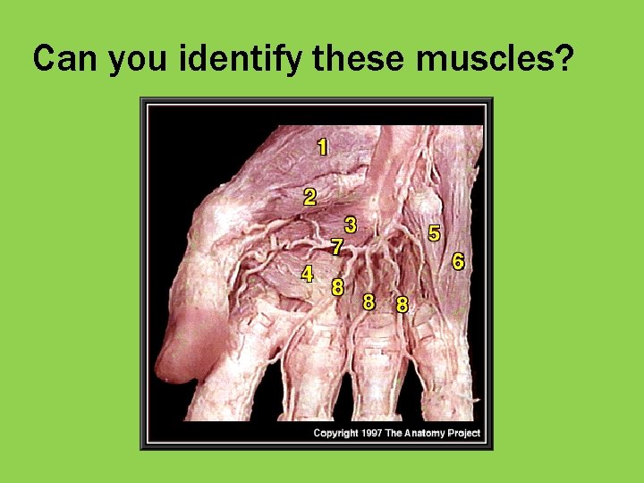
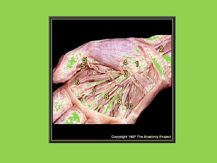
Can you identify these muscles?
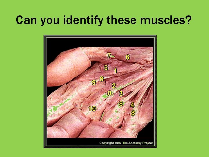
Can you identify these muscles?
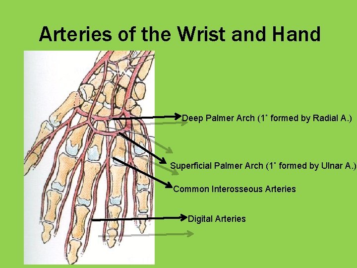
Arteries of the Wrist and Hand Deep Palmer Arch (1˚ formed by Radial A. ) Superficial Palmer Arch (1˚ formed by Ulnar A. ) Common Interosseous Arteries Digital Arteries
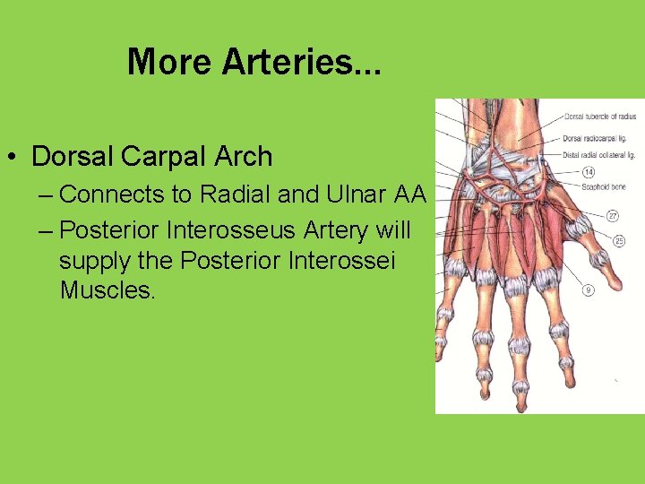
More Arteries… • Dorsal Carpal Arch – Connects to Radial and Ulnar AA – Posterior Interosseus Artery will supply the Posterior Interossei Muscles.
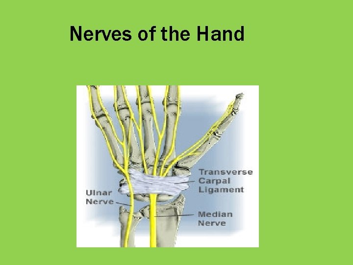
Nerves of the Hand
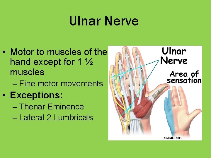
Ulnar Nerve • Motor to muscles of the hand except for 1 ½ muscles – Fine motor movements • Exceptions: – Thenar Eminence – Lateral 2 Lumbricals
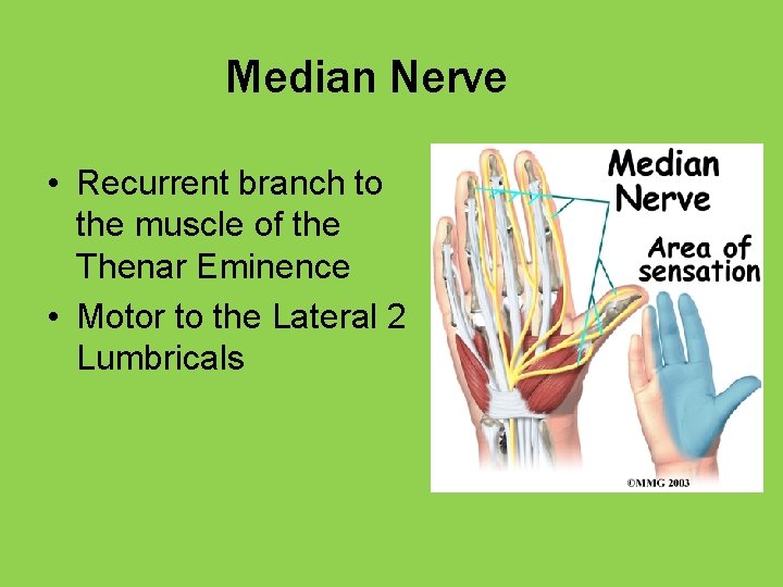
Median Nerve • Recurrent branch to the muscle of the Thenar Eminence • Motor to the Lateral 2 Lumbricals
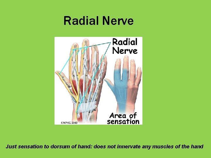
Radial Nerve Just sensation to dorsum of hand: does not innervate any muscles of the hand
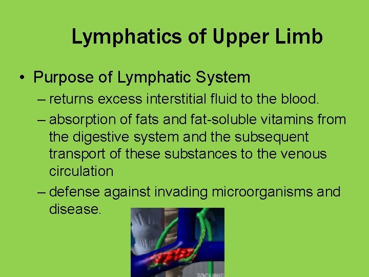
Lymphatics of Upper Limb • Purpose of Lymphatic System – returns excess interstitial fluid to the blood. – absorption of fats and fat-soluble vitamins from the digestive system and the subsequent transport of these substances to the venous circulation – defense against invading microorganisms and disease.
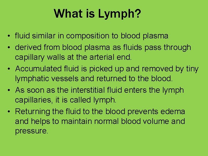
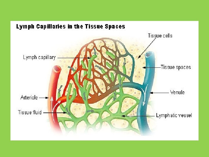
What is Lymph? • fluid similar in composition to blood plasma • derived from blood plasma as fluids pass through capillary walls at the arterial end. • Accumulated fluid is picked up and removed by tiny lymphatic vessels and returned to the blood. • As soon as the interstitial fluid enters the lymph capillaries, it is called lymph. • Returning the fluid to the blood prevents edema and helps to maintain normal blood volume and pressure.
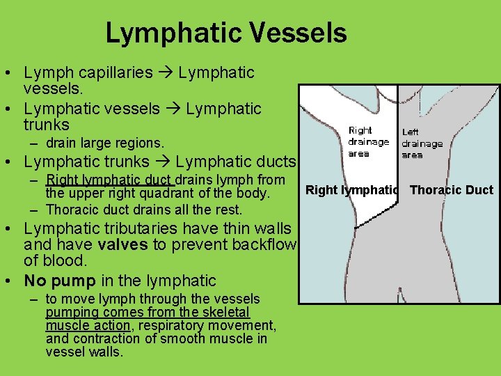
Lymphatic Vessels • Lymph capillaries Lymphatic vessels. • Lymphatic vessels Lymphatic trunks – drain large regions. • Lymphatic trunks Lymphatic ducts. – Right lymphatic duct drains lymph from the upper right quadrant of the body. – Thoracic duct drains all the rest. • Lymphatic tributaries have thin walls and have valves to prevent backflow of blood. • No pump in the lymphatic – to move lymph through the vessels pumping comes from the skeletal muscle action, respiratory movement, and contraction of smooth muscle in vessel walls. Right lymphatic Thoracic Duct
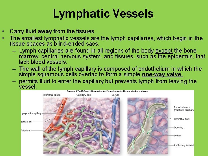
Lymphatic Vessels • Carry fluid away from the tissues • The smallest lymphatic vessels are the lymph capillaries, which begin in the tissue spaces as blind-ended sacs. – Lymph capillaries are found in all regions of the body except the bone marrow, central nervous system, and tissues, such as the epidermis, that lack blood vessels. – The wall of the lymph capillary is composed of endothelium in which the simple squamous cells overlap to form a simple one-way valve. – permits fluid to enter the capillary but prevents lymph from leaving the vessel.
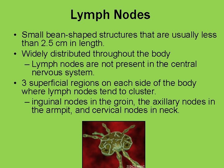
Lymph Nodes • Small bean-shaped structures that are usually less than 2. 5 cm in length. • Widely distributed throughout the body – Lymph nodes are not present in the central nervous system. • 3 superficial regions on each side of the body where lymph nodes tend to cluster. – inguinal nodes in the groin, the axillary nodes in the armpit, and cervical nodes in neck.
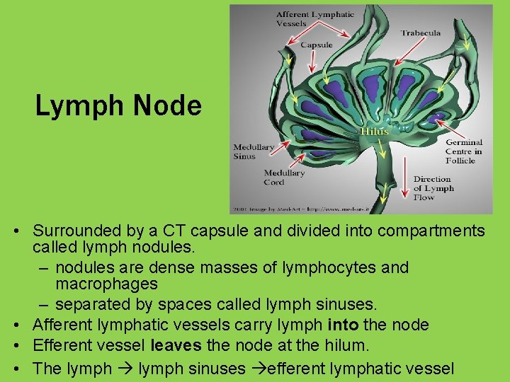
Lymph Node • Surrounded by a CT capsule and divided into compartments called lymph nodules. – nodules are dense masses of lymphocytes and macrophages – separated by spaces called lymph sinuses. • Afferent lymphatic vessels carry lymph into the node • Efferent vessel leaves the node at the hilum. • The lymph sinuses efferent lymphatic vessel
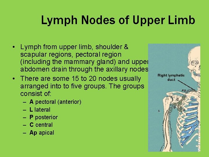
Lymph Nodes of Upper Limb • Lymph from upper limb, shoulder & scapular regions, pectoral region (including the mammary gland) and upper abdomen drain through the axillary nodes. • There are some 15 to 20 nodes usually arranged into to five groups. The groups consist of: – – – A pectoral (anterior) L lateral P posterior C central Ap apical
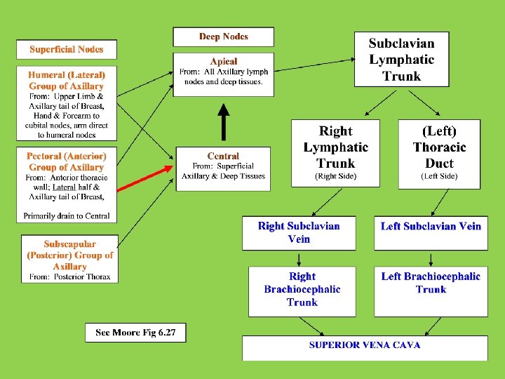
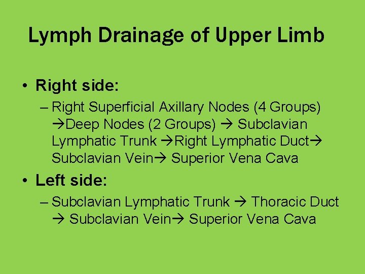
Lymph Drainage of Upper Limb • Right side: – Right Superficial Axillary Nodes (4 Groups) Deep Nodes (2 Groups) Subclavian Lymphatic Trunk Right Lymphatic Duct Subclavian Vein Superior Vena Cava • Left side: – Subclavian Lymphatic Trunk Thoracic Duct Subclavian Vein Superior Vena Cava
1. A patient complaining of difficulty picking up items. Physical examination shows weakness of grip in her left hand that she is unable to abduct, flex, and oppose her left thumb with her 5 th digit. There is loss of sensation on the palmar surface of the lateral side of the hand. Which one of the following nerves most likely has been damaged? A. B. C. D. Median Ulnar Musculocutaneous Radial
Review Questions
1. A patient complaining of difficulty picking up items. Physical examination shows weakness of grip in her left hand that she is unable to abduct, flex, and oppose her left thumb with her 5 th digit. There is loss of sensation on the palmar surface of the lateral side of the hand. Which one of the following nerves most likely has been damaged? A. B. C. D. Median Ulnar Musculocutaneous Radial
2. Lymph originating in the area of the skin overlying trapezius would most likely first be drained into which of the following groups of lymph nodes? A. B. C. D. Lateral group of axillary lymph nodes Subscapular group of axillary lymph nodes Pectoral group of axillary lymph nodes Apical group of axillary lymph nodes
3. An 18 -year-old high school basketball player had injured her right knee 2 weeks previously. The knee was immobilized and the athlete was fitted with crutches. After using the crutches for these last 2 weeks, she complains that the back of her right hand feels numb and that she finds it difficult to open (extend) the fingers of her right hand. Most likely the crutches were improperly fitted and put pressure on the brachial plexus. Which part of the brachial plexus most likely has been damaged? A. B. C. D. Lateral cord Medial cord Posterior cord Superior trunk
4. A patient with a severe tear of the medial (ulnar) collateral ligament of the wrist would likely display increased wrist movement in which of the following actions? A. B. C. D. Flexion Pronation Adduction Abduction
5. Lymph from the lateral group of axillary lymph nodes primarily drains into which of the following? A. B. C. D. Apical group of axillary lymph nodes Subclavian lymphatic trunk Central group of axillary lymph nodes Pectoral group of axillary lymph nodes
6. Which of the following carpal bones lies in the proximal row of 4? A. B. C. D. Trapezoid Scaphoid Hamate Capitate
7. A patient presents of general malaise, being tired, and with a fever. A physical examination is unremarkable except for enlarged lymph nodes palpated in the region of the lateral group of axillary lymph nodes. The only remarkable lab finding is an elevated with blood cell count. Most likely this patient has an infection in which one of the following places? A. B. C. D. Root of the neck in the region of the trapezius muscle Hand Chest/breast region Inferior border of the scapula
8. Which of the following muscles extend the metacarpo-phalangeal joint? A. B. C. D. Lumbrical Dorsal interosseous muscle Extensor digitorum Palmar interosseous muscle
9. A 14 -year-old boy falls while skateboarding and fractures the medial epicondyle of his right humerus. The bone is set and cast. After removal of the cast and a normal duration of rehabilitation his handwriting is noticeably more difficult to read than before the accident. What most likely is the cause of his handwriting deterioration? A. B. C. D. The fracture caused damage to the ulnar nerve The fracture caused damage to the radial nerve The fracture caused damage to the median nerve He had a stroke during his rehabilitation
10. Which muscle acts to abduct the digits 2 -5? A. B. C. D. Abductor pollicis longus Palmar interosseous Dorsal interosseous Opponens digiti minimi
11. Which muscle acts to flex the proximal interphalangeal joint? A. B. C. D. Flexor digitorum profundus Lumbrical Dorsal interosseous Flexor digitorum superficialis
12. Lymph from the lateral aspect of the breast would first be drained into which of the following lymph nodes? A. B. C. D. Central group of axillary lymph nodes Lateral group of axillary lymph nodes Apical group of axillary lymph nodes Pectoral group of axillary lymph nodes
12. A thrombus (blood clot) has occluded the brachial artery just proximal to the origin of the superior ulnar collateral artery. Trace the path a drop of blood would follow when flowing from the subclavian artery to the palmaris longus muscle, and then to the subclavian vein.
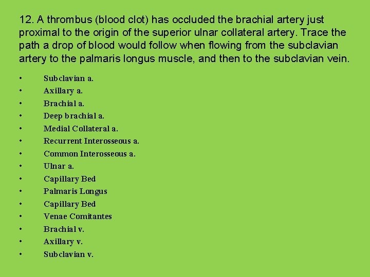
12. A thrombus (blood clot) has occluded the brachial artery just proximal to the origin of the superior ulnar collateral artery. Trace the path a drop of blood would follow when flowing from the subclavian artery to the palmaris longus muscle, and then to the subclavian vein. • • • • Subclavian a. Axillary a. Brachial a. Deep brachial a. Medial Collateral a. Recurrent Interosseous a. Common Interosseous a. Ulnar a. Capillary Bed Palmaris Longus Capillary Bed Venae Comitantes Brachial v. Axillary v. Subclavian v.