The Female Reproductive System Ovaries Uterine tubes All

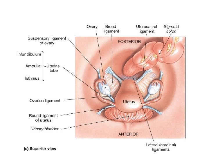
The Female Reproductive System • Ovaries • Uterine tubes All 3 enclosed within broad ligament. • Uterus • Vaginal canal and vagina • External genitalia

Female Reproductive System

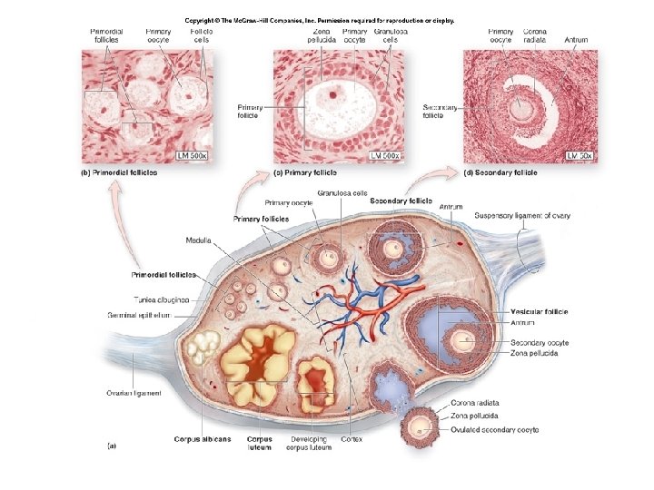
The Ovaries -> eggs, estrogen and progesterone. • Held in position by ovarian ligament and suspensory ligaments. • Ovarian artery and vein enter at ovarian hilus. • Has tunica albuginea. • Ovarian cycle and follicular development. (dictates uterine cycle)

ciliated and non-ciliated simple columnar epithelium

Frontal section of the Ovary

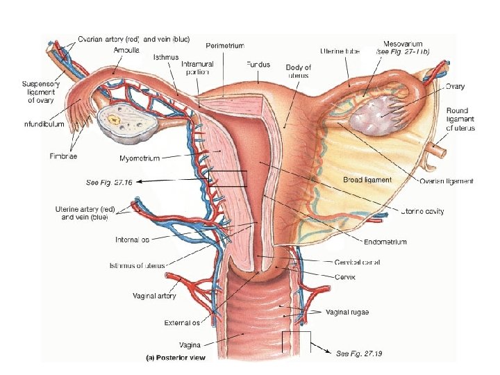
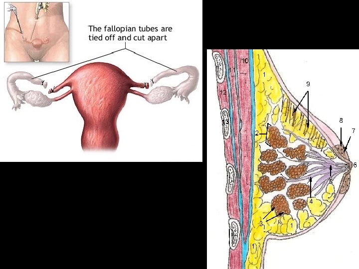
The Uterine Tubes • Infundibulum with fimbriae • Ampulla • Isthmus • Opens into uterine cavity • Lined with ciliated and non-ciliated simple columnar epithelium - aids in transport
 – Uterine cavity")
Gross Divisions of the Uterus • Fundus • Body (largest portion) – Uterine cavity • Cervix – Internal os – Cervical canal – External os
 • Endometrium - innermost layer – Contains endometrial (uterine) glands. –")
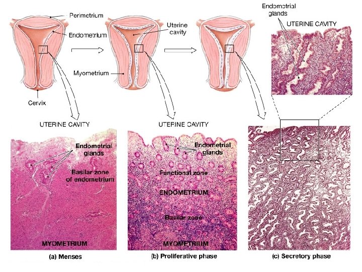
Uterine Wall (Layers) • Endometrium - innermost layer – Contains endometrial (uterine) glands. – Functional layer, top 1/3 (sheds). – Basal Layer, bottom 2/3 (remains). • Myometrium – muscular (thickest ~90%). • Perimetrium – outermost (superficial).

Histology of the Uterine Wall

Uterine Wall Histology

Ovarian Cycle

Uterine Cycle

Ovulation of Mature Follicle
. •")
The Vagina • Elastic muscular tube from uterus to external genitalia (has rugae). • Passageway. • Forms lower portion of birth canal. • Lined by stratified squamous epithelium.
 Labia majora – outer (larger) Labia minora –")
The External Genitalia • Vulva (pudendum) Labia majora – outer (larger) Labia minora – inner (smaller) Clitoris – erectile tissue (spongiosum) - Prepuce (hood, foreskin of clitoris) Vestibule (created by labia minora) Greater vestibular (Bartholin’s) gland. Mons pubis
- Slides: 24