THE EYE VISUAL RECEPTORS AND NEURAL FUNCTION OF


- Slides: 21
THE EYE: VISUAL RECEPTORS AND NEURAL FUNCTION OF THE RETINA LECTURER IN CHARGE BAMIDELE OLUBAYODE
RETINA • Retina is a delicate light-sensitive membrane that forms the innermost layer of eyeball. • It extends from the margin of optic disk to just behind ciliary body. • From ciliary body, it ends abruptly as a dentated border known as oraserrata. • Retina has the receptors of vision. It contains cones for day and colour vision while rods for night vision. • It contains neural architecture. Light must pass through the elements to strike the light sensitive rods and cones.
Adapted from: Faisal Mohammed
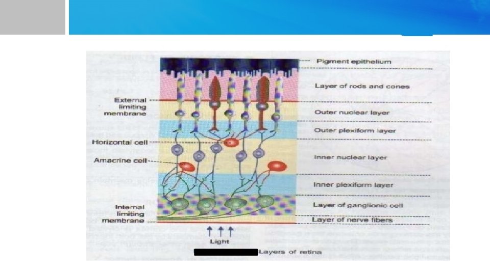
RETINA CONT'D • Structurally, retina is made up of 10 layers. Layers of retina from outside in: 1. Layer of pigment epithelium 2. Layer of rods and cones 3. Externallimitingmembrane 4. Outer nuclear layer 5. Outer plexiform layer 6. Inner nuclear layer 7. Inner plexiform layer 8. Ganglion cell layer 9. Layer of nerve fibers 10. Internal limiting membrane
MACULA LUTEA • Macula lutea is a small yellowish area, located a little lateral to the optic disk in retina. • It is also called yellow spot. Yellow color of macula lutea is due to the presence of a yellow pigment. • Macula lutea has fovea centralis in its center. • Fovea centralist is the region of most acute vision because it contains only cones. These cones have special structure and help in detecting detail. • I
MACULA LUTEA CONT'D • In the fovea centralist the neuronal cells and blood vessels are displaced to each side so that the light can strike the cones directly. • This is the area of greatest visual acuity. • Degeneration of macula lutea leads to blindness.
VISUAL PROCESS: IMAGE FORMING MECHANISM • Visual process is the series of actions that take place during visual perception. • During visual process, image of an object seen by the eyes is focused on retina, resulting in production of visual perception of that object. • When the image of an object in environment is focused on retina, the energy in visual spectrum is converted into electrical potentials (impulses) by rods and cones of retina via some chemical reactions. • Impulses from rods and cones reach the cerebral cortex through optic nerve and the sensation of vision is produced in cerebral cortex.
VISUAL PROCESS: IMAGE FORMING MECHANISM • The incoming light rays from an object are refracted and brought to a focus upon retina. • Image of the object falls on the retina in an inverted position and reversed side to side. However, the object is seen in an upright position. This is due to the role played by cerebral cortex. • Light rays are refracted by the lens and cornea. • Refractory power is measured in diopter (D). • A diopter is the reciprocal of focal length expressed in meters. • Focal length of cornea is 24 mm and refractory power is 42 D. Focal length of lens is 44 mm and refractory power is 23 D
NEURAL BASIS OF VISUAL PROCESS • Retina contains the visual receptors also known as light sensitive receptors or photoreceptors or electromagnetic receptors. • Visual receptors are rods and cones. There about 6 million cones and 12 million rods in the human eye. • Distribution of the rods and cones varies in different areas of retina. • Fovea has only cones and no rods. While proceeding from fovea towards the periphery of retina, the rods increase and the cones decrease in number. • At the periphery of the retina, only rods are present and cones are absent.
RODS • Rod cells are cylindrical structures with a length of about 40 to 60 µ and a diameter of about 2 µ. • They have high sensitivity and specialized for night vision. • They have more photopigment, high amplification, single photon detection. • They are saturated in day light. • Low response and integration time • They are more sensitive to scattered light. • Low acuity; highly convergent retinal pathways, not present in central fovea. • Achromatic: one type of rod pigment
CONES • Cone cells are the visual receptor with length of 35 µ to 40 µ and a diameter of about 5 µ. • They have low sensitivity, specialized for day vision. • They have less photopigment, less amplification and less divergence. • They are saturated with intense light. • Fast response, short integration time. • More sensitive to direct axial rays. • High acuity, less convergent retinal pathways, concentrated in central fovea. • Trichromatic: three types of cones each with a different pigment that is sensitive to a different part of the visible spectrum.
University of Jordan
Adapted from: Faisal Mohammed
PIGMENT LAYER OF RECTINAL • The pigment layer of retina contains the black pigment melanin. • Melanin prevents light reflection in the eye. • Absence of the pigment would cause rays of light entering the eye to be diffuse or scatter. • This is what happen in albinos that genetically lack melanin. • They have poor visual acuity owing to the scattering of light.
CHEMICAL BASIS OF VISUAL PROCESS • Chemical reactions involve the pigments in rods and cones lead to the development of electrical activity in retina and generation of impulses (action potentials), which are transmitted through optic nerve. • Rods and cones contain chemicals that decompose on the exposure to light. • This excites the nerve fibers leading from the eye. • The membranes of the outer segment of rods contains RHODOPSIN or VISUAL PURPLE. • Rhodopsin is a combination of a protein called scotopsin and a pigment, retinal which is a vitamin A derivatives. • The retinal is in the cis configuration. • Only the cis configuration can bind scotopsin to form rhodopsin.
LIGHT AND RHODOPSIN • When light is absorbed by rhodopsin it immediately begins to decompose. • Decomposition is the result of photoactivation of electrons in the retinal portion of rhodopsin which leads to a change from the cis form of the retinal to the trans form of the molecule. • Trans retinal has the same chemical structure but is a straight molecule rather than an angulated molecule. • This configuration does not fit with the binding ste on the scotopsin and the retinal begins to split away. • In the process of splitting away a number of intermediary compound are formed.
Adapted from: Faisal Mohammed
UUniversity of Jordan
MECHANISM FOR LIGHT TO DECREASE SODIUM CONDUCTANCE • c. GMP is responsible for the keeping of Na channel in the outer segment of the rods open. • Light activated rhodopsin ( metarhodopsin II) activates a G- protein, transduction. • Transduction activates c. GMP phosphorus at erase which destroys c. GMP. • Rhodopsin kinase deactivates rhodopsin which began the cascade and c. GMP regeneration reopening the Na channels.
THE END THANK YOU STAY SAFE