The Eye Eye Anatomy lens cornea sclera retina


- Slides: 28
The Eye
Eye Anatomy lens cornea sclera retina fovea pupil iris optic nerve
Step 1 Light rays pass through the clear cornea, which focuses them through the pupil.
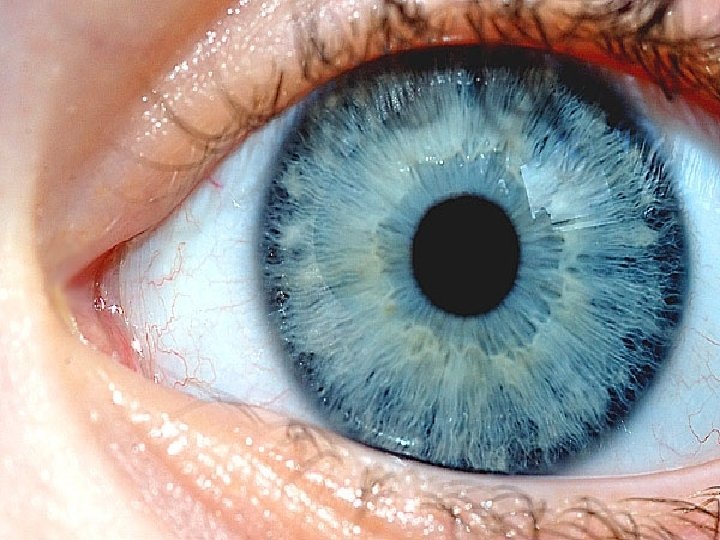
The Pupil The size of the pupil can be changed. This is done by a ring of muscles called the iris, which surrounds the pupil. The color of the iris muscles vary, which gives people different eye colors.
Dim Light Bright Light Iris muscles contract Iris muscles relax Pupil opens - dilates Pupil constricts Lets in more light Lets in less light
SEM – Iris Muscles
Step 2 Light passes through the lens, which further focuses the rays onto the retina.
The Lens The thickness of the lens can be changed. This lens can become thinner or thicker, depending on how far away an object is. This is done by ciliary muscles that pull the lens from above and from below.
Distant Objects Muscles contract Lens pulled thinner Focus shifts back Near Objects Muscles relax Lens made thicker Focus shifts forward
Step 3 The focused light rays hit a layer of lightsensitive cells found on the retina.
The Retina The retina is a layer of cells at the back of the eye that can detect light. Specialized cells on the retina called rod and cone cells detect light. Rods and cones sense different things about the light that hits them. Rod Cell Cone Cell
Rods and Cones Cone Cells At the centre of the retina 3 types: red, green & blue Detect color Rod Cells At the edges of the retina Only “see” black & white Detect brightness
The Fovea The fovea is a dip in the centre of the retina. Cone cells are concentrated in the fovea: most color vision occurs here. Cones concentrated at the fovea
The Fovea The fovea is a dip in the centre of the retina. Cone cells are concentrated in the fovea: most color vision occurs here. Rods found at the edges Cones concentrated at the fovea
From Eye To Brain Your brain “sees” differently than your eye. The optic nerve from the left eye connects to the right side of the brain, and vice versa. So, your brain sees everything reversed and then switches it again!
Summary 1. Light enters the eye through the cornea, a convex lens that focuses the light slightly. 2. It passes through a hole called the pupil, which is controlled by a ring of muscles called the iris. 3. The light passes through the convex lens, which focuses the light onto the retina. 4. The retina is a layer of light-sensitive cells: cones for color vision and rods for brightness 5. The rods and cones send electrical impulses to the brain along the optic nerve. 6. The brain interprets the signals as visual images.
Correcting Vision
Sight Defects Sometimes, people’s vision is not perfect. When light does not fall directly on the retina, images will appear blurry. This can happen in 2 major ways… Normal
Sight Defects Sometimes, people’s vision is not perfect. When light does not fall directly on the retina, images will appear blurry. The eye shape is not perfectly spherical. Long Short The lens muscles do not work This canproperly. happen in 2 major ways… Strong Weak
Nearsightedness occurs when light is focused in front of the retina. They see well near, but not far. Causes: Eye shape is too long Lens made too thick Fixed with a concave lens.
Nearsightedness
Farsightedness occurs when light is focused behind the retina. They see well far, but not near. Causes: Eye shape is too short Lens made too thin Fixed with a convex lens.
Farsightedness
Cataracts cause the lens to become cloudy. Cataracts are a genetic condition that run in families, especially African Americans. Cataracts cause a person to slowly lose their vision. Blindness can result.
Cataracts
Glaucoma High pressure in the eye causes glaucoma. Normally, fluid inside the eye is continually drained away by a drainage canal. eye fluid When this canal gets blocked, fluid builds up and pressure increases This pressure pushes on the lens, affecting vision
Glaucoma