The Endocrine System Prepared by Assoc Prof Dr

The Endocrine System Prepared by Assoc. Prof Dr Mohammad Iqbal Omar MBBS, DRM, M Med Program Kejuruteraan Bioperubatan Elektronik PPK Mekatronik, Uni. MAP iqbalomar@unimap. edu. my

Nervous and Endocrine Systems § Act together to coordinate functions of all body systems § Nervous system § Nerve impulses/ Neurotransmitters § Faster responses, briefer effects, acts on specific target § Endocrine system § Second-messenger system of the body § Hormone (chemical messengers )– mediator molecule released in one part of the body but regulates activity of cells in other parts § Slower responses, effects last longer, broader influence

§ 2 kinds of glands in body § Exocrine – ducted § Endocrine – ductless § Secrete products into interstitial fluid, diffuse into blood

§ Hormones control several major processes § Reproduction § Growth and development § Regulation of metabolism § Mobilization of body defenses § Maintenance of much of homeostasis

Hormone Overview § Hormones are produced by specialized cells § Cells secrete hormones into extracellular fluids § Blood transfers hormones to target sites § These hormones regulate the activity of other cells ( Target cells) § Endocrine gld. Hormone Target organs § Hormones = ‘to arouse’ in Greek

Hormonal action § Changes the permeability of the plasma membrane or its electrical state § Causes the synthesis of proteins (enzymes, regulatory proteins) § Activates or inactivates enzymes § Stimulate mitosis (cell growth or repair) § Promotion of secretory activity

The Chemistry of Hormones § Hormones are classified chemically as 1. Amino acid–based, which includes § Proteins § Peptides § Amines 2. Steroids—made from cholesterol e. g. . Sex hormones , hormones of adrenal cortex) 3. Prostaglandins—made from highly active lipids

Mechanisms of Hormone Action § Hormones affect only certain tissues or organs (target cells or target organs) § Target cells must have specific protein receptors § Hormone-binding alters cellular activity

The Chemistry of Hormones § Two mechanisms in which hormones act § Direct gene activation § Second-messenger system

1. Direct Gene Activation § Steroid Hormone Action & Thyroid hormone § Diffuse through the plasma membrane of target cells because they are lipid-soluble § Enter the nucleus § Bind to a specific protein within the nucleus § Bind to specific sites on the cell’s DNA § Activate genes that result in synthesis of new proteins

Steroid hormone Nucleus Cytoplasm Receptor protein Hormone-receptor complex DNA m. RNA New protein Plasma membrane of target cell Figure 9. 1 a

Steroid hormone Cytoplasm Nucleus Plasma membrane of target cell Figure 9. 1 a, step 1

Steroid hormone Cytoplasm Nucleus Plasma membrane of target cell Figure 9. 1 a, step 2

Steroid hormone Nucleus Cytoplasm Receptor protein Hormone-receptor complex Plasma membrane of target cell Figure 9. 1 a, step 3

Steroid hormone Nucleus Cytoplasm Receptor protein Hormone-receptor complex DNA Plasma membrane of target cell Figure 9. 1 a, step 4

Steroid hormone Nucleus Cytoplasm Receptor protein Hormone-receptor complex DNA m. RNA Plasma membrane of target cell Figure 9. 1 a, step 5

Steroid hormone Nucleus Cytoplasm Receptor protein Hormone-receptor complex DNA m. RNA New protein Plasma membrane of target cell Figure 9. 1 a, step 6

§ Steroid hormone

2. Second-Messenger System § Nonsteroid Hormone Action § Hormone binds to a membrane receptor § Hormone does not enter the cell § Sets off a series of reactions that activates an enzyme § Catalyzes a reaction that produces a second-messenger molecule § Oversees additional intracellular changes to promote a specific response
 Cytoplasm Enzyme ATP c. AMP Receptor protein Plasma membrane of")
Nonsteroid hormone (first messenger) Cytoplasm Enzyme ATP c. AMP Receptor protein Plasma membrane of target cell Second messenger Effect on cellular function, such as glycogen breakdown Figure 9. 1 b
 Cytoplasm Receptor protein Plasma membrane of target cell Figure 9.")
Nonsteroid hormone (first messenger) Cytoplasm Receptor protein Plasma membrane of target cell Figure 9. 1 b, step 1
 Cytoplasm Enzyme Receptor protein Plasma membrane of target cell Figure")
Nonsteroid hormone (first messenger) Cytoplasm Enzyme Receptor protein Plasma membrane of target cell Figure 9. 1 b, step 2
 Cytoplasm Enzyme ATP c. AMP Second messenger Receptor protein Plasma")
Nonsteroid hormone (first messenger) Cytoplasm Enzyme ATP c. AMP Second messenger Receptor protein Plasma membrane of target cell Figure 9. 1 b, step 3
 Cytoplasm Enzyme ATP c. AMP Receptor protein Plasma membrane of")
Nonsteroid hormone (first messenger) Cytoplasm Enzyme ATP c. AMP Receptor protein Plasma membrane of target cell Second messenger Effect on cellular function, such as glycogen breakdown Figure 9. 1 b, step 4

§ Non steroid Hormone

Control of Hormone Release § Hormone levels in the blood are mostly maintained by negative feedback § A stimulus or low hormone levels in the blood triggers the release of more hormone § Hormone release stops once an appropriate level in the blood is reached

Control of Hormone Release 1. Hormonal stimuli 2. Humoral stimuli 3. Neural stimuli

Hormonal Stimuli of Endocrine Glands § Most common stimuli § Endocrine glands are activated by other hormones § Examples: § Anterior pituitary hormones

Hormonal Stimuli of Endocrine Glands

Humoral Stimuli of Endocrine Glands § Changing blood levels of certain ions stimulate hormone release § Humoral indicates various body fluids such as blood and bile § Examples: § Parathyroid hormone § Calcitonin § Insulin

Humoral Stimuli of Endocrine Glands

Neural Stimuli of Endocrine Glands § Nerve impulses stimulate hormone release § Most are under the control of the sympathetic nervous system § Examples include the release of norepinephrine and epinephrine by the adrenal medulla

Neural Stimuli of Endocrine Glands

Major Endocrine Organs § Pineal gland § Hypothalamus § Pituitary gland § Thyroid gland § Parathyroid glands § Thymus gland § Adrenal glands § Pancreas § Gonads (Ovaries and Testes)

Major Endocrine Glands and Hormones

Major Endocrine Glands and Hormones

Major Endocrine Glands and Hormones

Major Endocrine Glands and Hormones

Pituitary Gland § Size of a pea § Hangs by a stalk from the hypothalamus in the brain § Protected by the sphenoid bone § Has two functional lobes § Anterior pituitary—glandular tissue § Posterior pituitary—nervous tissue § Often called the “master endocrine gland”

Hormones of the Anterior Pituitary § Six anterior pituitary hormones § Two affect non-endocrine targets § Growth hormone § Prolactin § Four stimulate other endocrine glands (tropic hormones) § Thyroid-stimulating hormone (thyrotropic hormone) (TSH) § Adrenocorticotropic hormone (ACTH) § Two gonadotropic hormones (Luteinizing hormone (LH) & Follicle stimulating hormone (FSH))

Hormones of the Anterior Pituitary § Characteristics of all anterior pituitary hormones § Proteins (or peptides) § Act through second-messenger systems § Regulated by hormonal stimuli, mostly negative feedback

Hormones of the Anterior Pituitary Figure 9. 4

Hormones of the Anterior Pituitary § Growth hormone § General metabolic hormone § Major effects are directed to growth of skeletal muscles and long bones § Plays a role in determining final body size § Causes amino acids to be built into proteins § Causes fats to be broken down for a source of energy

Hormones of the Anterior Pituitary GH : • protein synthesis • Breakdown of lipids • Glucose level • Somatic growth

Growth hormone
 disorders § Pituitary dwarfism results")
Hormones of the Anterior Pituitary § Growth hormone (GH) disorders § Pituitary dwarfism results from hyposecretion of GH during childhood § Gigantism results from hypersecretion of GH during childhood § Acromegaly results from hypersecretion of GH during adulthood

Growth Hormone Excess causes gigantism Too little causes dwarfism. This can be corrected today with synthetic hormone Injections.

Growth Hormone Gigantism Figure 9. 5 a

Growth Hormone Dwarfism Figure 9. 5 b
 § Stimulates and maintains milk production")
Hormones of the Anterior Pituitary § Prolactin (PRL) § Stimulates and maintains milk production following childbirth § Function in males is unknown § Adrenocorticotropic hormone (ACTH) Regulates endocrine activity of the adrenal cortex glucocorticoid secretion cortisol skin pigmentation
 § Influences growth and activity")
Hormones of the Anterior Pituitary § Thyroid-stimulating hormone (TSH) § Influences growth and activity of the thyroid gland ( T 4, T 3 secretions) § Gonadotropic hormones § Regulate hormonal activity of the gonads § Follicle-stimulating hormone (FSH) – Stimulates follicle development in ovaries – Stimulates sperm development in testes § Luteinizing hormone (LH) – Triggers ovulation of an egg in females – Stimulates testosterone production in males

Pituitary–Hypothalamus Relationship § Hormonal release is regulated by releasing and inhibiting hormones produced by the hypothalamus § Hypothalamus produces two hormones § These hormones are transported to neurosecretory cells of the posterior pituitary § Oxytocin § Antidiuretic hormone (ADH)

Hormones of the Posterior Pituitary § The posterior pituitary is not strictly an endocrine gland, but does release hormones § Oxytocin § Stimulates contractions of the uterus during labor, sexual relations, and breastfeeding § Causes milk ejection in a nursing woman
 § Inhibits urine production by")
Hormones of the Posterior Pituitary § Antidiuretic hormone (ADH) § Inhibits urine production by promoting water reabsorption by the kidneys § In large amounts, causes vasoconstriction leading to increased blood pressure § Also known as vasopressin

Hormones of the Posterior Pituitary Figure 9. 6

Summary of hormones of the Pituitary gland Anterior Pituitary Posterior Pituitary Hormone Target organ Major Physiological Effect Somatotropin HGH (human growth hormone) Liver, bones, and tissue Promotes growth (indirectly), control of protein, lipid and carbohydrate metabolism Thyroid-Stimulating Hormone TSH Thyroid Stimulates secretion of thyroid hormones Adrenocoricotropin (ACTH) Adrenal Cortex Stimulates secretion of glucocorticoids Prolactine Mammary Glands Milk production Luteinizing Hormone (LH) Ovary/Testes Control of reproductive function Follicle-Stimulating Hormone (FSH) Ovary/Testes Control of reproductive function Antidiuretic Hormone (ADH) Kidney Conservation of body water Oxytocin Ovary/Testes Stimulates milk ejection and uterine contractions

Thyroid Gland

Thyroid Gland § Thyroid hormone § Major metabolic hormone § Derived from Colloid of thyroid follicles § Every cell of body = target cells § Composed of two active iodine-containing hormones § Thyroxine (T 4)—secreted by thyroid follicles § Triiodothyronine (T 3)—conversion of T 4 at target tissues

Thyroid Gland Figure 9. 7 b

Thyroid Gland § Thyroid hormone disorders § Goiters § Thyroid gland enlarges due to lack of iodine § salt is iodized to prevent goiters § Cretinism § Caused by hyposecretion of thyroxine § Results in dwarfism during childhood

Thyroid Gland Figure 9. 8
 § Hypothyroidism § Myxedema § Results in physical and mental")
Thyroid hormone disorders (continued) § Hypothyroidism § Myxedema § Results in physical and mental sluggishness § Hyperthyroidism § Graves’ disease § Results in increased metabolism, heat intolerance, rapid heart beat, weight loss, and exophthalmos
")
Graves’ disease (Thyrotoxicosis)

Thyrotoxicosis

Normal Thyroid Gland

Graves’ disease

Thyroid Gland § Calcitonin § Decreases blood calcium levels by causing its deposition on bone § Antagonistic to parathyroid hormone § Produced by parafollicular cells § Parafollicular cells are found between the follicles

T 4 & T 3

Parathyroid Glands § Tiny masses on the posterior of the thyroid § Secrete parathyroid hormone (PTH)
 § Stimulate osteoclasts to remove calcium from bone § Stimulate the")
Parathyroid hormone (PTH) § Stimulate osteoclasts to remove calcium from bone § Stimulate the kidneys and intestine to absorb more calcium § Raise calcium levels in the blood

Hormonal Regulation of Calcium in Blood Calcitonin stimulates calcium salt deposit in bone Calcitonin Thyroid gland releases calcitonin Thyroid gland Rising blood Ca 2+ levels Imb ala nce Calcium homeostasis of blood 9– 11 mg/100 ml Imb ala nce Falling blood Ca 2+ levels Thyroid gland Osteoclasts degrade bone matrix and release Ca 2+ into blood Parathyroid glands PTH Parathyroid glands release parathyroid hormone (PTH)

Hormonal Regulation of Calcium in Blood Rising blood Ca 2+ levels Imb ala nce Calcium homeostasis of blood 9– 11 mg/100 ml Imb ala nce

Hormonal Regulation of Calcium in Blood Thyroid gland Rising blood Ca 2+ levels Imb ala nce Calcium homeostasis of blood 9– 11 mg/100 ml Imb ala nce

Hormonal Regulation of Calcium in Blood Calcitonin Thyroid gland releases calcitonin Thyroid gland Rising blood Ca 2+ levels Imb ala nce Calcium homeostasis of blood 9– 11 mg/100 ml Imb ala nce

Hormonal Regulation of Calcium in Blood Calcitonin stimulates calcium salt deposit in bone Calcitonin Thyroid gland releases calcitonin Thyroid gland Rising blood Ca 2+ levels Imb ala nce Calcium homeostasis of blood 9– 11 mg/100 ml Imb ala nce

Hormonal Regulation of Calcium in Blood Calcitonin Thyroid gland releases calcitonin Thyroid gland Rising blood Ca 2+ levels Calcium homeostasis of blood 9– 11 mg/100 ml Calcitonin stimulates calcium salt deposit in bone

Hormonal Regulation of Calcium in Blood Imb ala Falling blood Ca 2+ levels nce Calcium homeostasis of blood 9– 11 mg/100 ml Imb ala nce

Hormonal Regulation of Calcium in Blood Imb ala Falling blood Ca 2+ levels nce Calcium homeostasis of blood 9– 11 mg/100 ml Imb ala nce Thyroid gland Parathyroid glands

Hormonal Regulation of Calcium in Blood Imb ala Falling blood Ca 2+ levels nce Calcium homeostasis of blood 9– 11 mg/100 ml Imb ala nce Thyroid gland Parathyroid glands PTH Parathyroid glands release parathyroid hormone (PTH)

Hormonal Regulation of Calcium in Blood Imb ala Falling blood Ca 2+ levels nce Calcium homeostasis of blood 9– 11 mg/100 ml Imb ala nce Thyroid gland Parathyroid glands Osteoclasts degrade bone matrix and release Ca 2+ into blood PTH Parathyroid glands release parathyroid hormone (PTH)

Hormonal Regulation of Calcium in Blood Calcium homeostasis of blood 9– 11 mg/100 ml Thyroid gland Parathyroid glands Osteoclasts degrade bone matrix and release Ca 2+ into blood PTH Parathyroid glands release parathyroid hormone (PTH)

Hormonal Regulation of Calcium in Blood Calcitonin stimulates calcium salt deposit in bone Calcitonin Thyroid gland releases calcitonin Thyroid gland Rising blood Ca 2+ levels Imb ala nce Calcium homeostasis of blood 9– 11 mg/100 ml Imb ala nce Falling blood Ca 2+ levels Thyroid gland Osteoclasts degrade bone matrix and release Ca 2+ into blood Parathyroid glands PTH Parathyroid glands release parathyroid hormone (PTH)

Adrenal Glands § Sit on top of the kidneys § Two regions § Adrenal cortex—outer glandular region has three layers § Mineralocorticoids secreting area § Glucocorticoids secreting area § Sex hormones secreting area § Adrenal medulla—inner neural tissue region

Hormones of the Adrenal Cortex
 § Produced in outer adrenal")
Hormones of the Adrenal Cortex § Mineralocorticoids (mainly aldosterone) § Produced in outer adrenal cortex § Regulate mineral content in blood i. e. Na+ K+ § Regulate water and electrolyte balance § Target organ is the kidney (tubules) § Production stimulated by renin, Na+ K+ § Production inhibited by atrial natriuretic peptide (ANP) -hormone from heart

Hormones of the Adrenal Cortex
 § Produced in")
Hormones of the Adrenal Cortex § Glucocorticoids (include cortisone and cortisol) § Produced in the middle layer of the adrenal cortex § Promote normal cell metabolism § Blood glucose , Inflammation, Pain § Help resist long-term stressors § Released in response to increased blood levels of ACTH

Cortisone and Cortisol § Cortisol = 11 beta, 17 alpha, 21 -trihydroxy-4 -pregnene-3, 20 -dione, § Cortisone =17 alpha, 21 -dihydroxy-4 -pregnene-3, 11, 20 -trione. § The difference between cortisol and cortisone is that instead of the latter having a hydroxyl group at position 11, it has a ketone functional group instead § Cortisol is the more 'powerful' of the two, accounting for about 95% of the glucocorticoid activity in the body § Cortisone may be perceived as an inactive form of cortisol, although cortisone can be converted into cortisol if needed

CORTISOL

Hormones of the Adrenal Cortex § Sex hormones § Produced in the inner layer of the adrenal cortex § Small amounts are made throughout life § Mostly androgens (male sex hormones) are made but some estrogens (female sex hormones) are also formed

Adrenal Glands § Adrenal cortex disorders § Addison’s disease § Results from hyposecretion of all adrenal cortex hormones ( Mineralocorticoids , Glucocorticoids, Sex hormones ) § Bronze skin tone, muscles are weak, hypoglycemia, burnout, susceptibility to infection

Adrenal Glands § Adrenal cortex disorders § Hyperaldosteronism § May result from an ACTH-releasing tumor § Excess water and sodium are retained leading to high blood pressure and edema

Adrenal Glands § Adrenal cortex disorders § Cushing’s syndrome § Results from Pharmacological dose of glucocorticoids § “Moon face, ” “buffalo hump” on the upper back, high blood pressure, hyperglycemia, weakening of bones, depression of immune system

§ Masculinization § Results from hypersecretion of sex hormones § Beard and male distribution of hair growth

Secondary sexual features
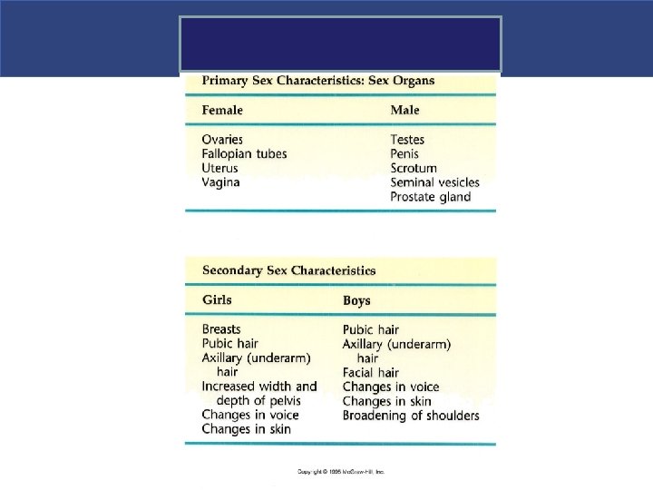
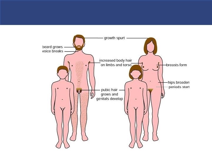

Male Secondary sexual features

Hormones of the Adrenal Medulla § Adrenal medulla is a modified sympathetic ganglion of autonomic nervous system § Produces two similar hormones (catecholamines) § Epinephrine (adrenaline) § Norepinephrine (noradrenaline) § Stimulated by sympathetic nervous system § Intensifies sympathetic responses
prepare the body to deal")
Hormones of the Adrenal Medulla § These hormones (catecholamines )prepare the body to deal with short-term stress (“fight or flight”) by § heart rate, § blood pressure, § blood glucose levels § Dilating small passageways of lungs

Epinephrine and norepinephrine

The Stress Response § Eustress in helpful stress / Distress is harmful § Body’s homeostatic mechanisms attempt to counteract stress § Stressful conditions can result in stress response or general adaptation syndrome (GAS) § Prolonged exposure to cortisol can result in wasting of muscles, suppression of immune system, ulceration of GI tract, and failure of pancreatic beta cells
 § Also called stress response § How")
Hormone Interactions § General Adaptation Syndrome (GAS) § Also called stress response § How body responds to stress-causing factors § Is divided into three phases: 1. Alarm phase ( Short Term response) 2. Resistance phase ( Long Term response) 3. Exhaustion phase Copyright © 2009 Pearson Education, Inc. , publishing as Pearson Benjamin Cummings Figure 18– 18

Role of Hypothalamus and adrenal glands in stress response

Roles of the Hypothalamus and Adrenal Glands in the Stress Response Short term Stress Hypothalamus

Roles of the Hypothalamus and Adrenal Glands in the Stress Response Short term Stress Hypothalamus Nerve impulses Spinal cord

Roles of the Hypothalamus and Adrenal Glands in the Stress Response Short term Stress Hypothalamus Nerve impulses Spinal cord Preganglionic sympathetic fibers Adrenal medulla

Roles of the Hypothalamus and Adrenal Glands in the Stress Response Short term Stress Hypothalamus Nerve impulses Spinal cord Preganglionic sympathetic fibers Adrenal medulla Catecholamines (epinephrine and norepinephrine) Short-term stress response

Roles of the Hypothalamus and Adrenal Glands in the Stress Response Short term Stress Hypothalamus Nerve impulses Spinal cord Preganglionic sympathetic fibers Adrenal medulla Catecholamines (epinephrine and norepinephrine) Short-term stress response 1. Increased heart rate 2. Increased blood pressure 3. Liver converts glycogen to glucose and releases glucose to blood 4. Dilation of bronchioles 5. Changes in blood flow patterns, leading to increased alertness and decreased digestive and kidney activity 6. Increased metabolic rate

Roles of the Hypothalamus and Adrenal Glands in the Stress Response Stress Hypothalamus More prolonged

Roles of the Hypothalamus and Adrenal Glands in the Stress Response Stress Hypothalamus Releasing hormone Corticotropic cells of anterior pituitary More prolonged

Roles of the Hypothalamus and Adrenal Glands in the Stress Response Stress More prolonged Hypothalamus Releasing hormone Corticotropic cells of anterior pituitary ACTH Adrenal cortex

Roles of the Hypothalamus and Adrenal Glands in the Stress Response Stress More prolonged Hypothalamus Releasing hormone Corticotropic cells of anterior pituitary ACTH Adrenal cortex Mineralocorticoids Long-term stress response

Roles of the Hypothalamus and Adrenal Glands in the Stress Response More prolonged Stress Hypothalamus Releasing hormone Corticotropic cells of anterior pituitary ACTH Mineralocorticoids Adrenal cortex Glucocorticoids Long-term stress response

Roles of the Hypothalamus and Adrenal Glands in the Stress Response More prolonged Stress Hypothalamus Releasing hormone Corticotropic cells of anterior pituitary ACTH Mineralocorticoids Adrenal cortex Glucocorticoids Long-term stress response 1. Retention of sodium and water by kidneys 2. Increased blood volume and blood pressure

Roles of the Hypothalamus and Adrenal Glands in the Stress Response More prolonged Stress Hypothalamus Releasing hormone Corticotropic cells of anterior pituitary ACTH Mineralocorticoids Adrenal cortex Glucocorticoids Long-term stress response 1. Retention of sodium and water by kidneys 2. Increased blood volume and blood pressure 1. Proteins and fats converted to glucose or broken down for energy 2. Increased blood sugar 3. Suppression of immune system

Roles of the Hypothalamus and Adrenal Glands in the Stress Response Short term More prolonged Stress Hypothalamus Nerve impulses Releasing hormone Corticotropic cells of anterior pituitary Spinal cord Preganglionic sympathetic fibers ACTH Adrenal medulla Mineralocorticoids Catecholamines (epinephrine and norepinephrine) Short-term stress response 1. Increased heart rate 2. Increased blood pressure 3. Liver converts glycogen to glucose and releases glucose to blood 4. Dilation of bronchioles 5. Changes in blood flow patterns, leading to increased alertness and decreased digestive and kidney activity 6. Increased metabolic rate Adrenal cortex Glucocorticoids Long-term stress response 1. Retention of sodium and water by kidneys 2. Increased blood volume and blood pressure 1. Proteins and fats converted to glucose or broken down for energy 2. Increased blood sugar 3. Suppression of immune system

Stress response The General Adaptation Syndrome.

Pancreatic Islets § The pancreas is a mixed gland has both endocrine and exocrine functions § Roughly 99% of cells produce digestive enzymes

Pancreas § The pancreatic islets produce hormones § Insulin—from beta cells § Allows glucose to cross plasma membranes into cells § Acts on all body cells, Hypoglycemic effect § Glucagon—from alpha cells § Allows glucose to enter the blood § These hormones are antagonists that maintain blood sugar homeostasis

Pancreatic Islets

Pancreatic Islets

Insulin-secreting cells of the pancreas activated; release insulin into the blood Uptake of glucose from blood is enhanced in most body cells Elevated blood sugar levels Stimulus: rising blood glucose levels (e. g. , after eating four jelly doughnuts) Blood glucose levels decline to set point; stimulus for insulin release diminishes Liver takes up glucose and stores it as glycogen Imb ala nce Homeostasis: Normal blood glucose levels (90 mg/100 ml) Imb ala nce Stimulus: declining blood glucose levels (e. g. , after skipping a meal) Low blood sugar levels Rising blood glucose levels return blood sugar to homeostatic set point; stimulus for glucagon release diminishes Liver breaks down glycogen stores and releases glucose to the blood Glucagon-releasing cells of pancreas activated; release glucagon into blood; target is the liver
")
Homeostasis: Normal blood glucose levels (90 mg/100 ml)
 Imb")
Stimulus: rising blood glucose levels (e. g. , after eating four jelly doughnuts) Imb ala nce Homeostasis: Normal blood glucose levels (90 mg/100 ml) Imb ala nce

Elevated blood sugar levels Stimulus: rising blood glucose levels (e. g. , after eating four jelly doughnuts) Imb ala nce Homeostasis: Normal blood glucose levels (90 mg/100 ml) Imb ala nce Figure 9. 15, step 3

Insulin-secreting cells of the pancreas activated; release insulin into the blood Elevated blood sugar levels Stimulus: rising blood glucose levels (e. g. , after eating four jelly doughnuts) Imb ala nce Homeostasis: Normal blood glucose levels (90 mg/100 ml) Imb ala nce

Insulin-secreting cells of the pancreas activated; release insulin into the blood Uptake of glucose from blood is enhanced in most body cells Elevated blood sugar levels Stimulus: rising blood glucose levels (e. g. , after eating four jelly doughnuts) Imb ala nce Homeostasis: Normal blood glucose levels (90 mg/100 ml) Imb ala nce

Insulin-secreting cells of the pancreas activated; release insulin into the blood Uptake of glucose from blood is enhanced in most body cells Elevated blood sugar levels Stimulus: rising blood glucose levels (e. g. , after eating four jelly doughnuts) Liver takes up glucose and stores it as glycogen Imb ala nce Homeostasis: Normal blood glucose levels (90 mg/100 ml) Imb ala nce

Insulin-secreting cells of the pancreas activated; release insulin into the blood Elevated blood sugar levels Stimulus: rising blood glucose levels (e. g. , after eating four jelly doughnuts) Uptake of glucose from blood is enhanced in most body cells Liver takes up glucose and stores it as glycogen Homeostasis: Normal blood glucose levels (90 mg/100 ml) Blood glucose levels decline to set point; stimulus for insulin release diminishes
 Imb ala nce")
Imb ala nce Homeostasis: Normal blood glucose levels (90 mg/100 ml) Imb ala nce Stimulus: declining blood glucose levels (e. g. , after skipping a meal)
 Imb ala nce")
Imb ala nce Homeostasis: Normal blood glucose levels (90 mg/100 ml) Imb ala nce Stimulus: declining blood glucose levels (e. g. , after skipping a meal) Low blood sugar levels
 Imb ala nce")
Imb ala nce Homeostasis: Normal blood glucose levels (90 mg/100 ml) Imb ala nce Stimulus: declining blood glucose levels (e. g. , after skipping a meal) Low blood sugar levels Glucagon-releasing cells of pancreas activated; release glucagon into blood; target is the liver
 Imb ala nce")
Imb ala nce Homeostasis: Normal blood glucose levels (90 mg/100 ml) Imb ala nce Stimulus: declining blood glucose levels (e. g. , after skipping a meal) Low blood sugar levels Liver breaks down glycogen stores and releases glucose to the blood Glucagon-releasing cells of pancreas activated; release glucagon into blood; target is the liver Figure 9. 15, step 11
 Stimulus: declining blood glucose levels (e.")
Homeostasis: Normal blood glucose levels (90 mg/100 ml) Stimulus: declining blood glucose levels (e. g. , after skipping a meal) Low blood sugar levels Rising blood glucose levels return blood sugar to homeostatic set point; stimulus for glucagon release diminishes Liver breaks down glycogen stores and releases glucose to the blood Glucagon-releasing cells of pancreas activated; release glucagon into blood; target is the liver

Insulin-secreting cells of the pancreas activated; release insulin into the blood Uptake of glucose from blood is enhanced in most body cells Elevated blood sugar levels Stimulus: rising blood glucose levels (e. g. , after eating four jelly doughnuts) Blood glucose levels decline to set point; stimulus for insulin release diminishes Liver takes up glucose and stores it as glycogen Imb ala nce Homeostasis: Normal blood glucose levels (90 mg/100 ml) Imb ala nce Stimulus: declining blood glucose levels (e. g. , after skipping a meal) Low blood sugar levels Rising blood glucose levels return blood sugar to homeostatic set point; stimulus for glucagon release diminishes Liver breaks down glycogen stores and releases glucose to the blood Glucagon-releasing cells of pancreas activated; release glucagon into blood; target is the liver
 § Type 1 D.")
Diabetes Mellitus § Abnormally high level of blood glucose (Hyperglycemia) § Type 1 D. M- Reduced production of insulin § Type 2 D. M- Reduced response by body to insulin ( insulin resistant ) § Symptoms - excessive thirst, excessive urination, loss of weight § Since cells can not used glucose , fats and even proteins are broken down to meet energy requirements of body which results in ketoacidosis, decreased ability to fight infection

Pineal Gland § Found on the third ventricle of the brain § Secretes melatonin § Helps establish the body’s wake and sleep cycles § More melatonin liberated during darkness than light § Believed to coordinate the hormones of fertility in humans

Location of Major Endrocrine Organs Figure 9. 3

Thymus Gland § Located posterior to the sternum § Largest in infants and children § Produces thymosin § Essential for T cell maturation § Important in developing the immune system

Gonads § Ovaries § Produce eggs § Produce two groups of steroid hormone § Estrogens § Progesterone § Testes § Produce sperm § Produce androgens, such as testosterone

Location of Major Endrocrine Organs Figure 9. 3

Hormones of the Ovaries § Estrogens § Stimulate the development of secondary female characteristics § Mature female reproductive organs § With progesterone, estrogens also § Promote breast development § Regulate menstrual cycle

Hormones of the Ovaries § Progesterone § Acts with estrogen to bring about the menstrual cycle § Helps in the implantation of an embryo in the uterus § Helps prepare breasts for lactation § Release is controlled by Ant. Pituitary hormones ( FSH & LH)

Hormones of the Testes § Produce several androgens § Testosterone is the most important androgen § Responsible for adult male secondary sex characteristics ( facial hair, heavy bones & muscles , voice changes) § Promotes growth and maturation of male reproductive system § Required for sperm cell production
 §")
Other Hormone-Producing Tissues and Organs § Parts of the small intestine (CCK, Secretin) § Parts of the stomach ( Gastrin) § Kidneys (Erythropoietin) § Heart ( Atrial Natriuretic Peptide) § Many other areas have scattered endocrine cells
")
Other Hormone-Producing Tissues and Organs Table 9. 2 (1 of 2)
")
Other Hormone-Producing Tissues and Organs Table 9. 2 (2 of 2)

Endocrine Function of the Placenta § Produces hormones that maintain the pregnancy § Some hormones play a part in the delivery of the baby § Produces human chorionic gonadotropin (h. CG) in addition to estrogen, progesterone, and other hormones

Developmental Aspects of the Endocrine System § Most endocrine organs operate smoothly until old age § Menopause is brought about by lack of efficiency of the ovaries § Problems associated with reduced estrogen are common § Growth hormone production declines with age § Many endocrine glands decrease output with age

Age-related changes in hormones & their effects § Age-related changes to the hormone system are generally not sufficient to prevent it from functioning adequately. The most serious hormone deficiencies tend to be pathological rather than age-related 1. Sex hormones Levels of the sex hormones estrogen and progesterone in women, and testosterone in men, fall. 2. Pancreas Reduced insulin production with age 3. Growth hormone (GH) levels fall- reduced muscle mass, Poor resistant to stress and infection 4. Reduced thyroid hormone production - hypothyroidism

Thank you
- Slides: 152