The Ear Hearing and Balance Exercise 17 Activities

The Ear, Hearing and Balance Exercise 17: Activities 9, 11, 12, 13 Page 204 -209

Objectives: 1. Distinguish the three main divisions of the ear 2. Identify the primary structures of the ear on models 3. Understand the physiology of the ear and how it perceives sound and senses equilibrium 4. Demonstrate some clinical screening tests for hearing and equilibrium 5. Using the microscope, analyze the microscopic structure of the cochlea
, vestibular nerve • External Ear: pinna/auricle, •")
• vestibule (utricle, saccule, maculae, otoliths), vestibular nerve • External Ear: pinna/auricle, • semicircular canals (ampulla, external auditory meatus, cupula, crista ampullaris), vestibular nerve tympanic membrane • microscopic anatomy of the • Middle Ear: auditory ossicles: cochlea (fig. 17. 10, scala vestibuli, malleus, incus, stapes, oval vestibular membrane, cochlear window, pharyngotympanic duct, spiral organ (of Corti), tectorial membrane, hair cells, (auditory) tube, round window basilar membrane, scala tympani, • Inner Ear: bony labyrinth, spiral ganglion of cochlear nerve, membranous labyrinth, perilymph, cochlear nerve) endolymph • ceruminous glands • cochlea (scala vestibuli, cochlear • acuity duct, spiral organ (of Corti), scala • Weber test • Rinne test tympani, basilar membrane, • Romberg test tectorial membrane, vestibular • balance test membrane), cochlear nerve • nystagmus Terms to know:

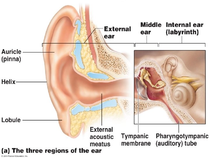
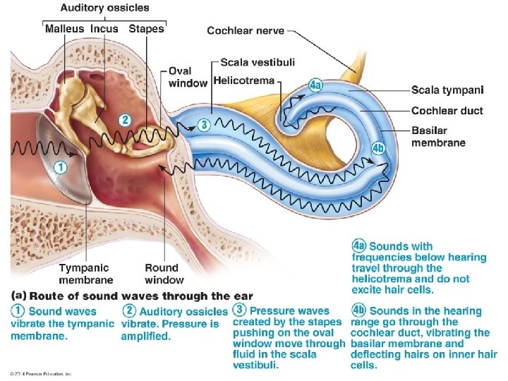
9: Identifying Structures of the Ear • Outer and middle ear for hearing only, inner ear for hearing and equilibrium • Outer ear – Auricle or pinna – External acoustic meatus • Ceruminous glands secrete earwax • Tympanic membrane (eardrum) vibrates to transmit sound waves from outer to middle ear
 is an air")
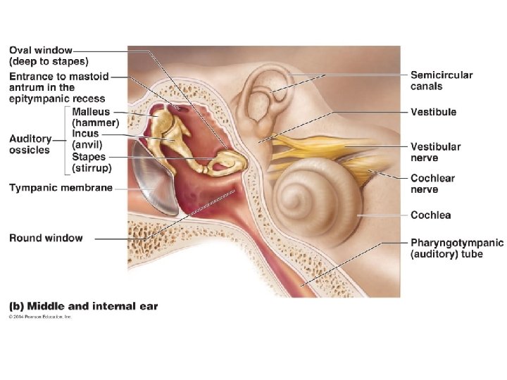
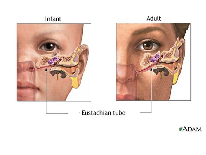
9: Identifying Structures of the Ear • Middle ear (tympanic cavity) is an air filled chamber in temporal bone – Contains auditory ossicles: malleus, incus, and stapes that transmit vibration to oval window – Pharyngotympanic (auditory) tube connects middle ear to nasopharynx • Used to equalize air pressure by “popping” your ears
 is")
9: Identifying Structures of the Ear • Inner ear (osseous or bony labyrinth) is filled with perilymph – Contains membranous labyrinth filled with endolymph • 3 subdivisions – Cochlea – Vestibule – Semicircular canals


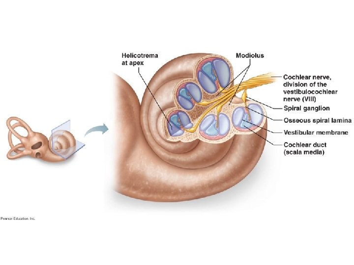
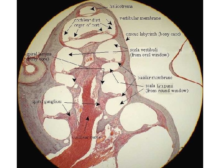
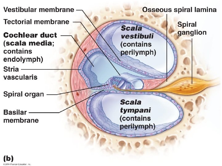
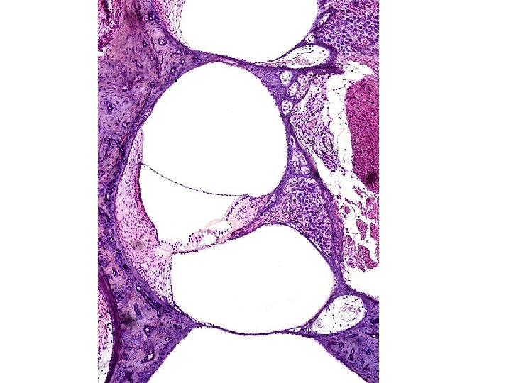
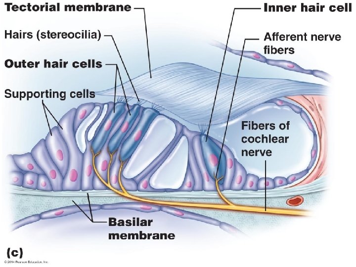
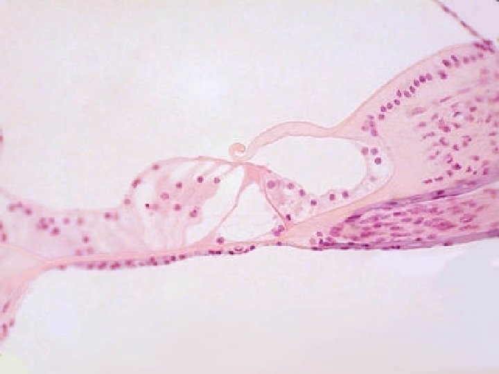
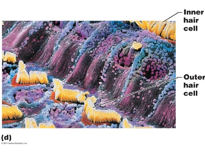
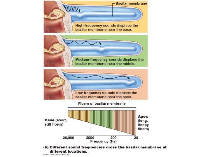
11: Examining the Microscopic Structure of the Cochlea • Stapes “footplate” vibrates oval window, which connects to scala vestibuli full of perilymph • Vibrations travel through the vestibular membrane to the endolymph filled 1. 5 inch long cochlear duct (scala media) below – Spiral organ (of Corti) contains “hair cells” (stereocilia) for hearing reception imbedded in tectorial membrane which stimulate the vestibulocochlear (VIII) nerve – High frequency sound waves resonate close to the oval window while low frequency sound waves peak closer to the apex of the cochlea • Vibrations travel through basilar membrane below to the lower chamber, the scala tympani and dampen out on the “round window”

12: Conducting Laboratory Test for Hearing • Acuity test-distance • Sound localization • Weber Test is used to determine conductive and sensorineural deafness • Rinne Test for comparing bone- and airconduction hearing

Anatomy of Equilibrium Apparatus and Mechanisms of Equilibrium • Vestibule contains the saclike utricle and saccule • Semicircular canals contain the membranous semicircular ducts • Both: – Are suspended in perilymph – Are filled with endolymph – Contain receptor “hair cells” that are activated by disturbance of cilia and stimulate the vestibulocochlear (VIII) nerve

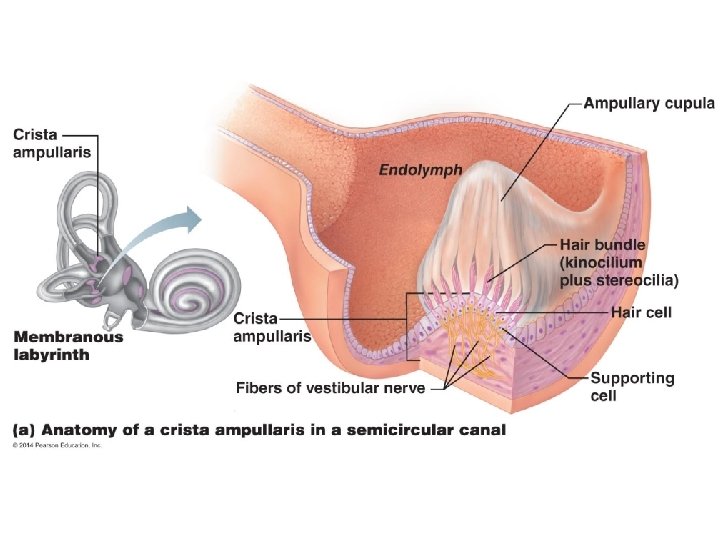
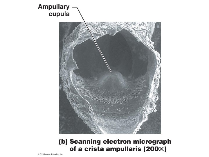
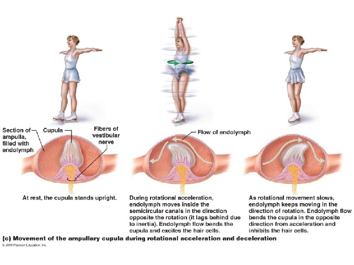
Semicircular Canals • ½ inch circumference • Oriented in the three planes of space • Enlarged ampulla at the base of each contains a tuft of receptor hair cells referred to as the crista ampullaris topped with a gelatinous “ampullary cupula” – Perceive dynamic equilibrium in response to changes in angular motion – As cupula is moved by flow of endolymph, hair cells cilia are stimulated, which in turn stimulated the vestibular portion of the vestibulocochlear (VIII) nerve

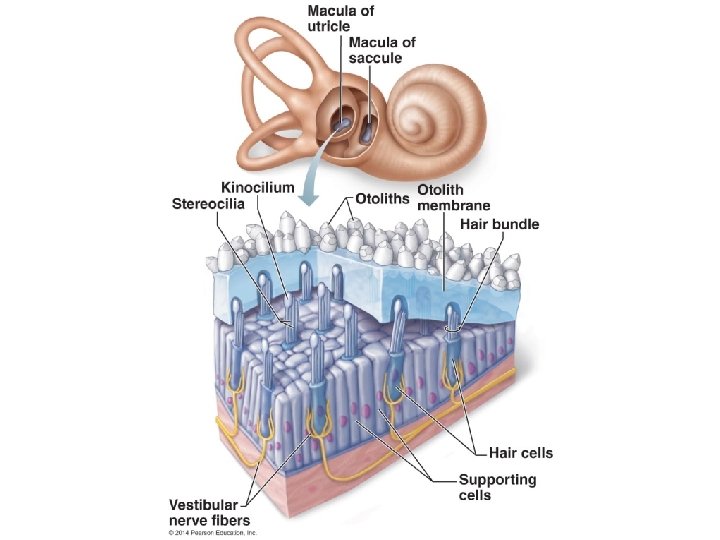
Vestibule • Utricle and saccule membranous sacs contain maculae, static equilibrium receptors that respond to gravity and linear changes in speed • Maculae “hair cell” cilia are covered in otolith membrane containing calcium carbonate otoliths – Otolith inertia bends cilia on hair cells, which stimulates the vestibular portion of the vestibulocochlear (VIII) nerve

13: Conducting Laboratory Test on Equilibrium • Balance Test – Nystagmus is the involuntary rolling of the eyes after rotation • Romberg Test to assess equilibrium and posture
")
For Review Complete pages 219 -222 #20 -32 All Mastering A&P assignments (8 -11) are due on Saturday at 9: 59 p. m.
- Slides: 31