The Digestive System The Gastrointestinal System consists of


 coordinate movements – innervated by the ANS")


 § Lasts")


 and Gastric")


 § Trypsinogen,")


- Slides: 81
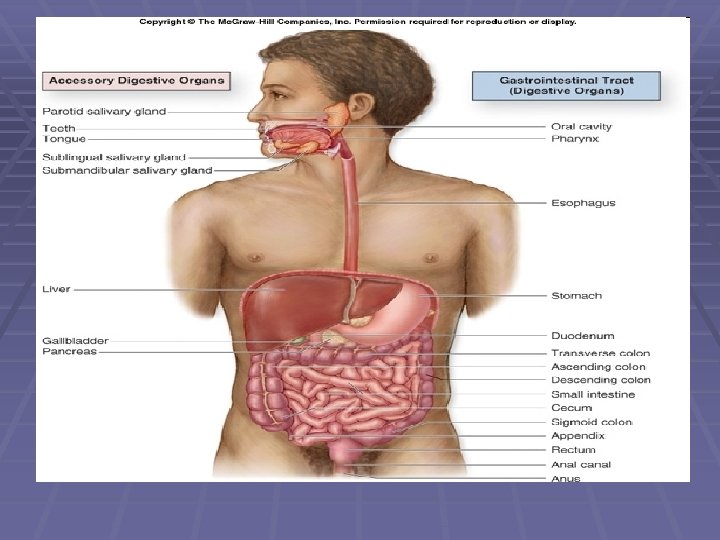
The Digestive System The Gastrointestinal System consists of the: v. Digestive Tract v. Accessory Organs • • • Oral Cavity Pharynx Esophagus Stomach Small Intestines Large Intestines Teeth Tongue Salivary Glands Liver Pancreas
General Functions 1. 2. 3. 4. 5. 6. Ingestion Mechanical Processing Digestion Secretion Absorption Excretion
The Digestive Organs and the Peritoneum § Peritoneal cavity found within the abdominopelvic cavity, lined by a serous membrane (mesothelium and areolar tissue) § Serous membrane – Serosa (visceral peritoneum) & Parietal Peritoneum § Peritoneal fluid separates the two layers
The Digestive Organs and the Peritoneum § MESENTERIES § Sheets of serous membrane that suspend portions of the digestive tract within the peritoneal cavity § Double sheets of peritoneal membrane § Provides access routes for blood vessels, lymphatic vessels and nerves § Stabilizes position of attached organs
Peritoneum and Mesenteries
Mesenteries
Mesenteries § Digestive tract & Accessory Organs suspended by dorsal and ventral mesenteries in embryonic life
Mesenteries § Ventral Mesentery > Lesser Omentum & Falciform Ligament § Dorsal Mesentery > Greater Omentum
Lesser and Greater Omentum
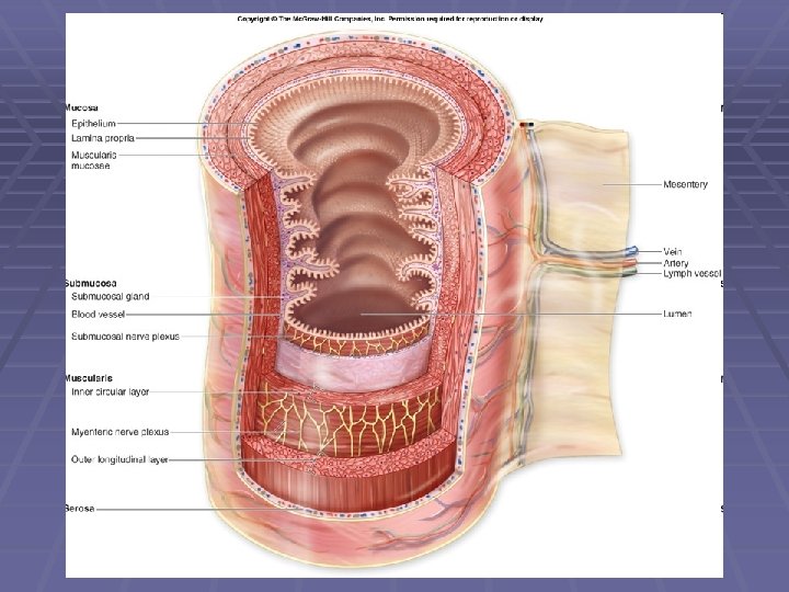
Layers of the wall of the Digestive Tract 1. 2. 3. 4. Mucosa Submucosa Muscularis Externa Serosa
Mucosa Epithelial Layer & Lamina Propria Epithelial Layer § The layer is folded to facilitate a large surface area for digestion. § The epithelium may be simple or stratified. § Stratified Squamous- Oral Cavity, Pharynx and Esophagus § Simple Columnar- Stomach, Small Intestine and almost all the length of the large intestine.
Lamina Propria • • Loose connective tissue Blood vessels Sensory nerve endings Lymphatic vessels Smooth muscle cells Scattered lymphoid tissues Secretory cells of mucus glands Muscularis Mucosae (circular and longitudinal)
Submucosa v. A layer of dense connective tissues that lies below the mucosa consisting of: • • Blood vessels Lymphatic Vessels Exocrine glands - buffers and enzymes Sub mucosal plexus/Plexus of Meissner • Sensory neurons, parasympathetic ganglionic neurons and sympathetic postganglionic fibers innervating mucosa and submucosa
Muscularis Externa § Dominated by smooth muscle cells § Forms an inner circular and outer longitudinal layer § Contraction facilitates peristalsis and segmentation
Muscularis Externa § Enteric Nervous System (ENS) coordinate movements – innervated by the ANS § Sympathetic fibers also innervate mucosa and Myenteric Plexus § Myenteric Plexus lies between circular and longitudinal muscle § Parasympathetic stimulation increases muscle tone and activity
Serosa/Adventitia § Found along most portions of tract except in the oral cavity, pharynx, esophagus and rectum. § The areas consist of a dense network of collagen fibers which attaches the digestive tract to the adjacent structures.
Control of Digestive Functions § Neural Mechanisms § CNS and Enteric Nervous System § Hormonal Mechanisms § Enteroendocrine cells § Local Mechanisms § Release of chemicals in interstitial fluid
Digestive Organs and Their Functions v Oral Cavity v Pharynx v Esophagus v Stomach v Small Intestine v Large Intestine
Oral Cavity Functions: ØAnalysis of material before swallowing ØMechanical processing by teeth, tongue and palate surfaces ØLubrication with mucus and salivary secretions ØLimited digestion of carbohydrates and lipids (Salivary Amylase & Lingual Lipase)
Salivary Glands
Salivary Gland v. Submandibular Glands -buffers, mucins & salivary amylase v. Sublinguals -contains mucus cells v. Parotid Glands -contains only serous cells –salivary amylase
Saliva ØFunctions Ø Helps keep oral surfaces clean Ø Moistening and lubricating of mouth and food. Ø Aids in tasting. Ø Aids in Swallowing. Ø Helps in the metabolism of carbohydrate. Ø Helps to maintain the calcium phosphate matrix of the teeth.
Four Major Components of Saliva: 1. 2. 3. 4. Mucus Amylase Lingual Lipase Alkaline Electrolyte Solution Note: Saliva contains thiocyanates and lysozymes that can attack and destroy the mouth bacteria. It also contains antibodies that destroy oral bacteria in certain people.
Control of Salivary Secretions § ANS control § PNS increases secretion § SNS produces small amounts of thick saliva § Other brain stem nuclei & higher centers § Chewing gum § Smelling or thinking about food § Irritating stimuli in esophagus, stomach & intestines
Pharynx
Esophagus § A hollow muscular tube that transfers solid food and liquids to the stomach. § Extends from cricoid cartilage, along posterior surface of trachea, through diaphragm to stomach § Esophageal/Cardiac Sphincter Muscles
Histology of Esophagus § Mucosa – nonkeratinized stratified squamous epithelium § Mucosa and submucosa thrown into folds § Submucosa contains mucus secreting glands
Esophagus
The Mechanism of Swallowing § Consists of three phases: § Oral Phase § Pharyngeal Phase § Esophageal Phase
The Mechanism of Swallowing § Oral Phase § Voluntary Phase § Hard palate compresses bolus § Tongue forces bolus into oropharynx § Soft palate elevated
The Mechanism of Swallowing § Pharyngeal Phase § Bolus enters pharynx § Tactile receptors on palatal arches and uvula stimulated by bolus § Swallowing center of medulla oblongata receives information § Pharyngeal muscles stimulated to contract § Larynx elevates § Respiratory centers inhibited
The Mechanism of Swallowing § Esophageal Phase § Bolus enters esophagus § Bolus pushed to stomach by peristalsis § Cardiac sphincter muscles open § Bolus enters stomach
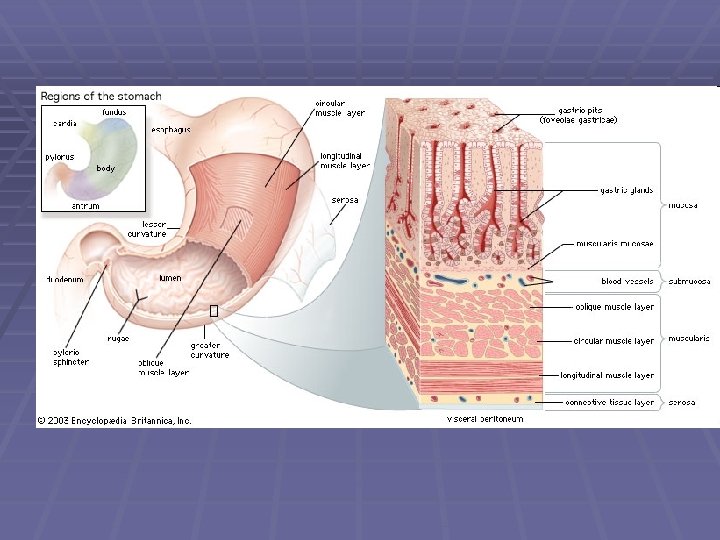
The Stomach Functions: ØBulk storage of ingested food. ØMechanical breakdown of ingested food. ØDisruption of chemical bonds in food. ØProduction of intrinsic factor (for vitamin B 12 absorption).
The Stomach
The Stomach Histology § Epithelium – simple columnar with goblet cells § Gastric pits open onto gastric surface § Gastric glands below gastric pits in fundus and body
Gastric Glands Consist of: Parietal Cells – Intrinsic factor & HCl Chief Cells Pepsinogen
Parietal Secretion Of HCl
Hydrochloric Acid § Functions: § Kills microbes. § Denatures proteins and inactivates most enzymes in food. § Breaks down plant cell walls and connective tissues in meat. § Provides an acidic environment for activation and function of pepsin.
Chief Cells § Secrete pepsinogen § Pepsinogen is converted to pepsin in the gastric lumen by HCl (p. H 1. 5 – 2. 0) § Rennin and Gastric Lipase produced in infants
Pyloric glands § Produce: § Mucus § Hormones by Enteroendocrine cells § Gastrin by G cells – stimulates parietal and chief cells and promotes gastric mixing § Somatostatin by D cells – inhibits gastrin release; overridden by neural & hormonal stimuli
Regulation of Gastric Activity § Controlled by CNS § Regulated by short reflexes of the ENS in wall of stomach § Regulated by hormones in the digestive tract
Regulation of Gastric Activity v There are three phases of Gastric Control: 1. 2. 3. Cephalic Phase Gastric Phase Intestinal Phase 1. Cephalic Phase -Begins before food enters the stomach, that is when we see, smell, think or taste food. 2. Gastric Phase -The presence of food in the stomach stimulates gastric secretions. 3. Intestinal Phase -Distension and presence of protein fragments in the duodenum trigger the release of gastrin.
Regulation of Gastric Activity § Cephalic Phase § Directed by CNS (Medulla) § Lasts one minute § Neural output via PNS and vagus nerves innervate submucosal plexus § Postganglionic parasympathetic fibers innervate mucous cells, chief cells, parietal cells and G cells § Gastric juice production accelerates (500 ml/hr) (Phase affected by emotional states)
Regulation of Gastric Activity Gastric Phase § Initiated by – stomach distension, increased p. H in stomach and undigested proteins in stomach § 3 – 4 hours
Mechanisms of Gastric Phase § Neural Response § Stretch receptors and chemoreceptors stimulated § Short reflexes triggered in submucosal and myenteric plexuses § Postganglionic fibers leaving submucosal plexus stimulate the release of Ach which stimulates chief and parietal cells § Myenteric plexus stimulation produce mixing waves
Mechanisms of Gastric Phase § Hormonal Response § Neural stimulation and the presence of peptides and amino acids stimulate Gastrin secretion § Gastrin accelerates parietal and chief cell secretion rates § Gastrin stimulates gastric motility
Mechanisms of Gastric Phase § Local Response § Distention of gastric wall stimulates the release of histamine in the lamina propria § Histamine binds to receptors on parietal cells
Regulation of Gastric Activity Intestinal phase Neural Responses § Chyme leaves the stomach relieving stomach distension § Stretch and chemo- receptors in duodenum stimulated by the presence of chyme § Enterogastric reflex – inhibits central and local stimulation of gastrin production & gastric contractions; also stimulating contraction of the pyloric sphincter § Local reflexes stimulate mucus production at the duodenum
Regulation of Gastric Activity Intestinal phase § Hormonal Responses § Cholecystokinin (CCK) and Gastric Inhibitory Peptide (GIP) secreted when lipids and carbohydrates enter the duodenum § CCK –inhibits acid and pepsin secretion § GIP – targets the pancreas and inhibits gastric secretion and reduces rate and force of gastric contractions
Regulation of Gastric Activity Intestinal phase § Hormonal Responses § Secretin secreted when p. H drops below 4. 5 § Secretin inhibits parietal and chief cell activity § Stimulates production of bicarbonate ions by the pancreas and bile by the liver
Regulation of Gastric Activity Intestinal phase § Hormonal Responses § Gastrin is produced by G cells in duodenum when partially digested proteins enter the duodenum § Accelerates acid and enzyme secretion in stomach
§ High fat, high protein meals slow gastric emptying § Large, low fat, low protein, wine and caffeine increases gastric emptying § Wine and caffeine increases gastric secretion and motility
Digestion and Absorption in the Stomach üProteins break down in the presence of pepsin to form polypeptides. üCarbohydrate and lipid digestion continue until p. H throughout the material in the stomach falls below 4. 5. üThe only substances absorbed by the stomach are fatsoluble (incl. alcohol and aspirin) in small amounts. NB: The stomach lacks the absorptive surfaces contained in the small intestine.
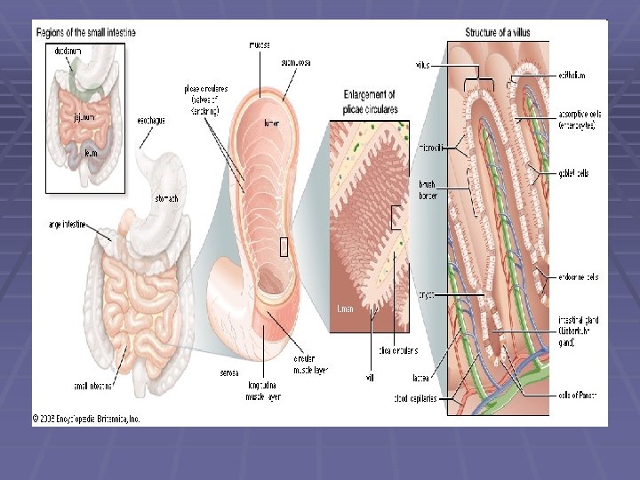
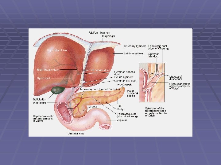
Small Intestines and Accessory Organs v Extends from the pyloric sphincter to the cecum. (Approx. 2. 5 cm in diameter & 7 m long) Ø Consists of three parts: § DUODENUM § JEJUNUM § ILEUM § NB -9 O% of absorption occurs in the small intestine. v Enzymes are secreted into the small intestines by the pancreas. Ø These are: § Pancreatic Amylase § Pancreatic Lipase § Nucleases § Proteolytic Enzymes § The liver and gall bladder secrete bile into the small intestine.
Histology v. Structural features of the Small intestines: § Plicae circulares § Villi § Microvilli v. Components of the villi: § Lamina propria has capillaries that transports gases and absorbed nutrients to the hepatic portal vein. § Lacteals transport substances too large to diffuse into the bloodstream eg. Chylomicrons (protein-lipid packages). § Muscularis mucosae and smooth muscle of intestinal villi move villi for maximum absorption of nutrients and squeeze lacteals therefore moving lymph out of villi
Histology v. Structure of Duodenum, Jejunum and ileum. ØDuodenum- has few plicae, numerous villi and mucous glands ØJejunum- has plicae and villi (prominent in its proximal portion). Øileum- has scattered villi but lacks plicae altogether; lymphoid tissue (Peyer’s Patches) at terminal portion
Intestinal Secretion § Intestinal secretions contain § Mucus § Brush border enzymes § Enterokinase § Maltase § Sucrase § Lactase § Dipeptidases § Peptidases
Intestinal Secretion § Intestinal secretion is controlled by § Local reflexes § Enterocrinin secretion § Parasympathetic stimulation
Intestinal Movements § Myenteric reflexes stimulate weak peristaltic contractions § PNS stimulation accelerates peristalsis and segmentation § Affects short segments of small intestine
Intestinal Movements § Two reflexes coordinate activities along the entire length of the small intestine: § Triggered by stretch receptors in stomach § Gastroenteric reflex stimulates motility and secretion along entire small intestine § Gastroileal reflex stimulates relaxation of ileocecal valve; enhanced by gastrin secretion
Digestion in the Small Intestine The Pancreas v. When acidic chyme enters the duodenum, pancreatic juice via the pancreatic duct enters as well. v. Functions of the pancreas v. Endocrine cells of the pancreatic islets produce insulin and glucagon v. Exocrine cells including acinar and epithelial duct cells produce pancreatic juice
Digestion in the Small Intestine The Pancreas ØPancreatic juice consists of: § Sodium bicarbonate (p. H 7. 5 -8. 8) § Buffer solution secreted when chyme enters duodenum and secretin produced § PANCREATIC ENZYMES: § CHOLECYSTOKININ stimulates their production
Digestion in the Small Intestine The Pancreas § PANCREATIC ENZYMES § Pancreatic amylase – carbohydrates to triand di- saccharides § Pancreatic lipase – lipids to fatty acids and monoglycerides § Nucleases – nucleic acids to simple sugars and nitrogen bases
Digestion in the Small Intestine The Pancreas § PANCREATIC ENZYMES (Proteolytic enzymes) § Trypsinogen, chymotrypsinogen, § § § procarboxypeptidase and proelastase In the duodenum enterokinase converts trypsinogen to trypsin Trypsin activates the other enzymes to chymotrypsin, carboxypeptidase and elastase These enzymes produce dipeptides, tripeptides and amino acids from proteins
Digestion in the Small Intestine The Liver § The liver has numerous functions which may be categorised as: § Metabolic regulation § Carbohydrate, lipid and amino acid metabolism, waste product removal, vitamin & mineral storage, drug inactivation § Hematological regulation § Bile production
Digestion in the Small Intestine The Liver § Functions of Bile § Emulsification of fats – increase surface area for pancreatic lipase to work § Facilitates absorption of lipids by intestinal epithelium
Digestion in the Small Intestine The Gallbladder § Stores bile § Concentrates bile
End of Digestion in Small Intestines § Brush border Enzymes complete digestion § Maltase – maltose to glucose § Sucrase – sucrose to glucose and fructose § Lactase – lactose to glucose and galactose § Dipeptidases and Peptidases – dipeptides and tripeptides to amino acids
Absorption in the Small Intestine v. Substances absorbed in the small intestines are: § Monosaccharides-absorbed by the duodenum and upper jejunum; co-transport with sodium § Amino Acids- absorbed by the end of the jejunum; co-transport with sodium and facilitated diffusion
Absorption in the Small Intestine § Fatty acids and Monoglycerides § absorbed in the duodenum, jejunum and ileum § diffusion in water-soluble micelles which diffuse into epithelial cells of villi § Fatty acids and monoglycerides combine to form triglycerides § Triglycerides combine with cholesterol, lipoprotein and phospholipids to form chylomicrons § Chylomicrons then diffuse into lacteals
Absorption in the Small Intestine § Vitamins - C and B by passive diffusion - B 12 with intrinsic factor active transport - A, D, E & K with micelles
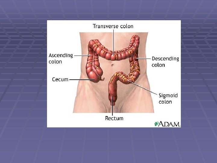
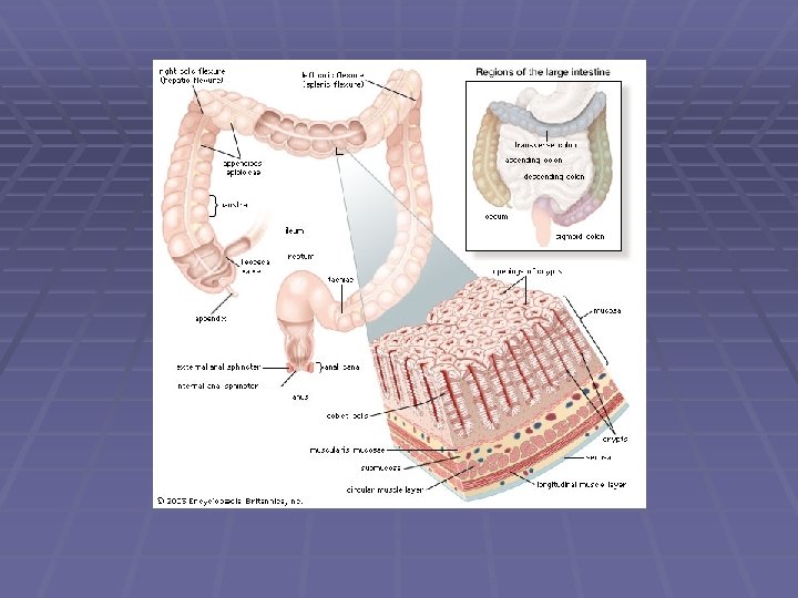
Large Intestine v. Parts of the Large Intestine • Cecum • Colon - Ascending, Transverse, Descending and Sigmoid • Rectum • Anal canal
Large Intestine § Simple columnar epithelium with goblet cells § Mucus § Protects intestinal wall and holds fecal matter together § Contains sodium bicarbonate that neutralizes acids produced by bacterial metabolism
Large Intestine § Produces large amounts of water and electrolytes when irritated § Movement of fecal matter is due to peristalsis, segmentation and contraction of longitudinal muscle bands